The Uterine And Cervical Ligaments
This article or area is currently under construction and may only be partially complete. Please come back soon to see the finished work! (14/06/2020)
Original Editor - Khloud Shreif
Top Contributors - Khloud Shreif, Lucinda hampton, Safiya Naz and Temitope Olowoyeye
Description[edit | edit source]
The structures of the internal genitalia/ female reproductive system is supported by the pelvic floor musculature, pelvic fascia, and ligaments on both sides of the uterus. These ligaments divided into, uterine ligaments that are soft and lax, having a limited role in supporting the uterus and internal genitalia, unlike the cervical ligaments which are tough, non-extensible, and condensed thinking of pelvic tissue.
Uterine Ligaments[edit | edit source]
Broad Ligament[1][edit | edit source]
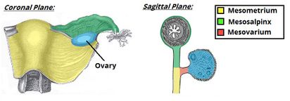
The broad ligament is a double flat peritoneum sheet extends from the uterus and fans out to the lateral pelvic wall. Its upper outer border form a ligament where ovarian vessels pass.
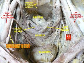
The broad ligament contains the round ligament, the fallopian tube, arteries, veins, lymphatics, nerve fibers, and loose connective tissue.
It has three subdivisions,
Mesometrium: the largest part of the ligaments surround the uterus.
Mesovarium: projects from the posterior surface of the broad ligament and enclose the ovary vascular supply not covering the ovary itself.
Mesosalpinx: trap the fallopian tube and originate from the mesovarium.
Round Ligament[edit | edit source]
A fibromuscular connective tissue attaches proximally at the superior and lateral side of the uterus at the cornu, runs forward and downward between the two sheets of the broad ligament. It runs down deep in the inguinal canal to enter the labia majora. its fibers blend with that of the labia majora and mons pubis to insert at mons pubis, helping to maintain the uterus anteverted[2].
Ovarian Ligament[edit | edit source]
It is a fibromuscular structure that lies with the broad ligament, connects the ovary with the uterus at the cornu.
Cervical Ligaments[edit | edit source]
Cardinal Ligaments/ Mackenrodt’s Ligaments[edit | edit source]
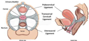
Ligaments of the cervix, connect the lateral side of the cervix and vagina to the lat pelvic wall and provide support to the uterus and vagina.
This ligament with uterosacral ligament and other pelvic musculature collaborate to support the pelvic organs and prevent prolapse.
During hysterectomy due to cervical cancer, the cardinal ligaments are involved and they may be removed as they are a common site for cancerous cells to ensure the disease is free.
Ureter and uterine vessels are related and near to the cardinal/ mackenrodt’s ligaments and may be injured during surgeries if the ligaments are manipulated[3].
Utero-sacral Ligaments[edit | edit source]
Known as sacrocervical ligaments or recto-uterine ligaments, extend from the posterior aspect of the cervix and vagina and directed backward to surround the rectum and insert in the base of the third sacral vertebrae.
Pubo-cervical Ligaments[edit | edit source]
Extend from the anterior of the cervix and vagina and directed forward to surround the urethra below the bladder and insert in the posterior aspect of the symphysis pubis.
References[edit | edit source]
- ↑ https://teachmeanatomy.info/pelvis/female-reproductive-tract/ligaments/
- ↑ Gossman W, Fagan SE, Sosa-Stanley JN, Peterson DC. Anatomy, abdomen and pelvis, uterus. InStatPearls [Internet] 2019 Jul 11. StatPearls Publishing.
- ↑ Eid S, Iwanaga J, Oskouian RJ, Loukas M, Tubbs RS. Comprehensive Review of the Cardinal Ligament. Cureus. 2018 Jun;10(6).
- ↑ AnatomyZone. Introduction to Female Reproductive Anatomy Part 2 - Ligaments - 3D Anatomy Tutorial. Available from: http://www.youtube.com/watch?v=Bf_sr2qRRWs[last accessed 14/6/2020]


