Glial Cells: Difference between revisions
No edit summary |
No edit summary |
||
| Line 47: | Line 47: | ||
In vivo imaging studies have revealed that in the resting healthy brain, microglia are highly dynamic, moving constantly to actively survey the brain parenchyma. | In vivo imaging studies have revealed that in the resting healthy brain, microglia are highly dynamic, moving constantly to actively survey the brain parenchyma. | ||
Microglia seem to be particularly involved in monitoring the integrity of synaptic function, optimizing different brain circuits to enable cognitive development (microglia cells eliminate previously-formed synapses that are no longer useful).<ref>Wake H, Moorhouse AJ, Nabekura J. [https://www.cambridge.org/core/journals/neuron-glia-biology/article/abs/functions-of-microglia-in-the-central-nervous-system-beyond-the-immune-response/B262044DDADE74974C8A268DD258B29A Functions of microglia in the central nervous system-beyond the immune response]. Neuron glia biology. 2011 Feb 1;7(1):47.Available from:https://www.cambridge.org/core/journals/neuron-glia-biology/article/abs/functions-of-microglia-in-the-central-nervous-system-beyond-the-immune-response/B262044DDADE74974C8A268DD258B29A (accessed 18.12.2020)</ref> | Microglia seem to be particularly involved in monitoring the integrity of synaptic function, optimizing different brain circuits to enable cognitive development (microglia cells eliminate previously-formed synapses that are no longer useful).<ref>Wake H, Moorhouse AJ, Nabekura J. [https://www.cambridge.org/core/journals/neuron-glia-biology/article/abs/functions-of-microglia-in-the-central-nervous-system-beyond-the-immune-response/B262044DDADE74974C8A268DD258B29A Functions of microglia in the central nervous system-beyond the immune response]. Neuron glia biology. 2011 Feb 1;7(1):47.Available from:https://www.cambridge.org/core/journals/neuron-glia-biology/article/abs/functions-of-microglia-in-the-central-nervous-system-beyond-the-immune-response/B262044DDADE74974C8A268DD258B29A (accessed 18.12.2020)</ref>[[File:Glymphatic system schematic.jpg|right|frameless|500x500px]] | ||
[[File:Glymphatic system schematic.jpg|right|frameless|500x500px]] | |||
== References == | == References == | ||
Revision as of 07:06, 21 January 2023
Original Editor - Lucinda hampton
Top Contributors - Lucinda hampton
Introduction[edit | edit source]
The brain is made up of more than just neurones. Numerous glial cells give support to the neurones, and in addition aid in the maintenance of homeostasis, and form myelin. Although there are about 86-100 billion neurons in the brain, glial cells are the most abundant cells in the central nervous system.
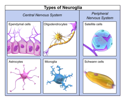
Glial cells exist in the both central nervous system (CNS) and the peripheral nervous system.[1] The most notable glial cells include oligodendrocytes, astrocytes, microglia, and ependymal cells in the CNS and in the Peripheral Nervous System, schwann cells, satellite cells, and enteric glial cells. Most glial cells are capable of mitotic division.[2][3][4]
Size plays a role with the macroglia (larger glial cells) insulating, protecting, and helping neurons to develop and migrate. and the microglia (smaller types of glia) have phagocytic properties, digesting foreign particles.[5]
Glial Cell Types[edit | edit source]
Astrocytes[edit | edit source]
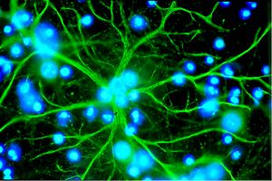
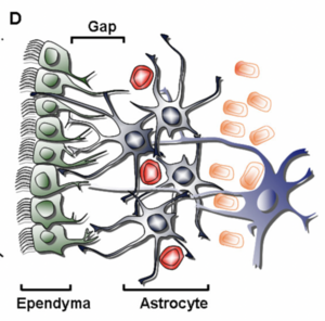
Astrocytes are the most abundant cells in the brain. They are star-shaped cells that provide physical and nutritional support for neurons. Functions include: clean up brain "debris"; transport nutrients to neurons; hold neurons in place; digest parts of dead neurons; regulate content of extracellular space; promote synaptic connections; clear excess neurotransmitters; ensure the continued function of neurons[6].
Astrocytes also
- Help to maintain the permeability of the blood-brain barrier where they sense glucose and ion levels inside the brain and regulate their flow into or out of it[5].
- Are involved in gliosis in response to injury.[4]
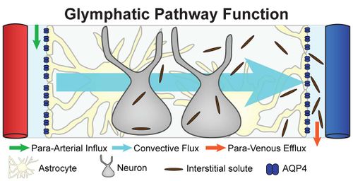
- Regulate extracellular fluid transport known as the glymphatic pathway (functionally represents the brain’s lymphatic system).[7]
Oligodendrocytes[edit | edit source]
Oligodendrocytes are the myelin-forming cells of the CNS. . Myelin is composed of layered phospholipid membranes and serves to support and insulate axons, allowing for faster impulse transduction. Saltatory conduction occurs as the impulses jump across sodium ion-rich nodes of Ranvier. One oligodendrocyte myelinates multiple axons. Oligodendrocytes are incapable of replication upon injury[8].
Ependymal Cells[edit | edit source]
Ependymal cells, which create cerebral spinal fluid (CSF), line the ventricles of the brain and central canal of the spinal cord. These cells are cuboidal to columnar and have cilia and microvilli on their surfaces to circulate and absorb CSF.
Depending on where they are located, ependymal cells also help to distribute neurotransmitters and hormones associated with the central nervous system. Also the microvilli of ependymal cells can absorb CSF and influence its flow and let certain substances in and out of the brain. Like the majority of glial cells, the ependyma also contributes to osmotic control within the brain via glucose and ion regulation[5].[8].
Radial Glia[edit | edit source]
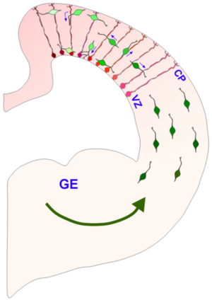
Radial glia are involved in neurogenesis and increased synapse stability, improved brain plasticity and play a role in neuroprotection. Radial glia also act as scaffolds along which new neurons can travel from their site of origin to their final destination in the brain. They are only found in specific areas of the CNS. This subgroup includes the Bergmann and Müller cells of the cerebellum and retina respectively. Radial cells modulate neurotransmission and optimize how information is processed. By forming a framework or scaffold on which other neurons can travel, radial glial cells are highly communicative.
They also produce all of the neurons in the cerebral cortex, being pivotal cell type in the developing central nervous system (CNS). They are thought to be a type of stem cell, meaning that they create other cells. In the developing brain, they're the "parents" of neurons, astrocytes, and oligodendrocytes.
Their role as stem cells, especially as creators of neurons, makes them the focus of research on how to repair brain damage from illness or injury. Later in life, they play roles in neuroplasticity as well[9][10].
Microglia[edit | edit source]
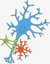
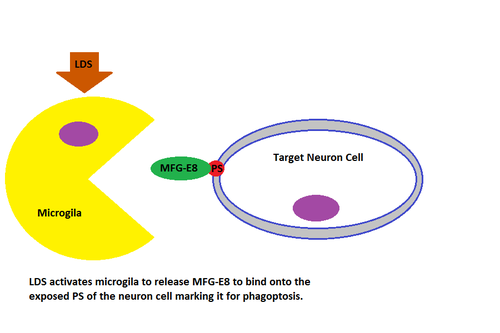
Microglia phagocytose and remove foreign or damaged material, cells, or organisms.[8] They are small, relatively sparse cells. They act as the brain's resident cleanup squad by phagocytosing apoptotic cells, plaques, and pathogens. Because they can prune and reshape synapses, microglia may also be influential in the pathogenesis of psychiatric illnesses[11]
With chronic stress, ageing and neurodegenerative disorders microglia may become more aggressive and less easy to regulate which makes them dangerous for the brain. In these cases, microglia can increase in numbers, unnecessarily kill nearby cells, and may contribute to making the brain even more inflamed by secreting inflammatory molecules[12].
In vivo imaging studies have revealed that in the resting healthy brain, microglia are highly dynamic, moving constantly to actively survey the brain parenchyma.
Microglia seem to be particularly involved in monitoring the integrity of synaptic function, optimizing different brain circuits to enable cognitive development (microglia cells eliminate previously-formed synapses that are no longer useful).[13]
References[edit | edit source]
- ↑ Radiopedia Glial cells Available from: https://radiopaedia.org/articles/glial-cells(last accessed 18.12.2020)
- ↑ Yuhas D, Jabr F. Know Your Neurons: What Is the Ratio of Glia to Neurons in the Brain?.Available from; https://blogs.scientificamerican.com/brainwaves/know-your-neurons-what-is-the-ratio-of-glia-to-neurons-in-the-brain/(last accessed 18.12.2020)
- ↑ Queensland Brain Institute Types of Glia Available:https://qbi.uq.edu.au/brain-basics/brain/brain-physiology/types-glia (accessed 21.1.2023)
- ↑ 4.0 4.1 Ludwig PE, Das JM. Histology, glial cells. InStatPearls [Internet] 2021 May 10. StatPearls Publishing.Available:https://www.ncbi.nlm.nih.gov/books/NBK441945/ (accessed 21.1.2023)
- ↑ 5.0 5.1 5.2 Biology Dic Glial cells Available:https://biologydictionary.net/glial-cells/ (accessed 10.5.2022)
- ↑ Washington Faculty neuroscience for kids Available from:https://faculty.washington.edu/chudler/glia.html (accessed 18.12.2020)
- ↑ radiopedia glymphatic pathway Available from:https://radiopaedia.org/articles/glymphatic-pathway?lang=gb (accessed 18.12.2020)
- ↑ 8.0 8.1 8.2 Ludwig PE, Das JM. Histology, Glial Cells. StatPearls [Internet]. 2020 Jun 19.Available from:https://www.ncbi.nlm.nih.gov/books/NBK441945/ (accessed 18.12.2020)
- ↑ Very Well Health What are glial cells and what do they do Available from:https://www.verywellhealth.com/what-are-glial-cells-and-what-do-they-do-4159734 (accessed 19.12.2020)
- ↑ Psychology Wiki. Radial Glia Available from: https://psychology.wikia.org/wiki/Radial_glial_cells (accessed 19.12.20200
- ↑ Scanlon ST. Depressing effects of microglia. Science. 2020 Apr 17;368(6488):279-80.Available from;https://science.sciencemag.org/content/368/6488/279.2.full (last accessed 18.12.2020)
- ↑ The Conversation Microglia: the brain’s ‘immune cells’ protect against diseases – but they can also cause them. Available from:https://theconversation.com/microglia-the-brains-immune-cells-protect-against-diseases-but-they-can-also-cause-them-139232 (last accessed 18.12.2020)
- ↑ Wake H, Moorhouse AJ, Nabekura J. Functions of microglia in the central nervous system-beyond the immune response. Neuron glia biology. 2011 Feb 1;7(1):47.Available from:https://www.cambridge.org/core/journals/neuron-glia-biology/article/abs/functions-of-microglia-in-the-central-nervous-system-beyond-the-immune-response/B262044DDADE74974C8A268DD258B29A (accessed 18.12.2020)
