Radius: Difference between revisions
Kim Jackson (talk | contribs) No edit summary |
No edit summary |
||
| (6 intermediate revisions by 3 users not shown) | |||
| Line 1: | Line 1: | ||
<div class="editorbox"> | <div class="editorbox"> | ||
'''Original Editor''' | '''Original Editor'''- [[User:Abbey Wright|Abbey Wright]] | ||
'''Top Contributors''' - {{Special:Contributors/{{FULLPAGENAME}}}} | '''Top Contributors''' - {{Special:Contributors/{{FULLPAGENAME}}}} | ||
| Line 6: | Line 6: | ||
== Description == | == Description == | ||
[[File:Radius whole body.png|thumb|Radius in relation to the whole body]] | [[File:Radius whole body.png|thumb|Radius in relation to the whole body]] | ||
The radius is one of the two bones that make up the forearm, the other being the [[ulna]]. It forms the radio- | The radius is one of the two bones that make up the forearm, the other being the [[ulna]]. It forms the radio-carpal joint at the wrist and the radio-ulnar joint at the [[elbow]]. It is similar to the [[tibia]] of the lower limb and is located in the lateral forearm when in the anatomical position. The radius bone is smaller than the [[ulna]] and has an upper end, a lower end, and a shaft. | ||
=== Structure === | === Structure === | ||
==== Proximal radius ==== | ==== Proximal radius ==== | ||
The proximal radius consists of the radial head, neck and tuberosity. | The proximal radius consists of the radial head, neck, and tuberosity. | ||
The radial head | The cylindrical radial head articulates with the capitellum of the [[humerus]] at the [[elbow]] joint <ref name=":0">Gray HFRS, Gray's Anatomy 15th edition, New York, NY: Barnes & Noble,2010. p126-128</ref> and is covered with hyaline cartilage. The circumference of the radial head is designed to fit perfectly into the socket created by the radial notch of the ulna and the annular ligament, forming what is known as the superior radioulnar joint.<ref>Chaurasia BD. Bd Chaurasia’s Human Anatomy Regional and Applied Dissection and Clinical. 2013. | ||
The neck and | </ref> This joint allows for the rotation of the head within the annular ligament, which in turn produces both supination and pronation of the forearm.<ref name=":1">Palastanga N, Soames R. Anatomy and Human Movement, structure and function 6th edition, London, UK: Churchill Livingstone Elsevier ,2011. p45</ref> | ||
The neck and tuberosity support the head and provide points of attachment for supinator brevis and [[Biceps Brachii|Biceps brachi]]<ref name=":0" />[[File:Overview of the radius and ulna - Kenhub.png|alt=Overview of the radius and ulna - anterior and posterior views|right|frameless|600x600px|Overview of the radius and ulna - anterior and posterior views]] | |||
==== Radial shaft ==== | ==== Radial shaft ==== | ||
The shaft of the radius is slightly curved into convex from the body. The majority of the shaft has three borders: anterior, posterior and interosseous. | The shaft of the radius is slightly curved into convex from the body. The majority of the shaft has three borders: anterior, posterior, and interosseous. | ||
Image: Overview of the radius and ulna - anterior and posterior views<ref >Overview of the radius and ulna - anterior and posterior views image - © Kenhub https://www.kenhub.com/en/library/anatomy/the-radius-and-the-ulna</ref> | |||
==== Distal radius<ref name=":1" /> ==== | ==== Distal radius<ref name=":1" /> ==== | ||
| Line 24: | Line 28: | ||
# Medial - consists of a concave ulnar notch to articulate with the ulnar head in pronation | # Medial - consists of a concave ulnar notch to articulate with the ulnar head in pronation | ||
# Posterior - convex and contains a prominent ridge called Lister's tubercle | # Posterior - convex and contains a prominent ridge called Lister's tubercle | ||
# Anterior - smooth and forms a distinct margin | # Anterior - smooth and forms a distinct margin, it extends from the anterior margin of radial tuberosity to the styloid process. | ||
# Distal articular surface - articulates laterally with scaphoid and medially with lunate[[File:Radius and ulna.jpg|none|thumb|561x561px|Annotated diagram of the radius and ulna]] | # Distal articular surface - articulates laterally with scaphoid and medially with lunate[[File:Radius and ulna.jpg|none|thumb|561x561px|Annotated diagram of the radius and ulna]] | ||
| Line 47: | Line 51: | ||
==== Proximal ==== | ==== Proximal ==== | ||
[[Biceps | [[Biceps Brachii|Biceps brachii]] attaches to the radial tuberosity. | ||
[[Supinator]], flexor pollicis longus and the flexor digitorum superficialis attach to the upper third part of the shaft of the radius. | [[Supinator]], flexor pollicis longus and the flexor digitorum superficialis attach to the upper third part of the shaft of the radius. | ||
==== Mid shaft ==== | ==== Mid shaft ==== | ||
Extensor | [[Extensor Pollicis Brevis]] muscle, [[abductor pollicis longus]] muscle, and [[Pronator Teres|pronator teres]] all attach to the mid shaft of the radius | ||
==== Distal ==== | ==== Distal ==== | ||
Latest revision as of 22:10, 21 December 2023
Original Editor- Abbey Wright
Top Contributors - Abbey Wright, Kim Jackson, Joao Costa, Chrysolite Jyothi Kommu, Amanda Ager and Pacifique Dusabeyezu
Description[edit | edit source]
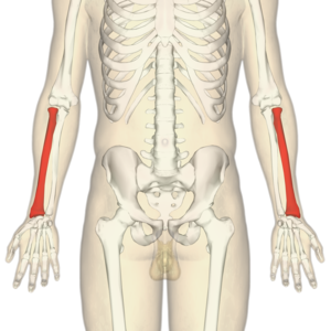
The radius is one of the two bones that make up the forearm, the other being the ulna. It forms the radio-carpal joint at the wrist and the radio-ulnar joint at the elbow. It is similar to the tibia of the lower limb and is located in the lateral forearm when in the anatomical position. The radius bone is smaller than the ulna and has an upper end, a lower end, and a shaft.
Structure[edit | edit source]
Proximal radius[edit | edit source]
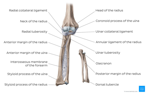
The proximal radius consists of the radial head, neck, and tuberosity.
The cylindrical radial head articulates with the capitellum of the humerus at the elbow joint [1] and is covered with hyaline cartilage. The circumference of the radial head is designed to fit perfectly into the socket created by the radial notch of the ulna and the annular ligament, forming what is known as the superior radioulnar joint.[2] This joint allows for the rotation of the head within the annular ligament, which in turn produces both supination and pronation of the forearm.[3]
The neck and tuberosity support the head and provide points of attachment for supinator brevis and Biceps brachi[1]
Radial shaft[edit | edit source]
The shaft of the radius is slightly curved into convex from the body. The majority of the shaft has three borders: anterior, posterior, and interosseous.
Image: Overview of the radius and ulna - anterior and posterior views[4]
Distal radius[3][edit | edit source]
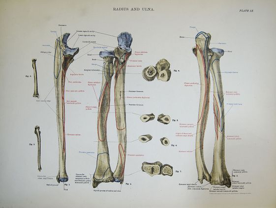
The distal radius has five surfaces:
- Lateral - which extends to form the styloid process
- Medial - consists of a concave ulnar notch to articulate with the ulnar head in pronation
- Posterior - convex and contains a prominent ridge called Lister's tubercle
- Anterior - smooth and forms a distinct margin, it extends from the anterior margin of radial tuberosity to the styloid process.
- Distal articular surface - articulates laterally with scaphoid and medially with lunate
Function[edit | edit source]
The radius' main functions are to articulate with the ulna and humerus at the elbow to provide supination and pronation. Then to articulate with the lunate and scaphoid to provide all the movements of the wrist.
Articulations[edit | edit source]
Elbow[edit | edit source]
The radius articulates with the ulna in a synovial pivot joint. The radial head rotates within the annular ligament and radial notch on the ulna to produce pronation of the forearm.[5]
The radius and ulna also articulate distally in reverse to their articulation at the elbow to produce supination. This movement occurs with the head of the ulna being received into the ulnar notch of the radius. [5]
Wrist[edit | edit source]
The radius articulates with the first row of carpel bones: mainly scaphoid laterally and the lunate medially to form the radio-carpel joint, triquetral only makes intermittent contact on ulnar abduction. The radio-carpel joint is an ellipsoid joint whereby the scaphoid, lunate and triquetral bones form a convex surface to articulate with the distal radius' concavity.
The radio-carpel joint has four movements: flexion, extension, radial and ulnar deviation.
Muscle attachments[edit | edit source]
Proximal[edit | edit source]
Biceps brachii attaches to the radial tuberosity.
Supinator, flexor pollicis longus and the flexor digitorum superficialis attach to the upper third part of the shaft of the radius.
Mid shaft[edit | edit source]
Extensor Pollicis Brevis muscle, abductor pollicis longus muscle, and pronator teres all attach to the mid shaft of the radius
Distal[edit | edit source]
Pronator quadratus muscle and the tendon of the supinator longus attach to the distal quarter of the radial shaft.
Clinical relevance[edit | edit source]
Colles fracture: is a fracture of the distal radius. Commonly caused by a fall on outstretched hand (FOOSH)
Radial head fracture: commonly caused from FOOSH this can be accompanied by dislocation of the radius and/or ulna which can complicate the management of this injury.[7]
Osteoarthritis of the wrist or elbow
Radial dysplasia or radial club hand can occur in paediatric patients as a congenital condition whereby there is a shortening of the radius or even complete lack of radius in the forearm. This can lead to thumb abnormalities and the need for surgical intervention to improve function.[8]
Resources[edit | edit source]
Muscle attachments source and diagram of muscular attachments
References[edit | edit source]
- ↑ 1.0 1.1 Gray HFRS, Gray's Anatomy 15th edition, New York, NY: Barnes & Noble,2010. p126-128
- ↑ Chaurasia BD. Bd Chaurasia’s Human Anatomy Regional and Applied Dissection and Clinical. 2013.
- ↑ 3.0 3.1 Palastanga N, Soames R. Anatomy and Human Movement, structure and function 6th edition, London, UK: Churchill Livingstone Elsevier ,2011. p45
- ↑ Overview of the radius and ulna - anterior and posterior views image - © Kenhub https://www.kenhub.com/en/library/anatomy/the-radius-and-the-ulna
- ↑ 5.0 5.1 Gray HFRS, Gray's Anatomy 15th edition, New York, NY: Barnes & Noble,2010. p231- 235
- ↑ Young Lae Moon. Forearm supination & pronation. Available from: https://www.youtube.com/watch?v=3F9d-qaXYp0 [Last accessed 26/03/2016]
- ↑ Cohen MS, Hill Hastings II. Acute elbow dislocation: evaluation and management. JAAOS-Journal of the American Academy of Orthopaedic Surgeons. 1998 Jan 1;6(1):15-23.
- ↑ Bednar MS, James MA, Light TR. Congenital longitudinal deficiency. The Journal of hand surgery. 2009 Nov 1;34(9):1739-47.


