Avascular Necrosis Femoral Head: Difference between revisions
No edit summary |
No edit summary |
||
| Line 5: | Line 5: | ||
</div> | </div> | ||
== Introduction == | == Introduction == | ||
Avascular necrosis of the femoral head is a pathologic process that results from interruption of blood supply to the bone. Etiopathogenesis is | [[File:Hip_X_ray_avascular_necrosis.jpg|alt=|thumb|Hip X ray avascular necrosis]] | ||
[[File:Avacular_necrosis_MRI.jpg|alt=|thumb|Avacular necrosis MRI]] | |||
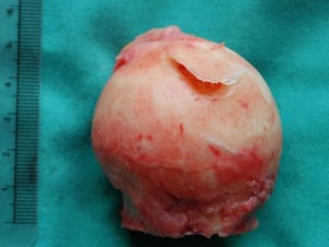
[[Avascular Necrosis|Avascular necrosis]] (AN) of the [[Femur|femoral]] head is a pathologic process that results from interruption of blood supply to the bone. Etiopathogenesis is not well understood. Femoral head ischaemia causes bone marrow and osteocytes death, resulting in the collapse of the necrotic segment eventually.[[Image:Head of femur avascular necrosis.jpg|thumb|right]] | |||
AN of the femoral head has often been described as a multifactorial disease. Risk Factors include: | |||
</ref> | |||
* Genetic predilection<ref>Adesina O, Brunson A, Keegan THM, Wun T. Osteonecrosis of the femoral head in sickle cell disease: prevalence, comorbidities, and surgical outcomes in California. Blood Adv. 2017;1(16):1287-1295.</ref> | |||
* Corticosteroid intake<ref>Xie XH, Wang XL, Yang HL, Zhao DW, Qin L. Steroid-associated osteonecrosis: Epidemiology, pathophysiology, animal model, prevention, and potential treatments (an overview). J Orthop Translat. 2015 Apr;3(2):58-70. </ref> | |||
* Alcohol | |||
* Smoking | |||
* Various chronic diseases <ref>Jaffré C, Rochefort GY. Alcohol-induced osteonecrosis--dose and duration effects. Int J Exp Pathol. 2012;93(1):78-9 | |||
</ref> eg Patients with [[Human Immunodeficiency Virus (HIV)|human immunodeficiency virus]] , (rare) complication of pregnancy<ref name="Mont" />. | |||
| | ||
| Line 15: | Line 23: | ||
It is very important that avascular necrosis is diagnosed early in the disease process since the success of the treatment is related to the stage at which the treatment starts.<br>There are several possible diagnostic modalities: | It is very important that avascular necrosis is diagnosed early in the disease process since the success of the treatment is related to the stage at which the treatment starts.<br>There are several possible diagnostic modalities: | ||
# Xrays: When standard anteroposterior and frog-leg lateral radiographs show obvious avascular necrosis of the femoral head, it is not necessary to perform an MRI. | |||
# Magnetic Resonance Imaging: This is the best method for cases that are radiographically occult or not obvious on radiographs. It has been found to be 99%sensitive and 98% specific for this disease<ref name="Mont" />. | |||
== History and Physical Examination == | == History and Physical Examination == | ||
| Line 20: | Line 31: | ||
Patients are often seen because of pain in the groin, but symptoms can also radiate to the [[knee]] or buttocks. On examination, there is usually a painful [[Range of Motion|range of motion]], especially on forced internal rotation<ref name="MA" />. It is also important to track any risk factors before the start of the examination. Investigators need to be wary of avascular necrosis in any patient who has pain in the [[Hip Anatomy|hip]], negative radiographic findings, and any of the risk factors, described above. The other hip must also be evaluated. | Patients are often seen because of pain in the groin, but symptoms can also radiate to the [[knee]] or buttocks. On examination, there is usually a painful [[Range of Motion|range of motion]], especially on forced internal rotation<ref name="MA" />. It is also important to track any risk factors before the start of the examination. Investigators need to be wary of avascular necrosis in any patient who has pain in the [[Hip Anatomy|hip]], negative radiographic findings, and any of the risk factors, described above. The other hip must also be evaluated. | ||
== Management == | |||
== Management | |||
The management of avascular necrosis of the femoral head ranges from conservative (non-operative) to invasive (operative). Factors such as patient's age, pain/discomfort threshold, severity of necrosis, intact or collapse of the articular surface, and comorbidities.<ref>Hsu H, Nallamothu SV. StatPearls. StatPearls Publishing; Treasure Island (FL), 2020. Hip Osteonecrosis. Available from:https://www.ncbi.nlm.nih.gov/books/NBK499954/ (Accessed 25 July 2020)</ref> | The management of avascular necrosis of the femoral head ranges from conservative (non-operative) to invasive (operative). Factors such as patient's age, pain/discomfort threshold, severity of necrosis, intact or collapse of the articular surface, and comorbidities.<ref>Hsu H, Nallamothu SV. StatPearls. StatPearls Publishing; Treasure Island (FL), 2020. Hip Osteonecrosis. Available from:https://www.ncbi.nlm.nih.gov/books/NBK499954/ (Accessed 25 July 2020)</ref> | ||
Conservative managements are physical therapy, restricted weight-bearing, alcohol cessation, discontinuation of steroid therapy, pain control medication, and targeted pharmacologic therapy, among others. | Conservative managements are physical therapy, restricted weight-bearing, alcohol cessation, discontinuation of steroid therapy, pain control medication, and targeted pharmacologic therapy, among others. | ||
'''1. Non-operative Treatment''' | |||
= | Observation or Protected Weight-Bearing: This method is believed to slow the progression of avascular necrosis so that the femoral head wouldn’t collapse. However, more than 80% of affected hips do progress to femoral head collapse and arthritis by four years after the diagnosis<ref name="Mont" />. There are various methods to reduce weight-bearing. The concept of this method is to reduce the forces on the hip joint. This (interventional) treatment has various modalities, such as a cane, crutch, walker, or two crutches.<br>Most studies have shown though that non-operative treatment yields poor results. The only condition for which protected weight-bearing might be effective is a type-A lesion<ref name="MA" />. | ||
Pharmacological Treatment: | |||
Anticoagulants, e.g. enoxaparin are used to prevent the progression of osteonecrosis due to hypercoagulability and thromboembolic events.<ref>Lai KA, Shen WJ, Yang CY, Shao CJ, Hsu JT, Lin RM. The use of alendronate to prevent early collapse of the femoral head in patients with nontraumatic osteonecrosis. A randomized clinical study. J Bone Joint Surg Am. 2005;87(10):2155-9. | * Vasodilators, e.g. iloprost (PGI2), are used to reduce intraosseous pressure thus, increased blood flow.<ref>Claben T, Becker A, Landgraeber S, Haversath M, Li X, Zilkens C, et al. Long-term Clinical Results after Iloprost Treatment for Bone Marrow Edema and Avascular Necrosis. Orthop Rev (Pavia). 2016;8(1):6150. </ref> | ||
* Statins act to decrease the differentiation of stem cells into fat cells, by reducing intraosseous pressure for better perfusion.<ref>Pritchett JW. Statin therapy decreases the risk of osteonecrosis in patients receiving steroids. Clin. Orthop. Relat. Res. 2001;(386):173-8. </ref> | |||
* Anticoagulants, e.g. enoxaparin are used to prevent the progression of osteonecrosis due to hypercoagulability and thromboembolic events.<ref>Lai KA, Shen WJ, Yang CY, Shao CJ, Hsu JT, Lin RM. The use of alendronate to prevent early collapse of the femoral head in patients with nontraumatic osteonecrosis. A randomized clinical study. J Bone Joint Surg Am. 2005;87(10):2155-9. | |||
</ref> | </ref> | ||
* Bisphosphonates, such as alendronate, prevent osteoclasts action thus reducing bone resorption.<ref>Agarwala S, Shah S, Joshi VR. The use of alendronate in the treatment of avascular necrosis of the femoral head: follow-up to eight years. J Bone Joint Surg Br. 2009;91(8):1013-8.</ref><ref name=":0">Immonen I, Friberg K, Grönhagen-Riska C, von Willebrand E, Fyhrquist F. Angiotensin-converting enzyme in sarcoid and chalazion granulomas of the conjunctiva. Acta Ophthalmol (Copenh). 1986;64(5):519-21.</ref> | |||
Bisphosphonates, such as alendronate, prevent osteoclasts action thus reducing bone resorption.<ref>Agarwala S, Shah S, Joshi VR. The use of alendronate in the treatment of avascular necrosis of the femoral head: follow-up to eight years. J Bone Joint Surg Br. 2009;91(8):1013-8.</ref><ref name=":0">Immonen I, Friberg K, Grönhagen-Riska C, von Willebrand E, Fyhrquist F. Angiotensin-converting enzyme in sarcoid and chalazion granulomas of the conjunctiva. Acta Ophthalmol (Copenh). 1986;64(5):519-21.</ref> | * Pain medication such as NSAIDs and Opioid care for pain modulation. Despite intra-articular steroid injections been fast-acting pain relieving agents, they are normally short-termed and their use can cause significant deterioration of avascular necrosis.<ref name=":0" /> | ||
Pain medication such as NSAIDs and Opioid care for pain modulation. Despite intra-articular steroid injections been fast-acting pain relieving agents, they are normally short-termed and their use can cause significant deterioration of avascular necrosis.<ref name=":0" /> | |||
=== [[Avascular Necrosis|Physical therapy]] === | === [[Avascular Necrosis|Physical therapy]] === | ||
Revision as of 06:42, 14 December 2022
Original Editor Anouk Toye
Top Contributors - Lucinda hampton, Anouk Toye, Kim Jackson, Uchechukwu Chukwuemeka, Aarti Sareen, Admin and 127.0.0.1
Introduction[edit | edit source]
Avascular necrosis (AN) of the femoral head is a pathologic process that results from interruption of blood supply to the bone. Etiopathogenesis is not well understood. Femoral head ischaemia causes bone marrow and osteocytes death, resulting in the collapse of the necrotic segment eventually.
AN of the femoral head has often been described as a multifactorial disease. Risk Factors include:
- Genetic predilection[1]
- Corticosteroid intake[2]
- Alcohol
- Smoking
- Various chronic diseases [3] eg Patients with human immunodeficiency virus , (rare) complication of pregnancy[4].
Diagnostic procedures[edit | edit source]
It is very important that avascular necrosis is diagnosed early in the disease process since the success of the treatment is related to the stage at which the treatment starts.
There are several possible diagnostic modalities:
- Xrays: When standard anteroposterior and frog-leg lateral radiographs show obvious avascular necrosis of the femoral head, it is not necessary to perform an MRI.
- Magnetic Resonance Imaging: This is the best method for cases that are radiographically occult or not obvious on radiographs. It has been found to be 99%sensitive and 98% specific for this disease[4].
History and Physical Examination[edit | edit source]
Patients are often seen because of pain in the groin, but symptoms can also radiate to the knee or buttocks. On examination, there is usually a painful range of motion, especially on forced internal rotation[5]. It is also important to track any risk factors before the start of the examination. Investigators need to be wary of avascular necrosis in any patient who has pain in the hip, negative radiographic findings, and any of the risk factors, described above. The other hip must also be evaluated.
Management[edit | edit source]
The management of avascular necrosis of the femoral head ranges from conservative (non-operative) to invasive (operative). Factors such as patient's age, pain/discomfort threshold, severity of necrosis, intact or collapse of the articular surface, and comorbidities.[6]
Conservative managements are physical therapy, restricted weight-bearing, alcohol cessation, discontinuation of steroid therapy, pain control medication, and targeted pharmacologic therapy, among others.
1. Non-operative Treatment
Observation or Protected Weight-Bearing: This method is believed to slow the progression of avascular necrosis so that the femoral head wouldn’t collapse. However, more than 80% of affected hips do progress to femoral head collapse and arthritis by four years after the diagnosis[4]. There are various methods to reduce weight-bearing. The concept of this method is to reduce the forces on the hip joint. This (interventional) treatment has various modalities, such as a cane, crutch, walker, or two crutches.
Most studies have shown though that non-operative treatment yields poor results. The only condition for which protected weight-bearing might be effective is a type-A lesion[5].
Pharmacological Treatment:
- Vasodilators, e.g. iloprost (PGI2), are used to reduce intraosseous pressure thus, increased blood flow.[7]
- Statins act to decrease the differentiation of stem cells into fat cells, by reducing intraosseous pressure for better perfusion.[8]
- Anticoagulants, e.g. enoxaparin are used to prevent the progression of osteonecrosis due to hypercoagulability and thromboembolic events.[9]
- Bisphosphonates, such as alendronate, prevent osteoclasts action thus reducing bone resorption.[10][11]
- Pain medication such as NSAIDs and Opioid care for pain modulation. Despite intra-articular steroid injections been fast-acting pain relieving agents, they are normally short-termed and their use can cause significant deterioration of avascular necrosis.[11]
Physical therapy[edit | edit source]
Electrical stimulation has been shown experimentally to enhance osteogenesis and neovascularization as well as to alter osseous turnover.[5]
Three different methods can be described:
- Non-invasive pulsed electromagnetic-field stimulation
- Direct-current stimulation of the necrotic area through the insertion of an electrode at the time of a core decompression
- Non-invasive direct-current stimulation by capacitive coupling after a core decompression
Electrical stimulation remains experimental for the treatment of avascular necrosis of the femoral head. Additional study is needed to define the optimum dosage, application, and timing of treatment. [5]
2. Operative Treatment[edit | edit source]
There are several possible ways to treat avascular necrosis of the hip: core decompression, Core decompression with electrical stimulation, Osteotomy, Non-vascularized bone-grafting, and Vascularized grafts. The joint preservation interventions are core decompression, osteotomy, bone grafts, and use of cellular therapies; while reconstructive interventions are arthroplasty.
Recommendations for treatment[edit | edit source]
All tables come from: MA Mont, LC Jones and DS Hungerford, Non-traumatic avascular necrosis of the femoral head: ten years later, J Bone Joint Surg Am. 2006;88:1117-1132 [4]
References[edit | edit source]
- ↑ Adesina O, Brunson A, Keegan THM, Wun T. Osteonecrosis of the femoral head in sickle cell disease: prevalence, comorbidities, and surgical outcomes in California. Blood Adv. 2017;1(16):1287-1295.
- ↑ Xie XH, Wang XL, Yang HL, Zhao DW, Qin L. Steroid-associated osteonecrosis: Epidemiology, pathophysiology, animal model, prevention, and potential treatments (an overview). J Orthop Translat. 2015 Apr;3(2):58-70.
- ↑ Jaffré C, Rochefort GY. Alcohol-induced osteonecrosis--dose and duration effects. Int J Exp Pathol. 2012;93(1):78-9
- ↑ 4.0 4.1 4.2 4.3 Mont MA, Jones LC, Hungerford DS. Non-traumatic avascular necrosis of the femoral head: ten years later. J Bone Joint Surg Am. 2006;88:1117-1132
- ↑ 5.0 5.1 5.2 5.3 Mont MA, Hungerford DS. Non-traumatic avascular necrosis of the femoral head. J Bone Joint Surg Am. 1995; 77:459-474
- ↑ Hsu H, Nallamothu SV. StatPearls. StatPearls Publishing; Treasure Island (FL), 2020. Hip Osteonecrosis. Available from:https://www.ncbi.nlm.nih.gov/books/NBK499954/ (Accessed 25 July 2020)
- ↑ Claben T, Becker A, Landgraeber S, Haversath M, Li X, Zilkens C, et al. Long-term Clinical Results after Iloprost Treatment for Bone Marrow Edema and Avascular Necrosis. Orthop Rev (Pavia). 2016;8(1):6150.
- ↑ Pritchett JW. Statin therapy decreases the risk of osteonecrosis in patients receiving steroids. Clin. Orthop. Relat. Res. 2001;(386):173-8.
- ↑ Lai KA, Shen WJ, Yang CY, Shao CJ, Hsu JT, Lin RM. The use of alendronate to prevent early collapse of the femoral head in patients with nontraumatic osteonecrosis. A randomized clinical study. J Bone Joint Surg Am. 2005;87(10):2155-9.
- ↑ Agarwala S, Shah S, Joshi VR. The use of alendronate in the treatment of avascular necrosis of the femoral head: follow-up to eight years. J Bone Joint Surg Br. 2009;91(8):1013-8.
- ↑ 11.0 11.1 Immonen I, Friberg K, Grönhagen-Riska C, von Willebrand E, Fyhrquist F. Angiotensin-converting enzyme in sarcoid and chalazion granulomas of the conjunctiva. Acta Ophthalmol (Copenh). 1986;64(5):519-21.


