The Vestibulocochlear Nerve (CN VIII): Difference between revisions
(Created page with "{{subst:New Page}}") |
No edit summary |
||
| (6 intermediate revisions by the same user not shown) | |||
| Line 1: | Line 1: | ||
<div class="editorbox"> | <div class="editorbox"> | ||
'''Original Editor '''- [[User: | '''Original Editor '''- [[User:Lucinda hampton|Lucinda hampton]] | ||
'''Top Contributors''' - {{Special:Contributors/{{FULLPAGENAME}}}} | '''Top Contributors''' - {{Special:Contributors/{{FULLPAGENAME}}}} | ||
</div> | </div> | ||
== Introduction == | == Introduction == | ||
[[File:CN 8.png|right|frameless|400x400px]] | |||
The vestibulocochlear nerve, also known as [[Cranial Nerves|cranial nerve]] eight (CN VIII), consists of the [[Vestibular System|vestibular]] and cochlear nerves. It is located in the internal auditory meatus (internal auditory canal). | |||
* The vestibular nerve is primarily responsible for maintaining body [[balance]] and eye movements, while | |||
* The cochlear nerve is responsible for hearing. | |||
CN VIII injuries are the result of pathological processes or injuries that commonly involve the cerebellopontine angle (CPA), the internal auditory canal (IAC), or the inner ear. In such cases, symptoms such as [[Benign Positional Paroxysmal Vertigo (BPPV)|vertigo]], nystagmus, tinnitus, and sensorineural [[Hearing in the Elderly|hear]]<nowiki/>ing loss may occur<ref>Bordoni B, Sugumar K, Daly DT. [https://www.ncbi.nlm.nih.gov/books/NBK537359/ Neuroanatomy, cranial nerve 8 (vestibulocochlear).] StatPearls [Internet]. 2020 Jan 12.Available from:https://www.ncbi.nlm.nih.gov/books/NBK537359/ (accessed5.2.2021)</ref> | |||
== | == Anatomy == | ||
[[File:Vestibulocochlear nerve.jpg|right|frameless]] | |||
The vestibulocochlear is one of the 12 cranial nerves, it runs between the pons (the middle of the [[brainstem]]) and the medulla oblongata (the lower part of the brainstem). | |||
* The vestibular part of the nerve then travels from the inner ear in a group of [[Neurone|nerve cells]] called the vestibular ganglion. | |||
* The cochlear part of the nerve travels from the cochlea in the inner ear in the spiral ganglion<ref name=":0">Very well health [https://www.verywellhealth.com/vestibulocochlear-nerve-5095249 CN VIII] Available from:https://www.verywellhealth.com/vestibulocochlear-nerve-5095249 (last accessed 5.2.2021)</ref>. | |||
Image 2: Tractography showing vestibulocochlear nerve (a procedure to demonstrate the neural tracts using special techniques of [[MRI Scans|MRI]], and computer-based image analysis<ref>Sensagent Tractography Available from:http://dictionary.sensagent.com/Tractography/en-en/ (accessed 5.2.2021)</ref>). | |||
== | == Function == | ||
[[File:Vestibulocochlear Nerve.jpg|right|frameless]] | |||
The function of the vestibulocochlear nerve is purely sensory. It has no motor function. It communicate sound and equilibrium information from the inner ear to the [[Brain Anatomy|brain]]. | |||
* The cochlea, the part of the inner ear where the cochlear part of the nerve originates, detects soundwaves. These then travel from the spiral ganglion to the brain. | |||
* The vestibular apparatus, where the vestibular part of the nerve originates, detects changes in the head’s position based on gravity. Then the position of the head communicates information about balance to the brain<ref name=":0" />. | |||
== | == Physiotherapy Implications == | ||
[[File:Balance-board-benefits.jpg|right|frameless]] | |||
A problem with the sensory information being relayed to the brain via the vestibulocochlear nerve is termed a peripheral vestibular disorder. | |||
Conditions of the vestibulocochlear nerve can affect balance and hearing and may cause vertigo, vomiting, ringing in the ears, a false sense of motion, motion sickness, or even hearing loss. | |||
Physiotherapists are commonly involved with the retraining of balance. See below links for more details. | |||
* [[Balance]] | |||
* [[Balance Training]] | |||
* [[Benign Positional Paroxysmal Vertigo (BPPV)]] | |||
* [[Vestibular Anatomy and Neurophysiology]] | |||
== References == | == References == | ||
<references /> | <references /> | ||
[[Category:Nerves]] | |||
[[Category:Neurology]] | |||
[[Category:Older People/Geriatrics]] | |||
[[Category:Vestibular System]] | |||
[[Category:Anatomy]] | |||
Latest revision as of 07:34, 5 February 2021
Original Editor - Lucinda hampton
Top Contributors - Lucinda hampton
Introduction[edit | edit source]
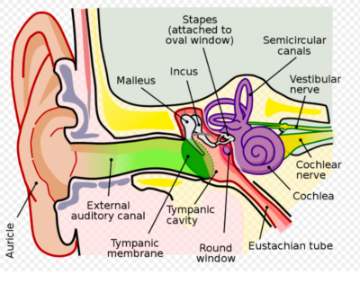
The vestibulocochlear nerve, also known as cranial nerve eight (CN VIII), consists of the vestibular and cochlear nerves. It is located in the internal auditory meatus (internal auditory canal).
- The vestibular nerve is primarily responsible for maintaining body balance and eye movements, while
- The cochlear nerve is responsible for hearing.
CN VIII injuries are the result of pathological processes or injuries that commonly involve the cerebellopontine angle (CPA), the internal auditory canal (IAC), or the inner ear. In such cases, symptoms such as vertigo, nystagmus, tinnitus, and sensorineural hearing loss may occur[1]
Anatomy[edit | edit source]
The vestibulocochlear is one of the 12 cranial nerves, it runs between the pons (the middle of the brainstem) and the medulla oblongata (the lower part of the brainstem).
- The vestibular part of the nerve then travels from the inner ear in a group of nerve cells called the vestibular ganglion.
- The cochlear part of the nerve travels from the cochlea in the inner ear in the spiral ganglion[2].
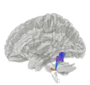
Image 2: Tractography showing vestibulocochlear nerve (a procedure to demonstrate the neural tracts using special techniques of MRI, and computer-based image analysis[3]).
Function[edit | edit source]
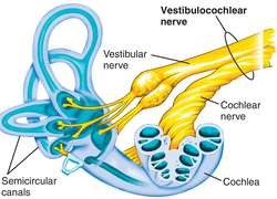
The function of the vestibulocochlear nerve is purely sensory. It has no motor function. It communicate sound and equilibrium information from the inner ear to the brain.
- The cochlea, the part of the inner ear where the cochlear part of the nerve originates, detects soundwaves. These then travel from the spiral ganglion to the brain.
- The vestibular apparatus, where the vestibular part of the nerve originates, detects changes in the head’s position based on gravity. Then the position of the head communicates information about balance to the brain[2].
Physiotherapy Implications[edit | edit source]
A problem with the sensory information being relayed to the brain via the vestibulocochlear nerve is termed a peripheral vestibular disorder.
Conditions of the vestibulocochlear nerve can affect balance and hearing and may cause vertigo, vomiting, ringing in the ears, a false sense of motion, motion sickness, or even hearing loss.
Physiotherapists are commonly involved with the retraining of balance. See below links for more details.
- Balance
- Balance Training
- Benign Positional Paroxysmal Vertigo (BPPV)
- Vestibular Anatomy and Neurophysiology
References[edit | edit source]
- ↑ Bordoni B, Sugumar K, Daly DT. Neuroanatomy, cranial nerve 8 (vestibulocochlear). StatPearls [Internet]. 2020 Jan 12.Available from:https://www.ncbi.nlm.nih.gov/books/NBK537359/ (accessed5.2.2021)
- ↑ 2.0 2.1 Very well health CN VIII Available from:https://www.verywellhealth.com/vestibulocochlear-nerve-5095249 (last accessed 5.2.2021)
- ↑ Sensagent Tractography Available from:http://dictionary.sensagent.com/Tractography/en-en/ (accessed 5.2.2021)


