Talus: Difference between revisions
No edit summary |
No edit summary |
||
| Line 26: | Line 26: | ||
[[File:Talus .png|right|frameless]] | [[File:Talus .png|right|frameless]] | ||
The talus is an incredible bone; despite its small size, it transmits considerable force during the normal gait cycle and even more significant force during impact activities. | The talus is an incredible bone; despite its small size, it transmits considerable force during the normal gait cycle and even more significant force during impact activities. | ||
* The talus is shaped like a truncated cone and is wider anteriorly than posteriorly. | |||
The talus is shaped like a truncated cone and is wider anteriorly than posteriorly. | * Approximately two-thirds of its surface area is covered with articular cartilage and it has a tenuous blood supply, similar to the scaphoid. | ||
* The talus has seven articular surfaces and is divided into the head, neck and body, and two processes, the posterior process and the lateral process (Fig. 1). | |||
Approximately two-thirds of its surface area is covered with articular cartilage and it has a tenuous blood supply, similar to the scaphoid. | * The posterior process is further subdivided into posterolateral and posteromedial tubercles.<ref>Majeed H, McBride DJ. [https://www.ncbi.nlm.nih.gov/pmc/articles/PMC5890124/ Talar process fractures: an overview and update of the literature]. EFORT open reviews. 2018 Mar;3(3):85-92. Available from:https://www.ncbi.nlm.nih.gov/pmc/articles/PMC5890124/ (last accessed 11.3.2020)</ref> | ||
The talus has seven articular surfaces and is divided into the head, neck and body, and two processes, the posterior process and the lateral process (Fig. 1). | |||
The posterior process is further subdivided into posterolateral and posteromedial tubercles.<ref>Majeed H, McBride DJ. [https://www.ncbi.nlm.nih.gov/pmc/articles/PMC5890124/ Talar process fractures: an overview and update of the literature]. EFORT open reviews. 2018 Mar;3(3):85-92. Available from:https://www.ncbi.nlm.nih.gov/pmc/articles/PMC5890124/ (last accessed 11.3.2020)</ref> | |||
== Function == | == Function == | ||
Revision as of 12:44, 11 March 2020
This article or area is currently under construction and may only be partially complete. Please come back soon to see the finished work! (11/03/2020)
Original Editor
Top Contributors - Lucinda hampton, Ewa Jaraczewska, Kim Jackson and Vidya Acharya
Introduction[edit | edit source]
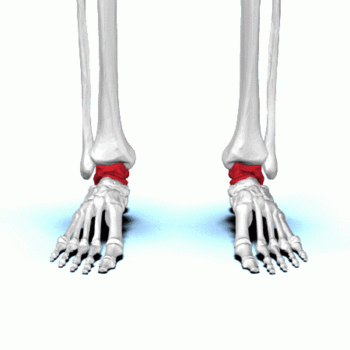
The talus is the second largest bone in the hindfoot region of the human body. Responsible for transmitting body weight and forces passing between the lower leg and the foot.
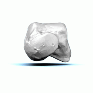
The Talus (left talus shown in image)
- Is a component of many multiple joints, including the talocrural (ankle), subtalar, and transverse tarsal joints.
- Does not have any direct muscular attachments and has a tenuous and limited blood supply,
- Serves as the site of attachment for many ligaments including the lateral ankle ligaments and medial deltoid ligament complex.
Talus fractures comprise about 1% of all foot and ankle fractures.
Nearly 70% of ankle injuries can cause varying degrees of chondral and osteochondral injuries to the talus.
It is important to be aware of the occurrence of both isolated and associated talar injuries and their extent of clinical and potentially long-term implications.[1]
Structure[edit | edit source]
The talus is an incredible bone; despite its small size, it transmits considerable force during the normal gait cycle and even more significant force during impact activities.
- The talus is shaped like a truncated cone and is wider anteriorly than posteriorly.
- Approximately two-thirds of its surface area is covered with articular cartilage and it has a tenuous blood supply, similar to the scaphoid.
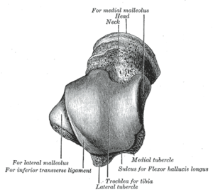
- The talus has seven articular surfaces and is divided into the head, neck and body, and two processes, the posterior process and the lateral process (Fig. 1).
- The posterior process is further subdivided into posterolateral and posteromedial tubercles.[2]
Function[edit | edit source]
Articulations[edit | edit source]
Muscle attachments[edit | edit source]
The talus does not serve as the site of any muscle attachment, which is significant because that means the talus does not have secondary sources of blood supply.
- If there is vascular compromise to the talus through traumatic injuries such as in the setting of displaced fractures, there is a relatively high risk of developing post-traumatic, avascular necrosis (AVN)[1]
Clinical relevance[edit | edit source]
Assessment[edit | edit source]
Treatment[edit | edit source]
Resources[edit | edit source]
References[edit | edit source]
- ↑ 1.0 1.1 Irfan A. Khan; Matthew Varacallo. Anatomy, Bony Pelvis and Lower Limb, Foot Talus ☀April 21, 2019. Available from: ☀https://www.ncbi.nlm.nih.gov/books/NBK541086/ (last accessed 11.3.2020)
- ↑ Majeed H, McBride DJ. Talar process fractures: an overview and update of the literature. EFORT open reviews. 2018 Mar;3(3):85-92. Available from:https://www.ncbi.nlm.nih.gov/pmc/articles/PMC5890124/ (last accessed 11.3.2020)


