Spinal cord anatomy: Difference between revisions
No edit summary |
No edit summary |
||
| Line 57: | Line 57: | ||
==== [[Reticulospinal Tract|Reticulospinal Tract]] ==== | ==== [[Reticulospinal Tract|Reticulospinal Tract]] ==== | ||
This tract begins in the caudal reticular formation in the pons and medulla. Provides both excitable and inhibitory effects on the interneurons in the spinal cord, and to a lesser extent, it also acts on the motor neurons. Its main action is to dampen down activity in the spinal cord. without this pathway, there is increased extensor tone observed. | This tract begins in the caudal reticular formation in the pons and medulla. Provides both excitable and inhibitory effects on the interneurons in the spinal cord, and to a lesser extent, it also acts on the motor neurons. Its main action is to dampen down activity in the spinal cord. without this pathway, there is increased extensor tone observed. | ||
'''Spinal Motorneurons''' | |||
Alpha and Gamma motorneurons (MNs) are both found in the ventral (anterior) horn. <br> | |||
''Alpha motorneurons'' are the largest motor neurons in the nervous system. They innervate skeletal muscle. | |||
''Gamma Motorneurons'' innervate intrafusal muscle fibres of the muscle spindle. | |||
Motor neurons are arranged somatotopically across the ventral horn. The more medially placed MNs innervate proximal muscles while laterally placed MNs innervate distal muscles. | |||
== References == | == References == | ||
Revision as of 20:57, 15 October 2018
- Please do not edit unless you are involved in this project, but please come back in the near future to check out new information!!
- If you would like to get involved in this project and earn accreditation for your contributions, [[[Special:Contact|please get in touch]]]!
Original Editor - Add a link to your Physiopedia profile here.
Top Contributors - Naomi O'Reilly, Lucinda hampton, Kim Jackson, Rucha Gadgil, Nikhil Benhur Abburi, Vidya Acharya, Aminat Abolade, Stacy Schiurring, Admin, Tarina van der Stockt, Ewa Jaraczewska and Jess Bell
Introduction[edit | edit source]
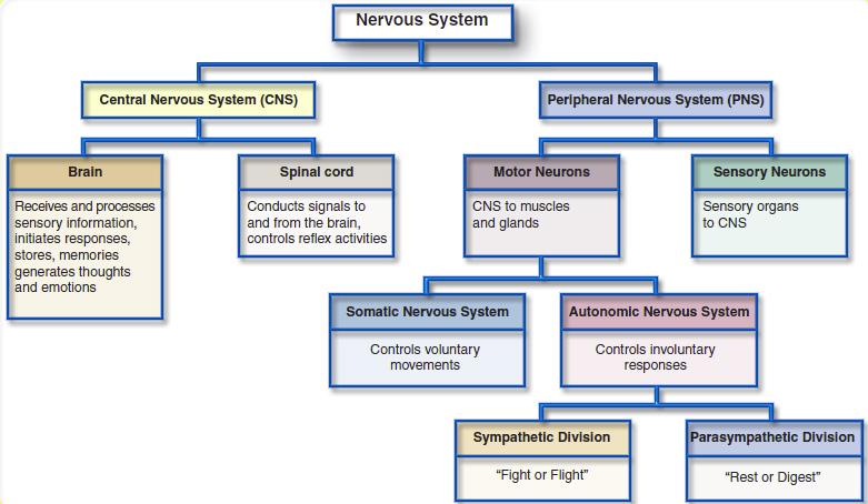
The Nervous System is divided into two main divisions.[1]
These are:
- Central Nervous System (CNS)
- Peripheral Nervous System (PNS)
Anatomy of the Spinal Cord[edit | edit source]
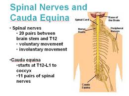
The spinal cord lies within the vertebral canal, extending from the foramen magnum to the lowest border of the first lumbar vertebra. It is enlarged at two sites, the cervical and lumbar region. The lower part of the spinal canal contains the lower lumbar and sacral nerves known as the Cauda Equina.
- Sensory Nerve Fibres enter the Spinal Cord via the Dorsal (Posterior) Root. The cell bodies for these neurons are situated in the Dorsal Root Ganglia.
- Motor and Preganglionic Autonomic Fibres exit via the Ventral (Anterior) Root.
This short video clip gives an overview of spinal cord anatomy.
| [2] |
Associated Pathways[edit | edit source]
Ascending Sensory Pathway[edit | edit source]
Spinothalamic Tract[edit | edit source]
- From Dorsal horn laminae I,III,IV,V. crosses midline in spinal cord, projects to brain stem and contr-lateral thalamus. Conveys pain and temperature.
Dorsal Column Medial Lemniscal Pathway[edit | edit source]
- Afferents from mechanoreceptors, muscle and joint receptors. terminates in dorsal column nuclei of medulla. Forms medial lemniscus at this level and synapses in ventroposterior nucleus of thalamus. Conveys proprioception, light touch and vibration.
Spinocerebellar Tract[edit | edit source]
- From spinal cord interneurons. It has two tracts a) Dorsal SCT relays via inferior cerebellar peduncle and b) VCT relays via superior cerebellar peduncle to the cerebellum. It conveys proprioceptive information and on-going activity in the spinal cord interneurons.
Descending Motor Pathways[edit | edit source]
Corticospinal (Pyramidal) Tract[edit | edit source]
From the motor cortex, premotor cortex, and somatosensory cortex. Has a role in sensory processing and fractionated finger movements.
Rubrospinal Tract[edit | edit source]
Originates from the magnocellular part of the red nucleus in the brain. It projects towards common structures with the CoST, particularly those involved with distal motor control. There is debate as to how significant this tract is.
Vestibulospinal Tract[edit | edit source]
Originates from Deiters nucleus in the medulla and innervates the extensor and axial muscles. It is involved in balance control and posture.
Reticulospinal Tract[edit | edit source]
This tract begins in the caudal reticular formation in the pons and medulla. Provides both excitable and inhibitory effects on the interneurons in the spinal cord, and to a lesser extent, it also acts on the motor neurons. Its main action is to dampen down activity in the spinal cord. without this pathway, there is increased extensor tone observed.
Spinal Motorneurons
Alpha and Gamma motorneurons (MNs) are both found in the ventral (anterior) horn.
Alpha motorneurons are the largest motor neurons in the nervous system. They innervate skeletal muscle.
Gamma Motorneurons innervate intrafusal muscle fibres of the muscle spindle.
Motor neurons are arranged somatotopically across the ventral horn. The more medially placed MNs innervate proximal muscles while laterally placed MNs innervate distal muscles.
References[edit | edit source]
References will automatically be added here, see adding references tutorial.
- ↑ Barker; Barasi; Neal. Neuroscience at a glance; Blackwell science Ltd; 1999
- ↑ Handwritten tutorials. Spinal Pathways 1 - Spinal Cord Anatomy and Organisation. Available from: http://www.youtube.com/watch?v=5B87zsAKmWc [last accessed 29/08/16]


