
Wilson's Disease
Original Editors - Students from Bellarmine University's Pathophysiology of Complex Patient Problems project.
Top Contributors - Rachel Rawson, Lexi Vessels, Angeliki Chorti, Aminat Abolade, Kim Jackson, Vidya Acharya, WikiSysop, Elaine Lonnemann and Kalyani Yajnanarayan
Definition/Description
Wilson’s disease (WD), also known as hepatolenticular degeneration, is an autosomal recessive genetic disorder that results from abnormal metabolism of copper.[1] The deposition of copper begins immediately at birth, with symptoms usually presenting in late adolescence. [2]
Aetiology/Causes

Wilson's Disease (WD) is a disorder that results in excess copper accumulation in target organs and impairing the normal function of particular organs (liver, brain, cornea, etc).[3] The exact cause of this disease is not fully understood, however, the gene that is currently believed to be responsible is the mutation of ATP7B in chromosome 13. [4] This gene, ATP7B, is a vitally important enzyme for copper transport in the body. The mutation in this gene is linked to abnormal excretion and ultimately deposition of copper in particular organs. It is believed that other factors may be involved with the development of the disease, such as other genetic variations, lipid metabolism, and possible environmental factors. Further research on the aetiology of Wilson's Disease is needed.[1]
Prevalence
Wilson’s disease is considered a rare condition. WD was originally reported (in 1984) to be affecting only one in 30,000 to one in 50,000, an estimate that is still applicable to most populations, however, it is now acknowledged that the genetic prevalence is 3-4 times higher in some instances than clinically based estimates. [5] This condition is reported as an autosomal recessive. [6] The gene frequency for WD has been found to be 56%, with a carrier frequency of 1 in every 90-150 people. [7] Wilson's gene has been described to present with more than 500 mutations, 380 of which have a key role in the pathogenesis of the disease. [8] According to recent evidence, an increase incidence of this gene mutation has been found in isolated populations like Costa Rica and Sardinia, an island off Italy. [5]

WD Classification & Coding
ICD-10: E83.0 (International Classification of Diseases, 10th edition)
ICD-11: 5C64.00 (Mortality & Morbidity statistics)
OMIM: 277900 (Genetic database)
ORPHA:905 (Rare Diseases database)
WD Characteristics
Clinical Presentation in WD according to reports and guidelines [8][9] [10] is as follows:
- Symptoms in WD usually vary.
- The main involvements are most commonly hepatic, neurological, ophthalmological, hemolytic and psychiatric due to these organ's large involvement in the accumulation of copper. These symptoms can present from asymptomatic to fatal if left untreated.
- Hepatic presentation can present as asymptomatic hepatomegaly, isolated splenomegaly, persistently elevated serum aminotransferase activity (AST, ALT), fatty liver, acute hepatitis, resembling autoimmune hepatitis, acute/chronic liver failure, and cirrhosis (compensated or decompensated). Other symptoms that may present are, Coomb's negative hemolytic anemia, low grade hemolysis and jaundice, abdominal swelling (ascites), vomiting blood, abdominal pain, oesophageal varices.
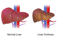
Liver Cirrhosis

- Neurological involvement can present with athetoid movements and unsteady gait. They can also present similar to Parkinson's, ataxia, pseudobulbar palsy, pseudosclerosis dominated by tremor, and a dystonic syndrome that often leads to contractures.
- Other central nervous system symptoms include pill rolling tremors in hands, facial and muscular rigidity, dysphagia, and dysarthria, dysautonomia, migraine headaches, insomnia, and seizures.
- Psychiatric characteristics can present in the hand-eye coordination, emotional and behavioral changes, and decrease in performance. Mental illness may include homicidal or suicidal behavior, depression, aggression, and insomnia.
- Wilson's can also have an ocular manifestation as a Kayser-Fleischer ring around the eye from copper deposits and sunflower cataracts in the lens, which are less commonly observed compared to the Kayser-Fleischer ring.
- Other systems involvement may include lunulae ceruleae (cutaneous), renal abnormalities such as aminoaciduria and nephrolithiasis, skeletal abnormalities (premature osteoporosis and arthritis), cardiomyopathy, dysrhythmias, pancreatitis, hypoparathyroidism, menstrual irregularities; infertility, repeated miscarriage.
- Symptomatic adolescents will tend to experience more symptoms related to liver pathologies, while older adults will experience neurological signs and symptoms.
Associated Co-morbidities
- Individuals with WD can present with a variety of different complications that are directly related to the excess of copper in targeted organs; however, in severe cases other systems can become affected as well. There can be musculoskeletal manifestations including muscle atrophy and wasting, contractures, deformities, osteomalacia, and pathological fractures.[3]
- Other less common characteristics that can present because of WD are osteoarthritis, abnormal kidney function tests, and cardiomyopathy.[1]
Diagnosis
Diagnosis of Wilson’s disease can be confirmed through a multitude of diagnostic tests. Typically:
- Ophthalmologic slit-lamp examination for Kayser-Fleischer rings
- Serum ceruloplasmin test: levels <0.1g/L will be seen in individuals with Wilson's disease.[8]
When Kayser-Fleischer rings are absent, a combination of tests (e.g. urinary and hepatic copper, genetic testing) and a diagnostic score based on these tests may be more suitable. [11]
Neurologic testing is warranted in presymptomatic and hepatic presentations. [8]
Liver biopsy is prescribed only when clinical signs and tests are inconclusive or if other liver pathologies are suspected. [12]
Differential Diagnosis
- Cirrhosis
- Haemochromatosis
- Portal Hypertension
- Hemorrhage associated with oesophageal Varices
- Hematochromatosis
- Essential Tremor
Medical Management
Wilson’s disease is a rare condition, but with proper management the progression of the disease can stop and even in some cases, symptoms can improve. Immediate treatment early on in the disease progression is crucial in order to prevent irreversible damage to the liver and other organs. [2]
The most important aspect of medical management for WD is compliance to pharmacological intervention. Treatment for Wilson’s disease is lifelong, and adherence to therapy provides a challenge for a considerable amount of patients, especially asymptomatic patients because there is failure to recognise the relationship between medication and the disease progression.[10] Patient education on medication compliance is therefore extremely important in this population.
Medications
Pharmacological treatment of WD is based around the removal of excess copper from the body, as well preventing the accumulation of it in target organs. [14] Chelation therapy drugs, such as Penicillamine and Trientine are used to remove excess copper from the affected organs; they act as binding agents and are removed through urinary excretion. Zinc salts are also used to block the intestinal absorption of copper, reducing its accumulation in the body. Zinc salts are shown to be very effective, and is an advantage over other pharmacological agents because of its lack of side effects.[15]
Patients who are asymptomatic should receive lower doses of zinc salts or chelators than patients who are symptomatic. [9] In the context of advanced liver disease, symptomatic patients may receive a combination of zinc salts and chelators, [16]although this approach may present with poor compliance. [17]
D-penicillamine (Cuprimine and Depen) was the first drug introduced to remove excess copper from the body through increased urine excretion. While it is commonly used amongst these patients, a disadvantage to this medication is the adverse side effects it brings with it, toxicity and worsening neurological symptoms.[9]
Initial Dose: 250-500 mg/d with 250 mg increments every 4-7 days in 2-4 divided doses Maintenance Dose: 750-1000 mg/d in 2 doses a day Pediatric Dose: 20mg/kg per day in 2-3 doses daily; maintenance therapy should reduce to 25-30% of dose
Administer drug one hour before or two hours after a meal to avoid inhibition.
Adverse side effects are common and include fever, cutaneous eruptions, neutropenia, lymphadenopathy, and proteinuria[9]
Trientene (Syprine and Trientine Dihydrochloride) is an alternative for patients who are intolerant to penicillamine. This drug acts in a similar manner, but without the serious side effects.[9]
Initial Dose: 750-1500 mg/d in 2-3 divided doses Maintenance Dose: 750-1000 mg/d in 2-3 divided doses Pediatric Dose: 20mg/kg per day in 2-3 doses daily; maintenance therapy should reduce to 25-30% of dose
Adverse side effects include possible neurotoxicity, dyspepsia, anemia, muscle cramps, and dystonia[9]
Zinc (Galzin) acts by inhibiting the absorption of copper in the intestinal tract. An advantage to this drug over chelators is the effectiveness and lack of adverse side effects.[9]
Zinc Dosage: Dose: 150 mg/d divided in 3 doses Pediatric dose: 75 mg/d divided in 3 doses
Adverse side effects are minimum and include gastric irritation, alcohol intolerance, headaches, excessive perspiration, and anemia.[9]
Patients with serious hepatitis or liver failure will not improve with pharmacological intervention, and may require a liver transplant.[9]
Additional therapy is necessary for treatment of this condition. Specifically, Physiotherapy can be beneficial to improve neurological symptoms, such as dystonia, dyscoordination, and difficulty with balance. Speech therapy can improve other symptoms like dysarthria and dysphagia.
Diet is also a crucial component in medical management. Patients should stick to a diet that is low in copper and avoid foods like certain meats, mushrooms, chocolate, shellfish, and nuts. Water should be tested for any copper content to avoid excessive copper intake.[10]
Physiotherapy Management
Physiotherapy can play a major role in the treatment of individuals with Wilson's disease. One of the most common manifestations of this condition is the neurological symptoms that present like a movement disorder. Patients may present with dystonia, dyscoordination, tremors and difficulty with balance and walking.[10]
Many patients may face problems if their dystonia worsens and a contracture develops. PT can provide patient education on positioning and stretching to prevent progression of the contracture, in addition to serial casting to reverse an acute contracture.[10]
Physiotherapy can be a useful tool in improving balance and coordination through the use of therapeutic activities and exercise, as well as maintaining range of motion and functionality.[10][18]
Patients who suffer from liver disease due to the effects of Wilson's disease may experience fatigue, muscle and joint pain, and a decrease in functional status. Physiotherapy can play a role in decreasing pain and improving the patient's functional capabilities with energy conservation in mind.[19]
While physiotherapy plays a crucial role in helping these individuals overcome their neurological symptoms, success with therapy is not possible unless the patient is consistently sticking to their medication regime.[9]
Case Reports/ Case Studies
Malik A, Khawaja A, Sheikh L. Wilson’s disease in pregnancy: case series and review of literature. BMC Res Notes. 2013 Oct 18;6:421. - four cases of successful pregnancy outcomes in three patients diagnosed with WD.
Grover S, Sarkar S, Jhanda S, Chawla Y. Psychosis in an adolescent with Wilson's disease: A case report and review of the literature. Indian J Psychiatry. 2014; 56(4): 395-8. - a case of a young boy with WD whose presentation of psychotic symptoms are less common.
Raju K, Bangalore G, Thuruvekere S, Pathavanalli V. Wilson's disease: A Clinical autopsy case report with review of literature. J Nat Sci Biol Med. 2015; 6(1): 248-52. - a clinical autopsy case in a 39 year-old female who had presented with clinical symptoms at 18 years of age.
Loudianos G, Incollu S, Mameli E, Lepori MB. Wilson’s disease in an adult asymptomatic patient: a potential role for modifying factors of copper metabolism. Ann Gastroenterol. 2016; 29(1):96-8. - this case describes the challenges of WD diagnosis through report of an asymptomatic WD patient diagnosed accidentally by genetic analysis.
Yang Y, Li X, Yang W. A New Dystonia Phenotype in Wilson Disease. JAMA Neurol. 2025 Jun 9. Epub ahead of print. - an interesting case report about a 36-year old male diagnosed with WD who was found with an unusual presentation of two opposite muscle tone manifestations at the same time.
Resources
Wilson Disease Association (WDA) International
EASL-ERN Clinical Practice Guidelines on Wilson's Disease, 2025 update
U.S. National Library of Medicine
References
- ↑ 1.0 1.1 1.2 Wu F, Wang J, Pu C, Qiao L, Jiang C. Wilson's disease: a comprehensive review of the molecular mechanisms. Int J Mol Sci. 2015 Mar 20;16(3):6419-31.
- ↑ 2.0 2.1 Pandey N, Blair K, John S. Kayser-Fleischer Ring. [Updated 2024 Jan 25]. In: StatPearls [Internet]. Treasure Island (FL): StatPearls Publishing; 2025 Jan-. Available from: https://www.ncbi.nlm.nih.gov/books/NBK459187/ [accessed 17/7/2025]
- ↑ 3.0 3.1 Goodman CC, Fuller KS. Goodman and Fuller’s Pathology E-Book: Implications for the Physical Therapist. Elsevier Health Sciences; 2020 Oct 9.
- ↑ Tao TY, Gitlin JD. Hepatic copper metabolism: insights from genetic disease. Hepatology. 2003 Jun;37(6):1241-7.
- ↑ 5.0 5.1 Sandahl TD, Laursen TL, Munk DE, Vilstrup H, Weiss KH, Ott P. The Prevalence of Wilson's Disease: An Update. Hepatology. 2020 Feb;71(2):722-32.
- ↑ Ferenci P. Wilson's disease. Clin Liver Dis. 1998 Feb;2(1):31-49, v-vi.
- ↑ Coffey AJ, Durkie M, Hague S, McLay K, Emmerson J, Lo C, Klaffke S, Joyce CJ, Dhawan A, Hadzic N, Mieli-Vergani G, Kirk R, Elizabeth Allen K, Nicholl D, Wong S, Griffiths W, Smithson S, Giffin N, Taha A, Connolly S, Gillett GT, Tanner S, Bonham J, Sharrack B, Palotie A, Rattray M, Dalton A, Bandmann O. A genetic study of Wilson's disease in the United Kingdom. Brain. 2013 May;136(Pt 5):1476-87.
- ↑ 8.0 8.1 8.2 8.3 European Association for the Study of the Liver. EASL-ERN Clinical Practice Guidelines on Wilson's disease. J Hepatol. 2025 Feb 22:S0168-8278(24)02706-5.
- ↑ 9.00 9.01 9.02 9.03 9.04 9.05 9.06 9.07 9.08 9.09 Rodriguez-Castro KI, Hevia-Urrutia FJ, Sturniolo GC. Wilson's disease: A review of what we have learned. World J Hepatol. 2015 Dec 18;7(29):2859-70.
- ↑ 10.0 10.1 10.2 10.3 10.4 10.5 Wilson Disease Association (WDA) International. [cited 2016 Mar 30]. Available from: http://www.wilsonsdisease.org/ [accessed 15/7/2025]
- ↑ Ferenci P, Caca K, Loudianos G, Mieli-Vergani G, Tanner S, Sternlieb I, Schilsky M, Cox D, Berr F. Diagnosis and phenotypic classification of Wilson disease. Liver Int. 2003 Jun;23(3):139-42.
- ↑ Ludwig J, Moyer TP, Rakela J. The liver biopsy diagnosis of Wilson's disease. Methods in pathology. Am J Clin Pathol. 1994 Oct;102(4):443-6.
- ↑ Goodman CC, Snyder TK. Differential Diagnosis for Physical Therapists: Screening for Referral. 5th Edition. St. Louis, Missouri: Elsevier Saunders, 2013.
- ↑ Aggarwal A, Bhatt M. The Pragmatic Treatment of Wilson's Disease. Mov Disord Clin Pract. 2014 Apr 10;1(1):14-23.
- ↑ Avan A, Członkowska A, Gaskin S, Granzotto A, Sensi SL, Hoogenraad TU. The Role of Zinc in the Treatment of Wilson's Disease. Int J Mol Sci. 2022 Aug 18;23(16):9316.
- ↑ Roberts EA, Schilsky ML; American Association for Study of Liver Diseases (AASLD). Diagnosis and treatment of Wilson disease: an update. Hepatology. 2008 Jun;47(6):2089-111.
- ↑ Jagadisan B, Dhawan A. Combination Treatment With Chelators and Zinc for Wilson Disease: A Double-edged Sword. J Clin Exp Hepatol. 2024 May-Jun;14(3):101372.
- ↑ Brewer G, Askari F. Wilson's disease: clinical management and therapy. J Hepatol. 2005;42 Suppl(1):S13-21.
- ↑ van den Berg-Emons RJ, van Ginneken BT, Nooijen CF, Metselaar HJ, Tilanus HW, Kazemier G, Stam HJ. Fatigue after liver transplantation: effects of a rehabilitation program including exercise training and physical activity counseling. Phys Ther. 2014 Jun 1;94(6):857-65.