Synkinesis
Original Editor - Wendy Walker
Lead Editors - Wendy Walker, Jess Bell, Kim Jackson, Tarina van der Stockt, Rishika Babburu, WikiSysop and Sheik Abdul Khadir
Introduction and Definition[edit | edit source]
Synkinesis (AKA aberrant regeneration) occurs after injury to the facial nerve and it is a common sequelae of facial palsy. The cause of the injury may be Bell's Palsy, Ramsay Hunt Syndrome (less common), surgical damage (eg. during surgical removal of an acoustic neuroma), trauma (skull fractures) or other conditions causing facial paralysis.
Synkinesis = "syn" meaning "together" and "kinesis" meaning "movement". Therefore, synkinesis means "moving together" or "mass movement". Thus, synkinesis is when an involuntary movement accompanies a voluntary movement.[1]
The type of synkinesis is commonly described by combining the names of the two involved muscle groups, with the first part referring to the voluntary motor group and the second part referring to the involuntary muscle group. For instance:
- Ocular-oral synkinesis is when voluntary eye contraction such as blinking or brow lifting elicits an involuntary mouth movement
- Oral-ocular synkinesis is when an involuntary eye contraction accompanies a volitional mouth movement such as smiling and lip puckering
Clinically Relevant Anatomy[edit | edit source]
The facial nerve is the seventh cranial nerve, and it controls the muscles of facial expression. More information on the anatomy of the facial nerve is available here.
Mechanism of Injury / Pathological Process[edit | edit source]
The unintentional or mass movements are thought to be caused by an undifferentiated regeneration of the facial nerve that occurs after it has been compressed or damaged.[2][3]
It is thought that synkinesis could be caused by four possible mechanisms:
- Aberrant regeneration[3][4][5][6][7] - "miss-wiring"
- Axons regrow from the facial nucleus to incorrect peripheral muscle groups
- It has generally been assumed that the site of the miss-wiring is the lesion site (i.e. where the nerve was damaged by crush / inflammation), but one 2004 study found that the regrowing axons are disorganised along their whole length, as well as at the lesion site[5]
- Ephaptic transmission[3][8] - electrical cross-talk between nerve branches
- Presumed to be due to the reduced myelin sheath of the nerve fibres, which means they are poorly insulated
- Nuclear hyperexcitability[6]
- This theory proposes that once the post-synaptic cell loses its input from the degenerated axons, it creates additional neurotransmitter receptors and, thus, becomes hypersensitive
- Because of this hypersensitivity, it responds to neurotransmitters provided by another nerve nearby
- Maladaptive cortical plasticity[3][9][10]
- A 2018 study using MRI found that there was cortical reorganisation in the primary sensorimotor area and the supplementary motor area in the brain
Many authors think that a combination of more than one, and possibly all, of these mechanisms is likely to be involved.
Clinical Presentation[edit | edit source]
While patients experience recovery and re-innervation of the affected side of the face after flaccid facial palsy, they also experience the involuntary linking of movements which are typical of synkinesis.
The effects which are most commonly observed are:[11][12]
- When moving the mouth (e.g. smile, lip pucker, when eating), the eye on the affected side moves towards partial (or occasionally full) closure, whereas the unaffected eye remains wide open = oral-ocular
- When raising the eyebrows or closing the eyes, the corner of the mouth on the affected side of the face raises = ocular-oral
It is important to recognise that synkinesis frequently starts to occur from the fifth or sixth month post onset of palsy, although in some instances it can present as early as the third month post onset, and generally increases for up to two years post onset.[13][14]
Appearance of Synkinesis when the face is at rest[edit | edit source]
When the person is not moving their face, synkinesis may still be visible[3]. The most common visible manifestations of synkinesis at rest are:
- Affected eye smaller in size than the other eye
- Affected nasolabial fold deeper than the contralateral side, and often it sits in a raised position
- The corner of the mouth may sit higher than the unaffected side
Appearance of Synkinesis when the face is moving[edit | edit source]
The effects of synkinesis are frequently more apparent on facial movements, and as a general rule more extreme/large ranges of movement cause greater asymmetry.
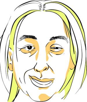
For instance, when smiling the affected side of the mouth will typically not move as far as the unaffected side, but the affected eye will become smaller, as shown in this illustration of left sided synkinesis:
The same pattern of reduced range of movement at the mouth/lips with increased eye closure can commonly be seen on the several facial postures, including lip pucker, raise top lip, show teeth.
When the person is chewing food, the synkinesis-behaviour at the ipsilateral eye, ie. moving towards closure/becoming smaller, can cause the person to report "I look like I'm winking when I eat".
Scoring / Measuring Synkinesis[edit | edit source]
The most commonly used measure of facial range of movement by surgeons and physicians is the House-Brackmann scale.[15] Unfortunately, this scale does not have a rating for the aberrant linking of movements which occurs in synkinesis.
The Sunnybrook Facial Grading System [FGS][16] is a more comprehensive scoring system for facial range of movement, and it has a section dedicated to rating the presence of synkinesis movements.[17] A 2015 systematic review of facial nerve grading systems identified the FGS as meeting the most criteria in regards to overall assessment of facial nerve function, sequelae and response to treatments. It was also identified as having the highest reliability.[18] This systematic review concluded: " The authors recommend widespread adoption of the Sunnybrook Facial Grading Scale as the current standard in reporting outcomes of facial nerve disorders."[18] The FGS is sensitive enough to show gains when facial range of movement increases, and also has an increased score if the person has a reduction in their synkinesis even in the absence of increased range of movement[6].
The Synkinesis Assessment Questionnaire [SAQ] consists of nine questions and has been shown to be both valid and reliable as a dedicated measurement of synkinesis.[19] It has also been shown to have good correlation with the synkinesis component of the FGS. A 2022 document, Pathogenesis, diagnosis and therapy of facial synkinesis: A systematic review and clinical practice recommendations by the international head and neck scientific group[20], makes a recommendation that the "SAQ should be used to assess the patient's view on their synkinesis severity."
Chuang and colleagues have also formulated a novel classification system with four categories:[21]
- Pattern I: Good smile (i.e. good range of movement) with mild synkinesis
- Pattern II:Acceptable smile with moderate to severe synkinesis
- Pattern III: Unacceptable smile (little or no range of movement) with severe synkinesis
- Pattern IV: Poor smile with mild synkinesis
This method of scoring synkinesis has not been assessed in inter- and intra-rater reliability studies, but it proved a useful tool to help decide on management in a study by Chuang et al.[21]
Management / Interventions[edit | edit source]
Physiotherapy Interventions[edit | edit source]
The following physiotherapy interventions have been shown to be effective in reducing or minimising synkinesis:
Non-Physiotherapy Interventions[edit | edit source]
Differential Diagnosis[edit | edit source]
Synkinesis is a clinical diagnosis. It is usually easy to diagnose as the patient will display clear linking of facial movements on the affected side only, and will have a history of facial palsy. Occasionally it can be confused with the following conditions:
- Facial dystonia
- Essential blepharospasm
- Essential hemifacial spasm
Resources[edit | edit source]
Facial Palsy UK has a comprehensive website, and this page explains more about synkinesis.
References[edit | edit source]
References will automatically be added here, see adding references tutorial.
- ↑ Ma ZZ, Lu YC, Wu JJ, Li SS, Ding W, Xu JG. Alteration of spatial patterns at the network-level in facial synkinesis: an independent component and connectome analysis. Ann Transl Med. 2021;9(3):240.
- ↑ Raslan A, Guntinas‐Lichius O, Volk GF. Altered facial muscle innervation pattern in patients with postparetic facial synkinesis, The Laryngoscope. 2019;130(5):E320-E326.
- ↑ 3.0 3.1 3.2 3.3 3.4 3.5 3.6 3.7 3.8 3.9 Guntinas-Lichius, Prengel et al. Pathogenesis, diagnosis and therapy of facial synkinesis: A systematic review and clinical practice recommendations by the International Head & Neck Scientific Group. Frontiers in Neurology, 9 Nov 2022.
- ↑ Moran CJ, Neely JG. Patterns of facial nerve synkinesis. Laryngoscope. 1996;106(12):1491–6.
- ↑ 5.0 5.1 Choi D, Raisman G. After facial nerve damage, regenerating axons become aberrant throughout the length of the nerve and not only at the site of the lesion: an experimental study. Br J Neurosurg. 2004;18 (1):45–8.
- ↑ 6.0 6.1 6.2 6.3 6.4 6.5 Husseman J, Mehta RP. Management of synkinesis. Facial Plast Surg. 2008;24:242-9
- ↑ Yamada H, Hato N, Murakami S, Honda N, Wakisaka H, Takahashi H, Gyo K. Facial synkinesis after experimental compression of the facial nerve comparing intratemporal and extratemporal lesions. Laryngoscope. 2010;120(5):1022-7.
- ↑ Sadjadpour K. Postfacial palsy phenomena: faulty nerve regeneration or ephaptic transmission? Brain Res. 1975;95(2-3):403–6.
- ↑ Wang Y, Wang WW, Hua XY, Liu HQ, Ding W. Patterns of cortical reorganization in facial synkinesis: a task functional magnetic resonance imaging study. Neural Regen Res. 2018;13(9):1637-42.
- ↑ Wang Y, Yang L, Wang WW, Ding W, Liu HQ. Decreased Distance between Representation Sites of Distinct Facial Movements in Facial Synkinesis-A Task fMRI Study. Neuroscience. 2019 Jan 15;397:12-17. doi: 10.1016/j.neuroscience.2018.11.036. Epub 2018 Nov 28.
- ↑ Beurskens CH, Oosterhof J, Nijhuis-van der Sanden MW. Frequency and location of synkineses in patients with peripheral facial nerve paresis. Otol Neurotol. 2010;31(4):671-5.
- ↑ Raslan A, Guntinas-Lichius O, Volk GF. Altered facial muscle innervation pattern in patients with postparetic facial synkinesis. Laryngoscope. 2020 May;130(5):E320-E326. doi: 10.1002/lary.28149. Epub 2019 Jun 25. PMID: 31237361.
- ↑ Fujiwara K, Furuta Y, Nakamaru Y, Fukuda S. Comparison of facial synkinesis at 6 and 12 months after the onset of peripheral facial nerve palsy. Auris Nasus Larynx. 2015;42(4):271-4.
- ↑ Pourmomeny, AA and Asadi1, S. Management of Synkinesis and Asymmetry in Facial Nerve Palsy: A Review Article. Iran J Otorhinolaryngol. 2014; 26(77): 251-6.
- ↑ House JW, Brackmann DE. Facial nerve grading system. Otolaryngol Head Neck Surg. 1985;93(2):146-7.
- ↑ Ross BG, Fradet G, Nedzelski JM. Development of a sensitive clinical facial grading system. Otolaryngol Head Neck Surg. 1996;114(3):380-6.
- ↑ Neely JG, Cherian NG, Dickerson CB, Nedzelski JM. Sunnybrook facial grading system: reliability and criteria for grading. Laryngoscope. 2010;120(5):1038-45.
- ↑ 18.0 18.1 Fattah AY, Gurusinghe ADR, Gavilan J, Hadlock TA, Marcus JR, Marres H et al. Facial nerve grading instruments: systematic review of the literature and suggestion for uniformity. Plast Reconstr Surg. 2015;135(2):569-79.
- ↑ Mehta RP, WernickRobinson M, Hadlock TA. Validation of the Synkinesis Assessment Questionnaire. Laryngoscope. 2007;117(5):923-6.
- ↑ Guntinas-Lichius O, Prengel J, Cohen O, Mäkitie AA, Vander Poorten V, Ronen O, Shaha A, Ferlito A. Pathogenesis, diagnosis and therapy of facial synkinesis: A systematic review and clinical practice recommendations by the international head and neck scientific group. Front Neurol. 2022 Nov 9;13:1019554. doi: 10.3389/fneur.2022.1019554. PMID: 36438936; PMCID: PMC9682287.
- ↑ 21.0 21.1 Chuang DC, Chang TN, Lu JC. Postparalysis facial synkinesis: clinical classification and surgical strategies. Plast Reconstr Surg Glob Open. 2015;3(3):e320.
- ↑ 22.0 22.1 22.2 22.3 Lapidus JB, Lu JC, Santosa KB, Yaeger LH, Stoll C, Colditz GA, Snyder-Warwick A. Too much or too little? A systematic review of postparetic synkinesis treatment. J Plast Reconstr Aesthet Surg. 2020 Mar;73(3):443-452. doi: 10.1016/j.bjps.2019.10.006. Epub 2019 Oct 11. PMID: 31786138; PMCID: PMC7067638.
- ↑ Brach JS, VanSwearingen JM, Lenert J, Johnson PC. Facial neuromuscular retraining for oral synkinesis. Plast Reconstr Surg. 1997;99(7):1922–31.
- ↑ Manikandan N. Effect of facial neuromuscular re-education on facial symmetry in patients with Bell's palsy: a randomized controlled trial. Clin Rehabil. 2007;21(4):338–43.
- ↑ Ross B, Nedzelski JM, McLean JA. Efficacy of feedback training in long-standing facial nerve paresis. Laryngoscope. 1991;101(7):744–50.
- ↑ Balliet R, Shinn JB, Bach-y-Rita P. Facial paralysis rehabilitation: retraining selective muscle control. Int Rehabil Med. 1982;4(2):67-74.
- ↑ Tavares H, Oliveira M, Costa R, Amorim H. Botulinum Toxin Type A Injection in the Treatment of Postparetic Facial Synkinesis: An Integrative Review. Am J Phys Med Rehabil. 2022 Mar 1;101(3):284-293. doi: 10.1097/PHM.0000000000001840. PMID: 35175961.
- ↑ Filipo R, Spahiu I, Covelli E, Nicastri M, Bertoli GA. Botulinum toxin in the treatment of facial synkinesis and hyperkinesis. Laryngoscope. 2012;122(2):266-70.
- ↑ Markey JD, Loyo M, 2017. Latest advances in the management of facial synkinesis. Curr Opin Otolaryngol Head Neck Surg. 2017;25(4):265-72.
- ↑ Guerrissi JO. Selective myectomy for postparetic facial synkinesis. Plast Reconstr Surg. 1991;87(3):459-66.
- ↑ van Veen MM, Dusseldorp JR, Hadlock TA, 2018. Long-term outcome of selective neurectomy for refractory periocular synkinesis. Laryngoscope. 2018;128(10):2291-5.
- ↑ Bran GM, Lohuis PJ. Selective neurolysis in post-paralytic facial nerve syndrome (PFS). Aesthetic Plast Surg. 2014;38(4):742-4.
- ↑ Zhang B, Yang C, Wang W, Li W. Repair of ocular-oral synkinesis of postfacial paralysis using cross-facial nerve grafting. J Reconstr Microsurg. 2010;26:375-80


