When refering to evidence in academic writing, you should always try to reference the primary (original) source. That is usually the journal article where the information was first stated. In most cases Physiopedia articles are a secondary source and so should not be used as references. Physiopedia articles are best used to find the original sources of information (see the references list at the bottom of the article).
If you believe that this Physiopedia article is the primary source for the information you are refering to, you can use the button below to access a related citation statement.
Our skin, which is part of the integumentary system, is the largest organ in the human body. However, it is often overlooked and underappreciated for the role it plays in overall health.[1] Many people only consider the role skin plays in our appearance and how we are perceived by the society. Facial expressions are an important form of non-verbal communication and can have a strong social influence.[2] While our skin is an important part of our outer appearance, it provides a greater contribution to human life and wellbeing than just aesthetics.[1]
This article will overview the anatomy and physiology of skin, skin's response to injury, normal tissue healing, the phases of acute wound healing, and the altered healing in chronic wounds.
Wound healing is complex and involves the coordination of many intricate processes. There are many factors that can impact wound healing, both positively and negatively.[3]
The Role of the Skin
Skin provides numerous functions vital to life and is important for overall health.[1] The skin's health and appearance can be an indicator of general health, and skin integrity failure often accompanies the failure of other organ systems within the body.[4]
Protection: Skin acts as a physical barrier to the external environment and provides protection for internal organs against external threats[1][5][6]
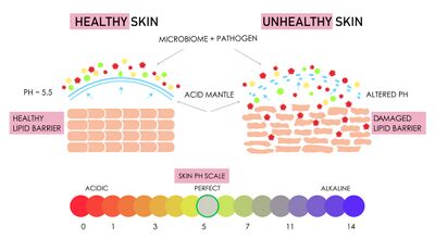
Immune function: Our skin contains a protective acidic barrier called the acid mantle. This acidic layer has a pH between 4.2 and 6.0, which creates a hostile environment for harmful invading organisms while maintaining a favourable environment for beneficial microbes.[1] The acid nature of skin is a key requisite for healthy skin. Skin pH can affect the synthesis and maintenance of a competent skin barrier, play a role in skin pigmentation, and ion homeostasis.[7]The skin's acid mantleSkin pH tends to be lower in people with darker skin because melanin by-products are acidic. Skin pH also tends to increase slightly as we age, which can contribute to an increased risk of infection and alter our healing ability.[1]
Specific immune cells and proteins contained in the dermis activate the immune response and attack invading microbes. These cells include: Langerhans cells, memory T cells, and lymphoid cells.[5][8]
The surface of our skin houses millions of bacteria, fungi and viruses that compose the skin microbiome and serves as a physical barrier to prevent the invasion of pathogens. As in our gut, the skin microbiome plays essential roles in the protection against invading pathogens, the education of the immune system,[9] and contributing back to the pH of the acid mantle.[1]
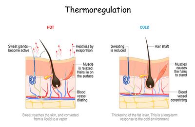
Thermoregulation and body homeostasisThermoregulation: Humans, and all mammals, can maintain a stable core body temperature via thermoregulatory responses.[10] By increasing blood flow to the skin through vessel dilation, body temperates are lowered by evaporative cooling of moist/sweaty surfaces to release body heat.[1][11] However, if water lost to evaporative cooling is not replaced, body fluid homeostasis will be challenged.[11]
Prevention of fluid loss: In addition to the physical barrier provided by skin, it also contains lipids, proteins, amino acids, and salts that work to maintain internal body homeostasis by attracting and holding onto water. Due to this mechanism, under normal circumstances, the outer layer of our skin is about 30% water.[1]
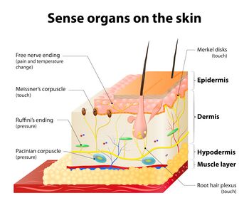
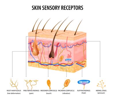
Synthesis of vitamin D: Vitamin D is recognised as a pro-hormone, also known as calciferol. There are two major forms of vitamin D: D2 which is human-made and fortified into foods (such as milk, cheese, yogurt, cereals, and juices) and D3 which is synthesised by the skin and from eating animal-based foods (fatty fish, fish liver oil, and egg yolk).[12] While vitamin D can be ingested through food or supplements, the skin and exposure to sunlight is the body's primary source of vitamin D.[13] Vitamin D is essential for calcium and phosphate absorption, bone formation, renal function, and our immune function.[1][12]Skin sensory organs are located throughout the layers of the skin.
Protection from ultraviolet radiation:[14] Ultraviolet radiation (UVR) can cause DNA photodamage, sunburn, and both local and systemic immunosuppressive properties.[15] Melanin and carotene give skin its colour and serve to reflect UVR as a protective mechanism to radiation damage.[1] Melanin also has antioxidant and radical scavenging properties.[15] Melanin is produced by melanocytes in response to increased sunlight, which is why populations that evolved in areas with more sun exposure tend to have darker skin.[1]
Interaction with the environment: Our skin gathers sensory information through free nerve endings, hairs, receptors to touch, temperature, and pain. In addition, through physiological processes like sweating or blushing, information is shared about our internal state to the outside world.[1]
Healing: Tissue restoration in response to injury.[1]
The following optional video provides an overview of the roles and functions of the skin.
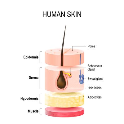
It is important to understand the layers of our skin so that we can understand how healing occurs differently based on depth. The skin has two principal layers, the epidermis and the dermis. The hypodermis is considered an extension of the skin by some sources, but not by others.[1]
The Epidermis
The epidermis is the outer layer of your skin, mostly made up of cells called keratinocytes. These cells fit tightly together to create a protective barrier. They also help fight off infections by using innate immune receptors (like Toll-like receptors or TLRs) and releasing antimicrobial peptides (AMPs) to protect your body.[8]
Composed of five layers
It is avascular
Its thickness varies based on location. For example, it is thickest on the heels and thinnest on the eyelids. Areas that have increased use from friction or weight bearing can build up thicker layers of skin (e.g., where a pencil rubs your writing finger or shoe rubs against your foot).
It has no nerves, but free nerve endings from the dermis do extend into the mid layers of the epidermis.
Five layers of the epidermis (most to least superficial):
Stratum corneum
Composed of 15 to 30 layers of keratinocytes called squames or corneocytes. These are dead keratinocytes. They contain a high concentration of keratin which provides a waterproof barrier for the skin, hair, and nails.
This layer is continually being shed from the body. Shed cells are replaced via the process of skin cell migration from the stratum basale. This process takes an average of 30 days, but this varies based on age and certain health conditions.[1]
Stratum lucidum
Contains two to three layers of keratinocytes and is not living. It can be penetrated or shaved off without awareness.
It is only found in areas of thick skin, like the palms of the hand and the soles of the feet. Present in calluses.[1]
Stratum granulosum
This layer contains the greatest concentration of free nerve endings that extend from the dermis. Free nerve endings are unencapsulated dendrites originating from a sensory neuron. They are the most common nerve endings in skin and provide sensory information about painful stimuli, hot and cold, and light touch. However, they are less sensitive to abrupt changes in stimulation.[17]
This is the most superficial layer of the epidermis which contains living cells.[1]
Stratum spinosum
Contains Langerhans cells and lymphocytes which play an important role in the immune system.[1]
Stratum basale
The only layer that undergoes continuous mitosis to produce new cells.[1]
Keratinocytes are constantly being produced in the stratum basale and they move up through the layers until they reach the outermost layer.[1] Keratinocytes are the most dominant cell type in the skin. They play a critical role in wound healing as they are structural cells and they perform important immune functions.[18]
Melanocytes are also produced in the stratum basale. They produce melanin, which contributes to the colour of skin. Humans have approximately the same amount of melanocytes. Therefore, skin colour is based on the amount of melanin that these melanocytes produce in response to their environment.[1]
This layer also contains Merkel cells which can perform both nervous and endocrine actions. They can synthesise and store locally produced hormones and neurotransmitters. They function as mechanoreceptors[1] for light and selective tactile perception, but not for hard touch and vibration; they are also involved in the transfer of nociceptive signals.[19]
The Dermis
Serving as the primary home for most skin-resident immune cells under homeostatic conditions, including T cells and myeloid cells like dermal dendritic cells and macrophages, the dermis is a fibrous layer composed of connective tissue.[8]
Located deep to the epidermis
Contains blood vessels and nerves which supply the epidermis via capillary loops and free nerve endings
Two layers of the dermis (most to least superficial):
Papillary layer
Interdigitates with the epidermis
The ridges of this layer give rise to our unique fingerprints.
Contains fibroblasts which are responsible for the production of collagen, elastin, and proteins. These qualities give skin strength and flexibility.
Contains mast cells which produce heparin and histamine, important factors in clot formation and the inflammatory response.
Contains macrophages which play an important role in the immune response, wound repair, cancer defence, salt balance, and hair regeneration. They are known for destroying foreign invaders through phagocytosis[1] (the process by which a phagocyte, a type of white blood cell, engulfs and digests foreign cells and removes dead cells[1][20]).
Contains leukocytes[1] which are crucial to the inflammatory response following an injury to the skin. Leukocytes are essential for clearing infection and normal wound healing.[21]
Reticular layer
Located between the papillary layer and the subcutaneous layer or hypodermis.
It is made up of collagen, blood vessels, nerve endings, T-cells, hair follicles and glands.
The hair follicles contain stem cells that produce keratinocytes that will become hair. They play an important role in wound healing by contributing epithelial cells for wound closure.
The T-lymphocytes are responsible for destroying pathogens and malignant cells.
Nerves located within the dermis detect sensations such as itching, touch, pressure, vibration, pain, and temperature.
Injuries which reach into the dermis can result in pain due to nerve exposure and or damage. There will be an absence of pain if the nerves are completely destroyed and or severed by an injury. [1]
The Hypodermis
Located below the dermis and contains subcutaneous tissue.
It is made up of loose connective tissue, adipose tissue. It is well vascularised and well innervated.
It helps to attach the skin to the muscles and bones through superficial fascia, and provides insulation and cushioning through fat storage.
Wounds
"A wound is an injury that breaks the skin or other body tissue. Wounds can be open, with broken skin and exposed body tissue, or closed when there is damage to tissue under intact skin."[22]
When there is injury to the skin, wounds and tissue loss can be categorised based on their depth and also the tissues involved.[23]
Loss of the superficial epidermis only, no dermal loss
Unlikely
Superficial abrasions
Stage one pressure injuries
Superficial burns (first-degree burns)
Local inflammatory process and epidermal replacement from keratinocyte migration upward
Erythema and
Mild and short-lived pain
Partial Thickness
Loss of the epidermis and part of the dermis
Yes
Stage two pressure injuries
Superficial and deep partial-thickness burns (second-degree burns)
Skin tears
Some deep abrasions
Re-epithelialisation as a result of epithelial cell migration from the wound edges towards the centre of the wound
Bleeding
Seeping of serous fluid
Blistering
Pain
Full Thickness
Loss of both the epidermis and dermis with extension into the subcutaneous tissue
Yes
Full-thickness burns (third-degree burns)
Stage three and four pressure injuries
Surgical incisions
Traumatic wounds
Necrotic wounds which require debridement
Via secondary intention (see below for definition)
May involve bone, tendon, ligament, or muscle
Bleeding
Seeping of serous fluid
Pain
Normal Tissue Healing
A clear understanding of the normal or expected healing process is important to recognise when improper healing is occurring in a wound. The process of tissue repair is incredibly complex, and involves many body systems and complex mechanisms.
There are four basic mechanisms by which healing takes place:[1]
Continuous cell cycling
Normal intact skin is replaced via keratinocyte production in the stratum basale, followed by upward migration through the layers of the epidermis.
Cell proliferation
Healthy cells undergo mitosis to repair damage. The new tissue formed through this process is called granulation tissue.
The structure and function of the replaced tissue cannot be duplicated, but it is similar to the original.
Results in scarring.
Regeneration
This type of healing can be performed by only a few types of tissue in the human body, including: the liver, kidney, gastrointestinal tract and the epidermis. No other tissue types can heal in this manner.
This type of healing involves a complete duplication of structure and function.
Fibroproliferative healing
This is a form of pathological healing where lost tissue is replaced by a fibrous scar.
Occurs in deep wounds, persistent inflammation, fibroproliferative diseases
The following optional video provides a time lapse of wound healing. Can you note the various stages of tissue healing?
The longer a wound is open, the more significant the residual scar will be. It is important to reduce healing time and provide the patient with realistic expectations.
The tissue is free from contamination with minimal tissue loss.
These wounds are closed with external force using sutures, staples, adhesive strips or glue.
The primary mechanism of healing is epidermal regeneration. Scarring should be minimal.
There should be no complications. Wound healing and closure occurs in about two weeks time.
Delayed primary intention
Surgical wounds, made by an incision. However, the wound is left open (did not approximate the wound edges) due to concern about contamination, active infection, or significant tissue loss which could result in the wound reopening (dehiscing).
The wound is closed with sutures, staples, grafting, or skin flap placement after it has undergone further healing, oedema management, infection control/antibiotics, or debridement of debris.
There is potential for significant scarring if closure is delayed or chronic inflammation sets in.
Secondary intention
This is the process that the majority of wound care practice revolves around.
Wounds extend into the subdermal tissue and heal as a result of the body's inflammatory response, formation of new granulation tissue to fill the empty space, and then closure via re-epithelialisation (migration of new skin cells over the surface of the wound).
The edges are approximated through wound contraction due to myofibroblasts.
Wounds typically take weeks to months to close, depending on size and other factors that may complicate the process.
Partial thickness wounds that heal by re-epithelialisation
Not often treated with skilled wound care practice.
Subdermal layers are not involved, so wound contraction is not required to approximate the wound edges.
Minimal to no granulation tissue, minimal to no scarring.
The duration to closure depends on depth, but ranges from one to two weeks.
The body goes through four phases of tissue healing with every wound, regardless of depth or severity. These phases are not consecutive, but they can overlap. An acute wound typically closes in about 21 days, but the entire healing process can last up to two years. A wound is considered chronic when healing is disrupted and the wound is still open after four weeks.
Blood vessels respond to injury immediately with vasoconstriction to prevent blood loss and further tissue injury.
Platelets and fibrin arrive at the site to form a clot which creates a temporary barrier to the external environment. Once dry, these clots form a scab. The scab is the body's temporary wound dressing that prevents blood loss and provides protection from the outside world while tissue repair and healing take place underneath.
This process begins within seconds and it continues for the first 12 hours after injury.
Inflammatory phase
This phase involves the destruction of any pathogens that may have entered the body, debris and necrotic tissue removal, and the stimulation of new blood vessel growth.
Neutrophils, mast cells, and macrophages play a big role in this phase.
The inflammatory phase occurs in the first 24 hours after injury and typically lasts for about one week. It is seen clinically as redness, swelling, heat, and pain. The process is essential to acute wound healing, but can become problematic if it persists for too long.
Proliferative phase
This phase of repair is characterised by new blood vessel formation or angiogenesis, connective tissue growth or fibroplasia, new skin cell formation or epithelialisation, and continued cleaning out of any remaining debris.
Neutrophils, mast cells, and macrophages are still present during this phase. However, the most predominant types are fibroblasts and endothelial cells, which are responsible for the growth of granulation tissue and capillaries. Fibroblasts initially produce type three collagen, which tends to be weaker and more disorganised than type one collagen. New tissue growth will be weaker than the original tissue and more susceptible to injury from outside sources.
Granulation tissue is bright red, beady, vascular tissue that fills in the wound cavity. It is made up of collagen, elastin, and blood vessels.
Within hours of injury, re-epithelisation begins with upward migration of the existing keratinocytes. Within a few days, the process continues with cell proliferation via mitosis.
The time it takes for a wound to be considered closed varies based on the circumferential size and also the depth. There will be a visible scar if the wound takes more than three to four weeks to re-epithelialise.
The proliferative phase begins four to six days after injury and can last from three weeks to two months. The wound may appear closed once it is re-epithelialised, but it is not considered healed until the next phase is complete.
Remodelling phase
During this phase, the wound contracts, granulation tissue settles, blood flow returns to pre-injury levels, and the wound tensile strength increases.
Myofibroblasts are responsible for wound contraction.
Fibroblasts, myofibroblasts, endothelial cells, and macrophages are the most predominant cells, their numbers slowly decrease as this phase progresses.
Type three collagen is slowly replaced by type one collagen, which has increased tensile strength.
The remodelling phase begins around two weeks after injury and lasts up to two years. The wound may appear healed before this process is complete.
Around six weeks after injury, the wound has about half its ultimate tensile strength. Once the remodelling phase is complete, that injured area will have approximately 80% of its original strength. These timelines are important to keep in mind when treating patients with chronic or repeated wounds.
The overlapping phases of wound healing
Chronic Wounds
Chronic wounds are those that do not follow this normal process of healing. They are not closed or making significant progress towards healing in three weeks due to a disruption in the healing process. These are the wounds most likely to be treated in wound care practice.[1]
The skin microbiome of chronic wounds often tips in favour of the harmful microbiome over the beneficial ones. These microbes can trigger the immune system to attack the body's cells required for healing rather than attacking the invaders. Chronic wounds may become stuck in one or more phases that persist for months or years; the inflammatory phase is the most common phase to be stuck in. It is common for chronic wounds to go through periods of healing, stagnation, regression, and reoccurrence.[1]
Chronic wounds typically have one or more of the following characteristics:[1]
Multifactorial aetiology
Advanced age or obesity
Have had a previous wound, injury or surgery, especially in the same area
Multiple co-morbidities particularly vascular diseases, diabetes, or auto-immune conditions
Poor tissue perfusion or oxygenation
Prolonged standing or dependent positioning
Mechanical forces, pressures, or recurrent trauma
Medications that can affect wound healing
Inadequate or inappropriate care
Malnutrition
Active infection, presence of a bacterial biofilm or changes in the skin microbiome
↑Sindhu PE, Taj T, Fakrudheen F, Sahana TG, Ahmed MG. Introduction to wound healing: Factors affecting, phases, and principles of wound healing. In Nanomaterials for Wound Healing 2025 May 30 (pp. 1-18). CRC Press.
↑Tripathi S, Manna T, Dey S, Majumder S, Manna M, Das A, Panda AK, Ghosh C. Classification of the Wound and Their Nature. In Functionalized Nanoparticles Hydrogels for Wound Healing 2026 Jan 2 (pp. 47-67). Singapore: Springer Nature Singapore.