
Principles of Amputation
Original Editor - Lucy Coughlan as part of the World Physiotherapy Network for Amputee Rehabilitation Project
Top Contributors - Admin, Aicha Benyaich, Tarina van der Stockt, Stacy Schiurring, Rachael Lowe, Jess Bell, Tony Lowe, Kim Jackson, Shaimaa Eldib, Sheik Abdul Khadir, Rucha Gadgil, Lauren Lopez, Simisola Ajeyalemi, Amrita Patro and Wanda van Niekerk
Introduction
Amputation is the surgical removal of a limb, or part of a limb.[1] It may be required following trauma, prolonged constriction of the limb, or may be performed as a planned procedure to control pain or a disease process in the affected limb, such as malignancy, infection, or gangrene. Prolonged constriction refers to sustained circumferential compression that cuts off blood supply to the limb; this may be congenital, as in constriction band syndrome, or acquired, such as a tourniquet or hair tourniquet left in place too long. In some cases, amputation is carried out as a preventative measure to avoid such problems developing, and some individuals are born with limb absence due to congenital disorders. Every care should be taken to ensure that the amputation is performed only when clinically indicated, that is when the limb is non-viable (e.g. gangrenous or grossly ischaemic), dangerous (e.g. malignancy or infection), or non-functional.[2]
This page provides an overview of the key principles guiding the decision to amputate, the goals and terminology of amputation surgery, and the surgical considerations, advantages, and disadvantages associated with each level of lower and upper limb amputation.
Assessment for Amputation
A decision to amputate a limb should be made through discussion with the interdisciplinary team, including the patient, wherever possible. In an emergency situation, the decision should be made based on medical need. A number of investigations may be used to assess limb viability and the underlying cause, examining the bones, soft tissues, and blood vessels.
| Investigation | What it assesses |
|---|---|
| Pulse examination and ankle-brachial index (ABI) |
|
| Toe-brachial index (TBI) |
|
| X-ray | Bones: fractures or bone disease |
| Computerised tomography (CT) scan | Detailed cross-sectional images of bone, soft tissue, and blood vessels |
| Magnetic resonance imaging (MRI) |
|
| Duplex/colour Doppler ultrasound |
|
| Angiography (CT, MR, or catheter) |
|
| Venogram | Detailed imaging of the venous system |
| Transcutaneous oxygen pressure (TcPO2) |
|
The choice of investigation depends on the suspected underlying cause: vascular tests such as ABI, TBI, duplex ultrasound, and angiography help establish whether blood supply to the limb remains intact, while X-ray, CT, and MRI are more broadly useful for assessing bone integrity, infection spread, or tumour extent.
Arterial Supply of the Limbs
Understanding the normal arterial supply of the limbs helps clinicians interpret vascular investigations, localise the level of an occlusion or injury, and anticipate how proximally an area of compromised blood flow may extend, all of which inform diagnosis and the eventual choice of amputation level.

Lower limb: the leg is supplied by a staged arterial chain running from the groin to the foot:
- External iliac artery, which becomes the femoral artery as it passes beneath the inguinal ligament
- Femoral artery, which gives off the profunda femoris (deep femoral) artery, the main supply to the thigh, then runs down the thigh and becomes the popliteal artery at the adductor hiatus
- Popliteal artery, which divides behind the knee into the anterior tibial artery and the tibioperoneal trunk
- Anterior tibial artery, which runs down the front of the leg and continues into the foot as the dorsalis pedis artery
- Tibioperoneal trunk, which divides into the posterior tibial artery and the fibular (peroneal) artery
- Posterior tibial artery, which passes behind the medial malleolus and divides into the medial and lateral plantar arteries supplying the sole of the foot
- Fibular (peroneal) artery, which supplies the lateral compartment of the leg and contributes to the collateral network around the ankle

Upper limb: the arm and hand are supplied by an arterial chain running from the shoulder to the digits:
- Subclavian artery, which becomes the axillary artery at the lateral border of the first rib
- Axillary artery, which becomes the brachial artery at the inferior border of teres major
- Brachial artery, which divides at the cubital fossa into the radial and ulnar arteries of the forearm
- Radial and ulnar arteries, which join in the hand to form the superficial and deep palmar arches
- Palmar arches, which give rise to the digital arteries supplying the fingers
In vascular disease, these arteries can become blocked or narrowed over time, reducing circulation to the affected limb. As the narrowing becomes more severe, blood supply can fail to meet the needs of the tissues even at rest, producing pain at rest and eventually tissue damage in the form of non-healing wounds, ulceration, or gangrene.[8] This tissue damage is the pathway through which vascular disease can lead to amputation. Atherosclerotic disease leading to amputation is much less common in the upper limb than in the lower limb. For more information, see Pathology Leading to Amputation.
In trauma,[9] one or more of these blood vessels may be ruptured beyond repair (e.g. in a road traffic collision, gunshot wound, or blast injury). In this situation, amputation is performed because the limb has no blood supply beyond the level of injury and is therefore non-viable.
A number of injury severity scores may be used alongside clinical investigations to establish the likelihood of successful limb salvage, including:
- Mangled Extremity Severity Score (MESS)[10][11]
- Nerve Injury, Ischemia, Soft-Tissue Injury, Skeletal Injury, Shock, and Age of Patient Score (NISSSA) developed by McNamara et al. as a modification of the MESS, adding assessment of nerve injury[12]
- Limb Salvage Index (LSI)[13]
Amputations are broadly classified as minor (e.g. toe and partial foot amputations) or major (e.g. amputation through or above the ankle).[14] Once a decision has been made to amputate, the level of amputation must be chosen carefully, as it has significant consequences.[15] The chosen level should allow all dead or diseased tissue to be fully resected and it must have an adequate blood supply for the wound to heal. Any infected or diseased tissue left behind may require further surgery. Each amputation level has its own prosthetic considerations, so the level should be chosen with function as well as healing in mind.[16] Residual limb length and shape also affect prosthetic fit and comfort.[17]
Goals of Amputation Surgery
Amputation surgery is reconstructive in intent, not simply ablative.[18] Key goals including controlling pain in both the short and long term and preserving as much residual limb length as adequate soft-tissue coverage allows.
The surgeon’s choice of muscle stabilisation technique is one way these goals are put into practice. Myodesis secures the muscle and its fascia directly to bone by suturing the muscle and its fascia to bone, typically through drill holes.[19] It can be performed at transtibial, transfemoral, and other amputation levels. By anchoring the muscle so that it cannot retract or slide over the bone end, myodesis maintains muscle length and function, helping to prevent deformity of the residual limb and supporting control of the prosthesis.[20] Myoplasty takes a different approach, attaching divided muscles to their opposing (antagonist) muscles rather than to bone.[19]
Wound healing should be achieved in a way that supports prosthetic fitting, but residual limb length should not be preserved at the expense of healthy tissue for wound closure.[21] Mobility of adjacent joints should be preserved and contractures avoided, as contractures increase energy expenditure during prosthetic gait and can compromise prosthetic fitting. The multidisciplinary team should be involved early, ideally before surgery where the clinical picture allows, so that rehabilitation and prosthetic goals inform surgical decisions.[14]
The traditional approach to nerve management is traction neurectomy. Nerves are placed under gentle traction and divided cleanly, so the cut end retracts proximally into well-vascularised tissue. This moves the resulting neuroma away from the end of the residual limb and the future prosthetic socket interface, aiming to reduce the risk of it becoming painful.[21][22] However, symptomatic neuromas remain a recognised complication of amputation.[22] More recent surgical techniques aimed at reducing neuroma-related pain and phantom limb pain include:
- Targeted muscle reinnervation (TMR): residual nerves from the amputated limb are joined to the motor nerve branch of a nearby muscle that no longer has a functional role. This gives the regenerating nerve fibres a new target.[22][23]
- Regenerative peripheral nerve interfaces (RPNI): the cut end of a nerve is wrapped in a small graft of muscle (or sometimes skin) taken from elsewhere in the body, which the nerve then reinnervates.[24]
Evidence for both techniques is growing but should still be regarded as an evolving area of practice rather than a universally established standard of care.
The following videos provide an overview of targeted muscle reinnervation and regenerative peripheral nerve interface procedures and potential benefits/outcomes:
Lower Limb Amputations
Lower limb amputation levels range from the toes to the hip, and the level is chosen based on the factors described in Assessment for Amputation above.
| Level | Description | Advantages | Disadvantages |
|---|---|---|---|
| Toe[14][20] | Through the phalanges or disarticulation at the metatarsophalangeal joint. Most common lower-limb amputation. | Usually only a mild effect on function |
|
| Ray[14][20] | The toe plus its corresponding metatarsal | Preserves the rest of the foot |
|
| Transmetatarsal[14][20] | Through the metatarsals | Excellent function achievable with good wound healing |
|
| Midfoot/hindfoot[20] |
Lisfranc and Chopart are midfoot disarticulations; Syme's is hindfoot level |
|
|
-
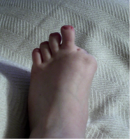 Great toe amputation
 -
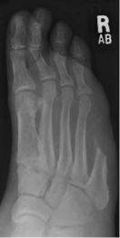 5th ray amputation
 -
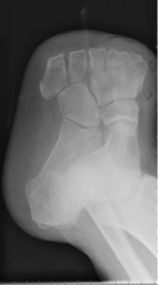 Transmetatarsal amputation
 -
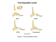 Midfoot/hindfoot amputation levels


| Level | Description | Advantages | Disadvantages |
|---|---|---|---|
| Transtibial (BKA) |
|
|
|
-
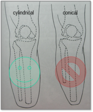 Cylindrical residual limb (fibula 5-10 mm shorter)
 -
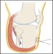 Long posterior muscular flap covering the distal end, attached ventrally to a shorter anterior flap (ventral suture)
 -
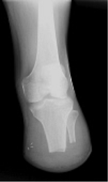
-
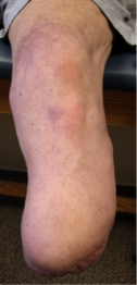

.png)


| Level | Description | Advantages | Disadvantages |
|---|---|---|---|
| Knee disarticulation[20] | Through the knee joint |
|
|


| Level | Description | Advantages | Disadvantages |
|---|---|---|---|
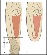
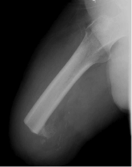
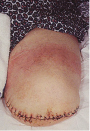
| Transfemoral (AKA) | Through the femur; current guidance favours preserving maximum length where soft tissue allows | Longer residual limb: better lever arm, muscular balance, and energy efficiency; ischial tuberosity-bearing prosthesis candidacy | Shorter residual limb: weaker adductors, flexion/abduction positioning, and substantially higher energy cost |
-
-
-
-
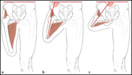 Very short trans-femoral residual limbs often end up in abduction because there is an imbalance between the adductor and abductor muscles.


| Level | Description | Advantages | Disadvantages |
|---|---|---|---|
| Hip disarticulation | Through the hip joint | Candidacy for an ischial tuberosity-bearing prosthesis | High risk of wound complications and mortality; usually wheelchair-dependent |
| Hemipelvectomy | Whole limb plus ipsilateral hemipelvis | Rare candidacy for a trunk/contralateral IT-bearing prosthesis | Rare, complex procedure; usually wheelchair-dependent |

Surgical and Residual Lower Limb Length Notes:
- Across all levels, nerves are dissected under gentle tension so the cut end retracts into well-vascularised tissue, reducing the risk of neuroma-related pain.
- Transtibial: below the ~12–17 cm ideal range, function becomes progressively more compromised; a residual limb under roughly 9 cm may require full fibula removal with additional muscle bulk, and under roughly 5 cm a more proximal level should usually be considered instead.[28] An ultra-short 5–6 cm stump is occasionally chosen deliberately to preserve the knee joint.
- Transfemoral: there is no single agreed optimum length; the literature ranges from roughly 9–17 cm above the knee joint line, or is expressed as around two-thirds of the contralateral femur length. Myodesis anchors the adductors (and sometimes hamstrings) to bone; myoplasty sutures the hamstrings and quadriceps together over the residual femur; a Gottschalk myodesis additionally fixes the adductor magnus trans-osseously to pad the distal femoral end.
Optional additional reading about lower limb amputations:
- Partial-Foot Amputations: Surgical Procedures
- Transtibial Amputation: Surgical Procedures and Immediate Postsurgical Management
- Knee Disarticulation: Surgical Procedures
- Transfemoral Amputation: Surgical Procedures
- Hip Disarticulation and Transpelvic Amputation: Surgical Procedures
- Lower Limb Amputation Postoperative Care
- Rotationplasty
Energy Expenditure by Amputation Level
Energy expenditure during prosthetic gait increases the more proximal the amputation level, and is generally higher after dysvascular than traumatic amputation, since people with traumatic amputation are typically younger with higher baseline fitness and can compensate more effectively.[29]
Upper Limb Amputations
Upper limb amputations are seen considerably less often than lower limb amputations, and trauma is the leading cause of upper limb amputation in adults,[9] in contrast to lower limb amputation, which is predominantly dysvascular in origin.
| Level | Description |
|---|---|
| Fingers | Partial or complete digit amputation |
| Partial hand | Through the metacarpals or carpus |
| Wrist disarticulation | Through the radiocarpal joint |
| Transradial | Through the forearm (radius and ulna) |
| Elbow disarticulation | Through the elbow joint |
| Transhumeral | Through the humerus |
| Shoulder disarticulation | Through the shoulder joint |
| Interscapulothoracic (forequarter) amputation | Removal of the whole upper limb together with the scapula and all or part of the clavicle |

Optional additional reading about upper limb amputations:
- Partial hand
- Wrist disarticulation
- Transradial
- Elbow disarticulation
- Transhumeral
- Shoulder disarticulation
- Scapulo-thoracic dissociation (forequarter)
A range of prosthetic options is available for the upper limb, including passive (cosmetic or functional), body-powered, externally powered (myoelectric), hybrid, and activity-specific devices. As with the lower limb, the amputation level strongly influences which prosthetic options are viable and how much residual native joint function remains for the person to control the device.[30]
Prosthesis rejection rates rise fairly consistently with more proximal amputation level across the literature, though the exact figures vary considerably between studies: transradial users report the lowest rejection rates, with transhumeral and shoulder disarticulation users reporting substantially higher rates, most often attributed to limited device functionality relative to the effort of control.[31][32] This is one of several reasons prosthetic and functional goals are best considered early and collaboratively with the patient.
Resources
Optional Additional Videos
Warning: video links to real surgical procedures
References
- ↑ Limb Loss Definitions. Fact Sheet. Amputee Coalition 2008. http://www.amputee-coalition.org/resources/limb-loss-definitions/ [accessed 31 August 2026]
- ↑ Clinical Practice Guideline for Rehabilitation of Lower Limb Amputation. Department of Veterans Affairs, Department of Defence. 2007
- ↑ McClary KN, Massey P. Ankle Brachial Index. InStatPearls [internet] 2023 January 16. StatPearls publishing.
- ↑ Casanegra AI, Liedl DA, McCarter C, Shuja F, Wennberg PW. Toe-brachial index: utility, futility, and diagnostic criteria. Angiology. 2026 Feb;77(2):221-8.
- ↑ Catella J, Long A, Mazzolai L. What is currently the role of TcPO2 in the choice of the amputation level of lower limbs? A comprehensive review. Journal of clinical medicine. 2021 Apr 1;10(7):1413.
- ↑ Arsenault KA, Al-Otaibi A, Devereaux PJ, Thorlund K, Tittley JG, Whitlock RP. The use of transcutaneous oximetry to predict healing complications of lower limb amputations: a systematic review and meta-analysis. Eur J Vasc Endovasc Surg. 2012 Mar;43(3):329-36.
- ↑ Nishio H, Minakata K, Kawaguchi A, Kumagai M, Ikeda T, Shimizu A, et al. Transcutaneous oxygen pressure as a surrogate index of lower limb amputation. Int Angiol. 2016 Dec;35(6):565-72.
- ↑ Bethel M, Annex BH. Peripheral arterial disease: A small and large vessel problem. Am Heart J Plus. 2023 Mar 23;28:100291.
- ↑ 9.0 9.1 Devinuwara K, Dworak-Kula A, O'Connor RJ. Rehabilitation and prosthetics post-amputation. Orthopaedics and Trauma. 2018 Aug 1;32(4):234-40.
- ↑ Johansen et al (1990) Objective Criteria Accurately Predict Amputation following Lower Extremity Trauma. J Trauma.30: 568-73
- ↑ Omoto R, Umemura Y, Hanaoka K, Yonao Y, Kai H, Oi K, et al. Re-evaluation of cutting criteria for limb amputation using the Mangled Extremity Severity Score. Trauma Surg Acute Care Open. 2025 Mar 28;10(1):e001694. d
- ↑ McNamara et al (1994) Severe Open Fractures of the Lower Extremity: A Retrospective Evaluation of the Mangled Extremity Severity Score (MESS). J Orthop Trauma 8: 81-7
- ↑ Russell et al (1991) Limb Salvage Versus Traumatic Amputation A Decision Based on a Seven-part Predictive Index Ann Surg. 213: 473-81
- ↑ 14.0 14.1 14.2 14.3 14.4 Guest F, Marshall C, Stansby G. Amputation and rehabilitation. Surgery (Oxford). 2019 Feb 1;37(2):102-5.
- ↑ Perkins ZB, De'Ath HD, Sharp G, Tai NR. Factors affecting outcome after traumatic limb amputation. Br J Surg. 2012 Jan;99 Suppl 1:75-86.
- ↑ Crowe CS, Impastato KA, Donaghy AC, Earl C, Friedly JL, Keys KA. Prosthetic and orthotic options for lower extremity amputation and reconstruction. Plast Aesthet Res. 2019;6:4.
- ↑ Isaacs-Itua A, Sedki I. Management of lower limb amputations. Br J Hosp Med (Lond). 2018 Apr 2;79(4):205-210.
- ↑ Potter BK. Editorial comment: Symposium: Recent advances in amputation surgery and rehabilitation. Clin Orthop Relat Res. 2014 Oct;472(10):2938-41.
- ↑ 19.0 19.1 Fabre I, Thompson D, Gwilym B, Jones K, Pinzur M, Geertzen JHB, et al. Surgical techniques of, and outcomes after, distal muscle stabilization in transfemoral amputation: a systematic review and narrative synthesis. Ann Vasc Surg. 2024 Jan;98:182-193.
- ↑ 20.0 20.1 20.2 20.3 20.4 20.5 Spires MC, Kelly BM, Davis AJ, editors. Prosthetic restoration and rehabilitation of the upper and lower extremity. Demos Medical Publishing; 2013 Dec 19.
- ↑ 21.0 21.1 Tintle S, Keeling J, Shawen S, Forsberg J, Potter B. Traumatic and trauma-related amputations part I general principles and lower-extremity amputations. Journal of Bone and Joint Surgery. American volume. 2010;92:2852-68.
- ↑ 22.0 22.1 22.2 Peters BR, Russo SA, West JM, Moore AM, Schulz SA. Targeted muscle reinnervation for the management of pain in the setting of major limb amputation. SAGE Open Med. 2020 Sep 15;8:2050312120959180.
- ↑ Cheesborough JE, Smith LH, Kuiken TA, Dumanian GA. Targeted muscle reinnervation and advanced prosthetic arms. Semin Plast Surg. 2015 Feb;29(1):62-72.
- ↑ Leach GA, Dean RA, Kumar NG, Tsai C, Chiarappa FE, Cederna PS, et al. Regenerative peripheral nerve interface surgery: anatomic and technical guide. Plast Reconstr Surg Glob Open. 2023 Jul 17;11(7):e5127.
- ↑ YouTube. What is Targeted Muscle Reinnervation? | Treating Phantom Limb Pain | The Institute for Advanced Reconstruction. Available from: https://www.youtube.com/watch?v=Le3Jbly3vCI [last accessed 31 August 2026]
- ↑ YouTube. What is R.P.N.I.? | Monoped Adventures. Available from: https://www.youtube.com/watch?v=Zd_IyEY4Ju0 [last accessed 31 August 2026]
- ↑ Unterfrauner I, Andronic O, Viehöfer AF, Wirth SH, Berli MC, Waibel FWA. Ulcer occurrence on adjacent toes and hallux valgus deformity after amputation of the second toe in diabetic patients. J Orthop Surg Res. 2023 Feb 13;18(1):99.
- ↑ Wheeless' Textbook of Orthopaedics: Below Knee Amputation.
- ↑ Waters RL, Mulroy S. The energy expenditure of normal and pathologic gait. Gait & posture. 1999 Jul 1;9(3):207-31.
- ↑ American Academy of Physical Medicine and Rehabilitation. Upper Limb Prosthetics. PM&R KnowledgeNow.
- ↑ Biddiss EA, Chau TT. Upper limb prosthesis use and abandonment: a survey of the last 25 years. Prosthetics and orthotics international. 2007 Sep;31(3):236-57.
- ↑ Resnik L, Ekerholm S, Borgia M, Clark MA. A national study of veterans with major upper limb amputation: survey methods, participants, and summary findings. PloS one. 2019 Mar 14;14(3):e0213578.