When refering to evidence in academic writing, you should always try to reference the primary (original) source. That is usually the journal article where the information was first stated. In most cases Physiopedia articles are a secondary source and so should not be used as references. Physiopedia articles are best used to find the original sources of information (see the references list at the bottom of the article).
If you believe that this Physiopedia article is the primary source for the information you are refering to, you can use the button below to access a related citation statement.
The foot and ankle structures are complex, consisting of many bones, joints, ligaments and muscles.[1] A clinician's ability to understand the anatomical structures of the foot and ankle is crucial for assessment and treatment, especially for clinicians working with clients with musculoskeletal conditions. A thorough evaluation ensures accurate diagnosis, appropriate treatment planning, and optimal patient outcomes.[2]
This Physiopedia page covers the essential components of foot and ankle assessment in physiotherapy practice. It follows a systematic approach from subjective history-taking through objective testing and special tests for common pathologies.
Subjective Assessment
The mnemonic L-M-N-O-P-Q-R-S-T ensures comprehensive gathering of patient history and helps develop hypothetical diagnoses while ruling out comorbid conditions.[3]
L: location of symptoms and level of functional impairment
The location of symptoms can help narrow differential diagnoses based on which tissues are affected. When the location of symptoms includes the anterior shin/tibia, consider medial tibial stress syndrome (shin splints). [4]Calf pain with gastrocnemius stretch may lead to the diagnosis of Achilles tendinopathy, particularly if pain increases during the push-off phase of gait. [5][6] Plantar fasciopathy may be characterised by heel pain, which is typically worse with first morning steps.[7][6]Paraesthesias along plantar foot indicate tarsal tunnel syndrome. [8] Morton's neuroma preesents with pain between 3rd and 4th metatarsals.[9]
M: medical factors (medications) and mechanism of injury
We need to find out what medications a patient is taking (including supplements) and what co-morbidities they may have. We also want to determine when and how the injury occurred, including foot position, if remembered:
Forced inversion or eversion: consider ankle sprain or possible fracture[10]
Inability to plantar flex after injury: consider Achilles tendon rupture[11]
Red flags: sudden onset, severe pain without incident or accident, neurological symptoms at time of injury
N: neurological symptoms
Neurological symptoms include numbness, tingling, paraesthesias, sharp or burning pain, and weakness.
When neurological symptoms are present, we need to determine whether they have a sock-like distribution, which may suggest involvement of multiple dermatomes or peripheral nerves. It is essential to consider that peripheral neuropathy (from diabetes, vitamin B12 deficiency, or certain medications) can present similarly.[12]
Clinical tip: Verify duration of symptoms and any changes if peripheral neuropathy is present in medical history.
O: occupation, including limitations
Are there any work- or activity-related factors that are relevant?[13][14] Is there a possibility of overuse?
P: palliating and provoking symptoms
Identify what makes symptoms better or worse. Assess tissue irritability by determining how long symptoms take to resolve after provocation and document positions, activities, or interventions that provide relief.[15]
Red flag: constant, unrelenting symptoms that neither improve nor worsen with any intervention
Q: quality of symptoms/pain
Describe the character of symptoms as well as the pattern and nature of pain. Find out whether symptoms are sharp, dull, stabbing, aching, or electric-shock-like, and whether there is any numbness, tingling, or a feeling of weakness/clumsiness.[3]
R: radiation of symptoms
Document radiation patterns carefully. Ask the following questions relating to radiating symptoms:[6]
What is the exact location of radiating symptoms?
How long did the radiating symptoms last?
Where are the symptoms related to activity or position?
Did symptoms include electric shock sensation, numbness, or tingling?
Red flag: sudden onset of severe pain without incident or accident
T: timing of symptoms
Document the timing of symptoms in 24 hours and in relation to activity. These symptoms include morning stiffness, night pain, and an activity-related pattern.[16]
Red flags: symptoms interrupting sleep, symptoms worse at night, constant symptoms throughout 24 hours.
Constitutional Symptoms (Screening)
Always screen for red flags, such as unexplained weight loss, fever or night sweats, general malaise, recent infections, and a history of cancer.[3]
Patient-Reported Outcome Measures
Standardised questionnaires help quantify impairments, track progress, and establish treatment goals. Choose questionnaires that best capture your patient's specific limitations and functional goals.[17] Review each tool to understand what impairments they assess, ensuring alignment with your patient's presentation.
Standing posture assessment: evaluate static positioning for weight distribution, calcaneal symmetry, foot arch, and overall alignment.[18]
Footwear examination: shoe wear patterns provide valuable insights into overpronation or oversupination, preferential weight-bearing areas, and asymmetrical wear between feet.[19][20]
Specific foot observations should include navicular height, alignment/ deformities in forefoot position, and varus/valgus positioning of the calcaneus.[3]
Oedema Measurement
Standard Circumferential Measurements use bony landmarks to ensure consistency:[3]
metatarsal heads level: 3 cm proximal
around malleoli
10 cm proximal to malleoli
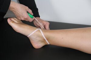
Figure-Eight Measurement may miss metatarsal swelling or areas proximal to malleoli:[3]
Figure-Eight oedema measurementPlace tape between tibialis anterior and lateral malleolus
Pull tape medially across the plantar surface toward the 5th metatarsal
Wrap around the medial malleolus back to the start point
Clinical importance: If swelling extends beyond the figure-eight pattern, use standard circumferential measurements at bony landmarks instead.
Functional Screening
Assess functional capacity with weight-bearing activities that include:[3][21]
Single-leg stance to assess balance and stability
Squatting to assess full weight-bearing dorsiflexion (adjust depth based on patient tolerance)
Sit-to-stand or its alternative for patients with significant pain
Sport-specific movements for athletes when appropriate
Safety consideration: Ensure activities are appropriate for the patient's current symptoms and functional level.
Palpation
Key palpation points at the foot and ankle are listed in Table 1.
Table 1. Key palpation points for the foot and ankle[21]
Bony Prominences
(assess for discomfort)
Medial and lateral malleoli
Talus
Navicular
Cuboid
Cuneiforms
Metatarsals and metatarsal heads
Soft Tissue and Muscle
(assess for discomfort and potential atrophy)
Fibularis muscles
Extensor hallucis longus
Tibialis anterior
Tibialis posterior
Plantar fascia
Range of Motion Testing
It is important to assess active and passive range of motion, including with over-pressure. For more information on the range of motion assessment, please see: Assessing Range of Motion.
Table 2 shows normative values for foot and ankle range of motion and typical end feel.
Table 2. Foot and ankle range of motion and end feel
Movement
Active range of motion
Typical end feel
Dorsiflexion
0-25 degrees
Firm
Plantarflexion
0-50 degrees
Firm
Supination*
0-20 degrees
Firm
Pronation*
0-5 degrees
Firm
Inversion*
0-30 degrees
Firm
Eversion*
0-18 degrees
Firm
*The literature presents vast ranges of subtalar motion ranging from 5° to 65°[22]
Abduction
a few degrees
Firm
Adduction
0-11 degrees
Firm
Flexion (interphalangeal joints)
0-45 degrees
Firm
Extension (interphalangeal joints )
0-70 degrees
Firm
Ankle kinematics can vary between individuals. Factors that affect ankle range of motion include activities of daily living (with geographical and cultural differences), age, gender, and the method used to assess range of motion.
Resisted Range of Motion
Assess the strength of the four compartments of the lower leg muscles ( anterior, superficialposterior, deep posterior, and lateral), as well as dorsal and plantar muscles of the foot.[23]
Clinical importance: weight-bearing dorsiflexion provides more functional information than non-weight-bearing assessment. Perform when patient tolerates weight-bearing.
Neurovascular Testing
Complete neurological screening includes dermatomes, myotomes, reflexes (Achilles reflex), and neural tension.[21]
Accessory Movements/Joint Mobility Testing
Joint mobilisation assessment evaluates joint play and helps identify restrictions that may contribute to functional limitations. Assessment should be completed in open-packed (resting) positions. The left and right sides must be compared for movement quality and end-feel. Documentation should include patient response to mobilisation.[24]
Table 3 identifies which accessory movements you can assess at the foot and ankle:[24]
Table 3. Accessory movements of the foot and ankle
Type of movement
When to test/use
Technique
Calcaneal Inversion and Eversion
to increase subtalar joint mobility
to reduce pain in the subtalar joint
Tibiotalar Posterior Glide
to assess and improve ankle dorsiflexion
Metatarsophalangeal Joint Glides
to assess extension via dorsal glide
to assess flexion via plantar glide
to restore the normal range of motion to the metatarsophalangeal joint
Interphalangeal Joint Glides
to assess extension via dorsal glide
to assess flexion via plantar glide
Additional joints to assess include tibiofibular joint, subtalar joint, and midtarsal joints.[24]
Assessment for Common Foot and Ankle Pathologies
Special tests help confirm clinical hypotheses developed during history and objective examination.
Positive Test: Affected foot falls into neutral or dorsiflexion. The test is negative if the foot remains in slight plantar flexion with the Achilles tendon intact.
Non-Weight-Bearing Position: Patient sitting or supine
Technique:
Stabilise calcaneus with one hand
Maximally extend the first metatarsophalangeal joint with the other hand
Weight-Bearing Position: Patient standing on the step with toes hanging off the edge
Technique:
Therapist maximally extends the first metatarsophalangeal joint
Positive Test: Reproduction of the patient's plantar pain near the calcaneus
Summary
The foot and ankle assessment requires systematic evaluation from subjective history through objective testing and special tests. Using the L-M-N-O-P-Q-R-S-T mnemonic ensures comprehensive data gathering, while standardised outcome measures quantify functional limitations.
Thorough documentation should include a clear description of the mechanism of injury, specific location and quality of symptoms, functional limitations and goals, objective measurements with comparison to the contralateral side, results of special tests, clinical reasoning for diagnosis, as well as treatment plan and prognosis.
During the assessment, clinicians should always consider red flag symptoms requiring immediate referral, peripheral neuropathy affecting assessment reliability, patient tolerance for weight-bearing activities, and appropriate test selection based on acuity of injury.
↑Yavuz U, Celayir A, Karaismailoglu B. Clinical Anatomy of the Foot and Ankle. In: Clinical and Radiological Examination of the Foot and Ankle: The Path to Definitive Diagnosis 2024 Aug 20 (pp. 33-51). Singapore: Springer Nature Singapore.
↑Frain J. Exploring Symptoms: An Evidence-based Approach to the Patient History. John Wiley & Sons; 2025 Jan 13.
↑Lee I, Jeon HG, Ha S, Jeong H, Lee SY. How Medial Tibial Stress Syndrome Is Affected by Alignment, Range of Motion, Strength, and Gait Biomechanics: A Systematic Review and Meta-Analysis. Journal of Sport Rehabilitation. 2024 Nov 22;34(2):134-55.
↑Traweger A, Scott A, Kjaer M, Wezenbeek E, Scattone Silva R, Kennedy JG, Butler JJ, Gomez-Florit M, Gomes ME, Snedeker JG, Dakin SG. Achilles tendinopathy. Nature Reviews Disease Primers. 2025 Mar 27;11(1):20.
↑Haq II, Banerjee AA, Arshad Z, Iqbal AM, Bhatia M. The management of tarsal tunnel syndrome: A scoping review. Journal of Clinical Orthopaedics and Trauma. 2024 Jul 1;54:102489.
↑Matthews BG, Thomson CE, Harding MP, McKinley JC, Ware RS. Treatments for Morton's neuroma. Cochrane Database of Systematic Reviews. 2024(2).
↑Wall R, Plant C, Chapman AW. Ankle fractures in athletes: epidemiology, anatomy, and management considerations. Orthopaedics and Trauma. 2024 Feb 1;38(1):2-6.
↑Şahin E, Tanrıöver A. General Examination of the Ankle. In Clinical and Radiological Examination of the Foot and Ankle: The Path to Definitive Diagnosis 2024 Aug 20 (pp. 83-96). Singapore: Springer Nature Singapore.
↑Malliaras P, Silbernagel KG, de Vos RJ, Bourke J, Sancho I, Hanlon SL, Agergaard AS, Bahr R, Bittencourt NF, Bordalo M, Brorsson A. Diagnostic domains, differential diagnosis and conditions requiring further medical attention that are considered necessary in the assessment for Achilles tendinopathy: a Delphi consensus study. British Journal of Sports Medicine. 2025 Jul 1;59(13):891-901.
↑Kandasamy S, Wukich DK. Foot and Ankle Pain. In Musculoskeletal Pain: Evidence-Based Clinical Evaluation and Management 2025 May 25 (pp. 341-355). Cham: Springer Nature Switzerland.
↑Young CC, Niedfeldt MW, Morris GA, Eerkes KJ. Clinical examination of the foot and ankle. Primary care: Clinics in office practice. 2005 Mar 1;32(1):105-32.
↑Ball P, Johnson GR. Technique for the measurement of hindfoot inversion and eversion and its use to study a normal population. Clinical Biomechanics 11(3):165–169, 1996