
De Quervain's Tenosynovitis
Original Editor - Elizabeth Dallas, Boris Alexandra, Robin Tacchetti
Top Contributors - Wanda van Niekerk, John Winkelhaus, David Cameron, John Fite, Robin Leigh Tacchetti, Robin Tacchetti, Laura Ritchie, Admin, Boris Alexandra, Kim Jackson, Aurelie Canas Perez, Julie Schuermans, Elizabeth Dallas, Lucinda hampton, Corin Arundale, Redisha Jakibanjar, Rachael Lowe, Jess Bell, Stacy Schiurring, Tarina van der Stockt, Jorge Rodríguez Palomino, WikiSysop, Claire Knott, Kai A. Sigel, Arturo Quiroz Marnef, Anas Mohamed, Chrysolite Jyothi Kommu, Naomi O'Reilly, Evan Thomas and 127.0.0.1 - Corin Arundale, David Cameron, John Fite, Bryan Purkey, John Winkelhaus
Definition/Description
De Quervain's Tenosynovitis is a painful condition, historically termed an inflammatory tenosynovitis, caused by tendons on the side of the wrist at the base of the thumb. Pain, which is the main complaint, gets worse with abduction of the thumb, a grasping action of the hand, and an ulnar deviation of the wrist. Thickening and swelling can also be present. [1] [2][3]
Clinical Pearl: Tendon terminology
- Tendinitis – active tendon inflammation, typically acute
- Tendinosis – chronic, non-inflammatory tendon degeneration (the more accurate term for De Quervain's)
- Tendinopathy – umbrella term for tendon pain/dysfunction where the underlying pathology isn't confirmed histologically
- Tenosynovitis – pathology of the tendon sheath rather than the tendon body; anatomically correct for De Quervain's, though "-itis" overstates the inflammatory component
These terms are often used loosely and interchangeably in clinical practice; management doesn't hinge on which one is used.[4][5]
Aetiology

The most common cause is chronic overuse. Activities such as golfing, playing the piano, fly fishing, carpentry, or activities by office workers and musicians can lead to chronic overuse injuries. The classic patient population is mothers of newborns who are repeatedly lifting their baby with their thumbs radially abducted and wrists going from ulnar to radial deviation.
Repetitive gripping, grasping or wringing of objects can cause inflammation of the tendons and tendon sheaths which narrows the first dorsal compartment limiting motion of the tendons.[6] If left untreated, the inflammation and progressive narrowing (stenosis) can lead to scarring that further limits thumb motion. [2][3]
This deposition of the fibrous tissues causes thickening of the tendon sheath, and this can entrap the abductor pollicis longus and extensor pollicis brevis tendons and cause pain. More recent histopathological analysis has identified inflammatory markers, including neutrophil elastase and macrophages, within De Quervain's tissue, suggesting an inflammatory component may still play a role, particularly in earlier stages of the condition.[7]
Relevant Clinical Anatomy

De Quervain's syndrome affects the extensor pollicis brevis (EPB) tendon and the abductor pollicis longus (APL) tendon. These muscles are located on the dorsal side of the forearm and go to the lateral side of the thumb through a fibrous-osseous tunnel made of the processus styloideus radii and the extensor retinaculum. [8]
Extensor pollicis brevis (EPB)
- Origin: ½ dorsal side of the radius, the membrana interossea
- Insertion: base of the proximal phalanx of the thumb
- Function: wrist joint radial abduction; thumb extension
- Innervation: posterior interosseus branch of N. radialis
- Artery: A. interossea posterior
Abductor pollicis longus (APL)
- Origin: dorsal side of the radius and the ulna, the membrana interossea
- Insertion: base of ossis metacarpi
- Function: wrist joint radial abduction; thumb abduction
- Innervations: posterior interosseus branch of N. radialis
- Artery: A. interossea posterior
Epidemiology
Estimated prevalence is 0.5% in men and 1.3% in women. Peak prevalence is usually among individuals between the ages of 40 -50 years.[9] It occures more commonly found in people with a history of medial or lateral epicondylitis. New mothers or child care providers often experience bilateral symptoms, but these symptoms usually subside once the child is lifted less often.[10] In industrial settings, studies have shown a point prevalence of 8% when wrist pain and a positive Finkelstein’s test is present.[2]
Differential Diagnosis
- Osteoarthritis of the first carpometacarpal joint(main differential diagnosis for De Quervain's Tenosynovitis[11])
- Trigger thumb
- Wartenberg's syndrome (superficial radial nerve neuritis)
- Scaphoid or radial styloid fractures
- Intersection syndrome
Clinical Presentation
Examination
The evaluation of a patient with signs and symptoms of De Quervain’s Tenosynovitis begins with a thorough history followed by a physical examination.
History
- Overuse injury vs acute trauma
- Prior history of symptoms
- Repetitive movements of the upper extremity with work or activities of daily living (ADL)
- Hand dominance
- Pregnant or currently in the post-partum stage
- Pain. The primary complaint is radial sided wrist pain (base of thumb and dorsolateral aspect of the wrist near the radial styloid process) that radiates up the forearm with grasping or extension of the thumb, described as a “constant aching, burning, pulling sensation."[12] Aggravated by repetitive lifting, gripping, or twisting motions of the hand (such as opening a jar lid).[12]
Physical Examination
- On palpation, some key, significant findings will be tenderness over the base of the thumb and/or first dorsal compartment extensor tendons on the thumb side of the wrist, particularly over the radial styloid process[2]
- Swelling in the anatomical snuffbox
- Decreased carpometacarpal (CMC) abduction range of motion (ROM) of the first digit
- Palpable thickening of the extensor sheaths of the first dorsal compartment and crepitus of the tendons moving from the extensor sheath [13]
- Other possible findings include: weakness and paraesthesia in the hand[3] and a positive provocative Eichhoff manoeuvre. During this test, the thumb is flexed and held inside a fist. The patient actively deviates the wrist towards the ulnar side. This causes sharp pain along the radial wrist at the first dorsal compartment.[10]
Clinical Pearl: Eichhoff manoeuvre vs Finkelstein test
- Eichhoff manoeuvre – patient makes a fist around the thumb, then actively deviates the wrist ulnarly; this is the version most commonly performed in clinical practice
- Finkelstein test – the original description; a graded, largely examiner-led sequence: active ulnar deviation first, then overpressure if needed, then passive thumb flexion into the palm by the examiner
- The two are frequently confused and used interchangeably, a mix-up traced back to a 1958 paper that mislabelled the Eichhoff manoeuvre as "Finkelstein's test"
- The Eichhoff version tends to produce more false positives than the original Finkelstein sequence, so bear this in mind when interpreting a positive result
Please see this optional Physiopedia Page for more information on this topic.
Treatment Tiers
Generally, there are three tiers of treatment for De Quervain's[11]:
Tier 1: Conservative management, including: splinting, ultrasound, multimodal hand therapy, and activity modification.
Tier 2: Corticosteroid injection
Tier 3: Surgery
Tiers 1 and 2 can be combined dependent on patient presentation and willingness to get a corticosteroid injection.[11]
Non-Surgical Treatment
The aim of non-surgical management is to reduce pain and swelling. Interventions can include:
Patient education regarding avoiding repetitive or aggravating movements[11]
Non-steroidal anti-inflammatory drugs (NSAIDs)
Ice/heat packs
Physical therapy[14]
Occupational therapy

Thumb splinting. Literature supports the use of a forearm brace including the thumb to reduce ulnar deviation and thumb movement.[11][15][16] Clinicians do not agree on the frequency and duration of splint use; some think it should be worn continually for four to six weeks; others recommend wearing it only as needed for pain.[17] Weiss and colleagues[18] found that a 19% improvement was observed when splints were used, but when splint use was combined with NSAIDs, the improvement was 57%. Cavaleri et al.[9] reported that combined orthosis/corticosteroid injection approaches are more effective than either intervention alone in the treatment of de Quervain's disease.
Ultrasound may improve treatment outcomes.[19] Therapeutic ultrasound. Ferrara et al.[20] reported that therapeutic ultrasound may effectively control pain. However, the studies in this systematic review were heterogenous, with poor sample sizes and wide variations in outcome measures. Ultrasound-guided injections. McDermott et al.[21] found that ultrasound-guided injections were beneficial for De Quervain's tenosynovitis. Their results were slightly better than was previously reported in the literature and they reported no adverse reactions.[21] Kume et al.[22] found that ultrasound-guided injections which target the Extensor Pollicis Brevis with septation was more effective than manual injection.
Corticosteroid injection has been reported to be be effective.[15] One or two injections are usually sufficient for pain-relief.[23] If there is no significant improvement in symptoms following two corticosteroid injections, surgical management may be considered. Surgery is usually done in an outpatient setting and the anaesthetic may be local, regional or general.[10] Patients with moderate to severe symptoms usually require cortisone injections in combination with splinting.[11]
In individuals with persistent symptoms the most commonly non-surgical management includes: splinting, systemic anti-inflammatories, and corticosteroid injection.[9][19] [24]
Surgical Treatment
Surgery is rare and is usually selected in cases where non-surgical treatment has failed and the patient experiences persistent inflammation affecting their function.[25] The goal of surgery is to open the dorsal compartment covering to make more room for the irritated tendons. The opening allows pressure relief of the tendons, to ultimately restore free tendon gliding.[26]
Post-operative care is usually straight forward with a simple dressing and no complicated wound care necessary. Individuals are encouraged to start with early use of the hand for light activities of daily living. Sutures are removed after 14 days and individuals are allowed to continue with normal activities. Mild swelling and tenderness around the surgical site may be present for a few months.[10]
Rehabilitation Management
Ice/Heat Packs. Heat can help relax and loosen tight musculature, and ice can be used to help relieve inflammation of the extensor sheath
Strengthening. The progression of exercise therapy is as follows: isometric, eccentric, concentric inner range, theraputy, radial nerve glides.[11]
Patients can start with strengthening exercises once their pain has settled to a manageable level. The strengthening programme should be graded and very gradual. Only progress patients through the strengthening programme if they are able to tolerate their current strength programme for at least 1 week. It is important to progress slowly and that patients strengthen in a pain-free range of motion, to avoid aggravation of symptoms.[11] Below are some examples of strengthening exercises.
-
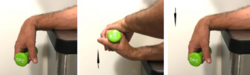 Eccentric wrist extension
 -
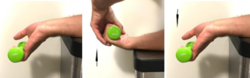 Eccentric wrist flexion
 -
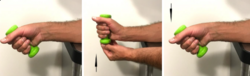 Eccentric wrist radial deviation


Mobilisation. Mobilisation with movement has shown effectiveness in decreasing the pain, improving range of motion, and improving the function of a patient with De-Quervain's tenosynovitis. The therapist provided a manual radial glide of the proximal row of carpals, then asked the patient to move her thumb into radial abduction-adduction.[36] Mobilisation with movement performed for 3 sets of 10 repetitions and followed by eccentric hammer curl exercise with theraband and high voltage electrical stimulation was shown to be effective at 6 months follow-up.[37] Savva et al.[38] investigated the analgesic effect of joint mobilisation in tendinopathy and concluded that the literature on joint mobilisations in tendinopathies such as De Quervain's remains limited since the effect of these techniques have been sparsely reported in a few retrospective case-series and case studies.
Taping. Taping can also be used to decrease pain and improve function.[40]
Ultrasound. Therapeutic ultrasound has also better outcomes in pain reduction and healing. [42]
Education ideas for mothers or care takers with De Quervain's
Red Flags
Red flags to look out for in patients with De Quervain's can include:[11]
- Signs of infection such as unresolved redness or swelling
- High levels of pain (≥ 7/10) after 4 weeks of conservative treatment
- High demand workplace or someone who is forced to continue with aggravating activities
Conclusion
Effective management of DeQuervain’s tenosynovitis will involve a highly individualised, impairment driven approach for the patient in question. Early splinting during the acute phase will prevent aggravation of the tissues, and allow the patient to perform activities essential to self-care and employment. The patient will need to be educated on the tissue healing timetables, as well as why it is important to avoid activities that are aggravating to their symptoms. The progression of exercise therapy is from isometric to eccentric to concentric inner range. Patients should be pain-free before progressing to the next level of strengthening.
References
- ↑ Pagonis T, Ditsios K, Toli P, Givissis P, Christodoulou A. Improved corticosteroid treatment of recalcitrant de Quervain tenosynovitis with a novel 4-point injection technique. The American journal of sports medicine. 2011 Feb;39(2):398-403.
- ↑ 2.0 2.1 2.2 2.3 Ashurst JV, Turco DA, Lieb BE. Tenosynovitis caused by texting: an emerging disease. Journal of Osteopathic Medicine. 2010 May 1;110(5):294-6.
- ↑ 3.0 3.1 3.2 González-iGlesias J, Huijbregts P, Fernández-de-Las-Peñas C, Cleland JA. Differential diagnosis and physical therapy management of a patient with radial wrist pain of 6 months' duration: a case report. journal of orthopaedic & sports physical therapy. 2010 Jun;40(6):361-8.
- ↑ Maffulli N. Overuse tendon conditions: time to change a confusing terminology. Arthroscopy. 1998 Nov;14(8):840-3.
- ↑ Canosa-Carro L, Bravo-Aguilar M, Abuín-Porras V, Almazán-Polo J, García-Pérez-de-Sevilla G, Rodríguez-Costa I, López-López D, Navarro-Flores E, Romero-Morales C. Current understanding of the diagnosis and management of the tendinopathy: An update from the lab to the clinical practice. Disease-a-Month. 2022 Oct 1;68(10):101314.
- ↑ Charzewski P, Starzyk A. Comprehensive insights into de quervain’s tenosynovitis: from etiology to rehabilitation. Quality in Sport. 2025 Feb 3;38:58308-.
- ↑ Kuo YL, Hsu CC, Kuo LC, Wu PT, Shao CJ, Wu KC, Wu TT, Jou IM. Inflammation is present in de Quervain disease—correlation study between biochemical and histopathological evaluation. Annals of plastic surgery. 2015 May 1;74:S146-51.
- ↑ Katechia D, Gujral S. De Quervain's tenosynovitis. InnovAiT. 2017 Sep;10(9):505-9.
- ↑ 9.0 9.1 9.2 Cavaleri R, Schabrun SM, Te M, Chipchase LS. Hand therapy versus corticosteroid injections in the treatment of de Quervain's disease: A systematic review and meta-analysis. Journal of Hand Therapy. 2016 Jan 1;29(1):3-11.
- ↑ 10.0 10.1 10.2 10.3 Satteson E, Tannan SC. De Quervain Tenosynovitis. StatPearls [Internet]. 2021 Aug 8.
- ↑ 11.0 11.1 11.2 11.3 11.4 11.5 11.6 11.7 11.8 Kate Thorn. De Quervain's Tenosynovitis. Plus Course. 2021
- ↑ 12.0 12.1 Walker MJ. Manual physical therapy examination and intervention of a patient with radial wrist pain: a case report. Journal of orthopaedic & sports physical therapy. 2004 Dec;34(12):761-9.
- ↑ Anderson M, Tichenor CJ. A patient with de Quervain's tenosynovitis: a case report using an Australian approach to manual therapy. Physical therapy. 1994 Apr 1;74(4):314-26.
- ↑ Földvári-Nagy L, Takács J, Hetthéssy JR, Mayer ÁA, Szakács N, Szávin-Pósa Á, Lenti K. Treatment of De Quervain's tendinopathy with conservative methods. Orvosi Hetilap. 2020 Mar 1;161(11):419-24.
- ↑ 15.0 15.1 Cuenca-Zaldívar JN, Martínez-Pozas O, Riba E, Rouafi H, Ucero-Lozano R, Selva-Sarzo F, Sinatti P, Comte NC, Romero EA. Conservative treatments for De Quervain's tenosynovitis: A systematic review and network meta-analysis. Journal of Hand Therapy. 2025 Nov 26.
- ↑ Jafarian FS, Sadeghi Demneh E. Effect of Orthotic Interventions on Pain Intensity and Hand Function in Subjects with De Quervain Tenosynovitis: A Systematic Review and Meta-Analysis. Archives of Rehabilitation. 2026 Sep 10;27(3):0-.
- ↑ Huisstede BM, Gladdines S, Randsdorp MS, Koes BW. Effectiveness of conservative, surgical, and postsurgical interventions for trigger finger, Dupuytren disease, and De Quervain disease: a systematic review. Archives of physical medicine and rehabilitation. 2018 Aug 1;99(8):1635-49.
- ↑ Weiss AP, Akelman E, Tabatabai M. Treatment of de Quervain's disease. The Journal of hand surgery. 1994 Jul 1;19(4):595-8.
- ↑ 19.0 19.1 Abi-Rafeh J, Kazan R, Safran T, Thibaudeau S. Conservative management of de Quervain stenosing tenosynovitis: review and presentation of treatment algorithm. Plastic and reconstructive surgery. 2020 Apr 15;146(1):105-26.
- ↑ Ferrara PE, Codazza S, Cerulli S, Maccauro G, Ferriero G, Ronconi G. Physical modalities for the conservative treatment of wrist and hand's tenosynovitis: A systematic review. InSeminars in arthritis and rheumatism 2020 Dec 1 (Vol. 50, No. 6, pp. 1280-1290). WB Saunders.
- ↑ 21.0 21.1 McDermott JD, Ilyas AM, Nazarian LN, Leinberry CF. Ultrasound-guided injections for de Quervain’s tenosynovitis. Clinical Orthopaedics and Related Research®. 2012 Jul;470(7):1925-31.
- ↑ Kume K, Amano K, Yamada S, Amano K, Kuwaba N, Ohta H. In de Quervain’s with a separate EPB compartment, ultrasound-guided steroid injection is more effective than a clinical injection technique: a prospective open-label study. Journal of Hand Surgery (European Volume). 2012 Jul;37(6):523-7.
- ↑ Sobba WD, Jacobi S, Sánchez-Navarro G, Tedesco L, Ayalon O, Azad A, Hacquebord JH. Utility of patient-reported outcomes in prognosis of corticosteroid injection treatment success for trigger finger and de Quervain’s stenosing tenosynovitis. HAND. 2026 Apr;21(4):595-603.
- ↑ Başar B, Aybar A, Basar G, Başar H. The effectiveness of corticosteroid injection and splint in diabetic de Quervain's tenosynovitis patients: A single-blind, randomized clinical consort study. Medicine. 2021 Sep 3;100(35).
- ↑ Asano K, Iwatsuki K, Yokoyama H, Mabuchi M, Yamamoto M. Patient-specific factors associated with conservative treatment failure in de Quervain tenosynovitis. Journal of Clinical Orthopaedics and Trauma. 2025 Apr 1;63:102923.
- ↑ Saaiq M. Management Outcome of de Quervain’s Disease with Corticosteroid Injection Versus Surgical Decompression. Archives of Bone and Joint Surgery. 2021 Mar;9(2):167.
- ↑ Rehab my Patient. Abductor pollicis longus strengthening. Available from: https://www.youtube.com/watch?v=3_8SIqWZ8w4&t=9s[last accessed 10 November 2021]
- ↑ Rehab my Patient. Isometric thumb extension. Available from: https://www.youtube.com/watch?v=JlNiPRTe4Rw [last accessed 10 November 2021]
- ↑ Rehab my Patient. Radial deviation isometric. Available from: https://www.youtube.com/watch?v=rSzba5Cq6SM [last accessed 10 November 2021]
- ↑ Rehab my Patient. Thumb and finger band strengthening Available from: https://www.youtube.com/watch?v=5kp45nPJxa8 [last accessed 11 November 2021]
- ↑ Rehab my Patient.Thumb strengthening with band part 1 Available from: https://www.youtube.com/watch?v=0me9b2Kjkbc [last accessed 6/6/2009]
- ↑ Rehab my Patient. Thumb strengthening with band part 2 Available from: https://www.youtube.com/watch?v=IgokBrYeIy8 [last accessed 11 November 2021]
- ↑ Rehab my Patient. Wrist flexion with a band. Available from: https://www.youtube.com/watch?v=qSLGFWWQjfU[last accessed 11 November 2021]
- ↑ Rehab my Patient. Wrist band strengthening. Available from: https://www.youtube.com/watch?v=YetNUU3sCH4 [last accessed 11 November 2021]
- ↑ Rehab my Patient. Radial deviation band Available from: https://www.youtube.com/watch?v=a_92x8kpOqo [last accessed 11 November 2021]
- ↑ Backstrom KM. Mobilization with movement as an adjunct intervention in a patient with complicated de Quervain's tenosynovitis: a case report. Journal of Orthopaedic & Sports Physical Therapy. 2002 Mar;32(3):86-97.
- ↑ Rabin A, Israeli T, Kozol Z. Physiotherapy Management of People Diagnosed with de Quervain's Disease: A Case Series. Physiotherapy Canada. 2015 Aug;67(3):263-7.
- ↑ Savva C, Karagiannis C, Korakakis V, Efstathiou M. The analgesic effect of joint mobilization and manipulation in tendinopathy: a narrative review. Journal of Manual & Manipulative Therapy. 2021 Mar 28:1-2.
- ↑ Bob & Brad | KDe Quervain's Syndrome-How to Stop It When It Just Keeps Hurting! Available from: https://youtu.be/eRCE501w0-s [last accessed 26/10/2021]
- ↑ Kaçmaz İE, Koca A, Basa CD, Zhamilov V, Reisoğlu A. Efficacy of Kinesiologic Taping in de Quervain's Tenosynovitis: Case Series and Review of Literature. Medical Journal of Bakirkoy. 2019 Sep 1;15(3).
- ↑ STRENGTHTAPE®| Kinesiology Tape | De Quervain. Available from: https://youtu.be/n-m9RT7sdUA [last accessed 26/10/2021]
- ↑ Goel R, Abzug JM. De Quervain's tenosynovitis: a review of the rehabilitative options. Hand. 2015 Mar;10(1):1-5.
- ↑ Grampians Health Ballarat| Looking after your baby when you have De Quervain's. Available from: https://www.youtube.com/watch?v=sILt995Pgqo [last accessed 18/10/2023]