|
|
| (30 intermediate revisions by 9 users not shown) |
| Line 1: |
Line 1: |
| Be the first to edit this page and have your name permanently included as the original editor, see the <a href="Editing pages">editing pages tutorial</a> for help.
| | <div class="editorbox"> '''Original Editor '''- '''Original Editor '''- [[User:Sarah Harnie|Sarah Harnie]] |
|
| |
|
| {| cellspacing="5" cellpadding="2" style="border: 1px solid rgb(163, 177, 191); margin: 15px 0pt 0pt; width: 300px; vertical-align: top; float: right; background-color: rgb(227, 228, 250); color: rgb(0, 0, 0);" | | '''Top Contributors''' - {{Special:Contributors/{{FULLPAGENAME}}}} </div> |
| |-
| |
| | style="color: rgb(0, 0, 0);" |
| |
| Sarah Harnie -
| |
| | |
| Lead Editors - Bouzarpour Faryân - Amir Adam - Evelynn Van Hautegem - Alynn De Maeyer
| |
| | |
| |}
| |
| | |
| == Search strategy ==
| |
| | |
| <br>We searched the website of the central library of the university (VUB) and used search engines such as: google scholar, PubMed, Web of Science, Science direct to use. We also used ResearchGate to find scientific articles. Used keywords: “thoracic disc syndrome”, “thoracic disc herniation”,”thoracic disc prolaps” whether or not combined with “description”, “symptoms”, “treatment”, “diagnosis”, “examination”, “physical therapy” <br>We only used articles of which the full text were available using the university platform. <br><br>
| |
|
| |
|
| == Definition/Description == | | == Definition/Description == |
| | [[File:Sam-burriss-zHSX9o2 B7Y-unsplash.jpg|right|frameless]] |
| | Symptomatic thoracic discogenic pain syndrome (TDPS) is a rare phenomenon making it challenging to diagnose. The rarity of TDPS is attributed to the particular orientation, structure, and function of the thoracic spine in the vertebral column. |
| | * The lordotic nature of the cervical and lumbar spine allows the imaginary line of gravity to run through, allowing them to bear most of the weight of the of the axial skeleton as compared to the thoracic and sacral spine. |
| | * Consequently, they are subject to a higher percentage of degenerated discs and subsequent discogenic pain syndrome. |
| | The majority of the thoracic disc herniation is asymptomatic, or the patient presents with nonspecific symptoms like chest wall pain, epigastric pain, upper extremity pain, and sometimes, a pain in the groin or the lower extremity. |
| | * While the rare nature, coupled with the atypical presentation, may lead to delay in diagnosis. |
| | * Treatment of thoracic discogenic pain syndrome is conservative but sometimes surgical. Surgical interventions, with surgical intervention associated with many complications<ref name=":3" />. |
|
| |
|
| The term ‘thoracic syndrome’ refers to all pathological clinical manifestations due to functional (physiopathological) disturbances and degenerative changes of the thoracic motion segments.<ref name="1">Juergen Kraemer, 2009, Intervertebral Disk Diseases: Causes, Diagnosis, Treatment and Prophylaxis , Thieme , Stuttgart, 375p. (LoE 5)</ref><br>Thoracic disc disease accounts for only 2% of all cases of disc disease and tends to be less serious than disc disease elsewhere in the spine.<ref name="1" /><br>Symptomatic degenerative disc disease is much less common in the thoracic spine than in the cervical and lumbar regions because very little motion is associated with the thoracic spine compared to the neck and low back.<ref name="1" />,<ref name="2">Jed S. Vanichkachorn, MD and Alexander R. Vaccaro, MD. Thoracic Disk Disease: Diagnosis and Treatment. The American Academy of Orthopaedic Surgeons. 2000. 8:159-169.</ref><br>It most often affects the lower thoracic spine, between T9 and T12, because of the greater mobility of these vertebrae.<ref name="3">http://www.mdguidelines.com/degeneration-thoracic-or-thoracolumbar-intervertebral-disc</ref><br>Thoracic disc is most seen in the third to fifth decades, and is equally seen in men and women. <br>The herniation of the thoracic disc is relatively uncommon.It is estimated that only 4-5% of all disc herniations take place in the thoracic spine. Even though, it causes a significant problem in health care, because it can often be misdiagnosed and can cause severe morbidity such as irreversible lower extremity weakness ranging in severity from difficulty walking to complete paraplegia. In the study of M. Scott Linscott, was the duration of the symptoms mostly described chronic (lasting longer than 12 weeks) 69% of the cases, 26% of the patients studied had the symptoms for less than 6 weeks (acute) and 5 % of the patients had the duration of the symptoms lasting from 6-12 weeks (sub-acute). (18)<br><br>
| | == Pathophysiology == |
| | | [[Thoracic Anatomy|Thoracic]] discogenic pain syndrome may be a radicular or myelopathic pain. |
| == Clinically Relevant Anatomy<br> ==
| | * The radicular pain is mostly secondary to posterolateral herniations that compress spinal nerves as they exit through the intervertebral foramen. Radicular pain will usually radiate towards the dermatome of the nerve roots innervated by the exiting nerve. |
| | | * Myelopathic pain is seen in central herniations. The herniated disc compresses the spinal cord, leading to sensory and/or motor problems in the corresponding compressed area and below. This is particularly more severe in the thoracic spinal cord since the spinal canal in this region is smaller compared to the cervical and lumbar region. Hence, a slight compression will lead to symptoms<ref name=":3" />. |
| Clinically relevant for this condition are the thoracic spine (T1-T12) and the intervertebral discs between the vertebrae. The thoracic spine, starts from the first thoracic vertebrae just under the last cervical vertebrae and extends down about five inches past the angulus inferior of the shoulder blades. At this point the thoracic spine connects with the lumbar spine. In contrast to the lumbar and cervical spine, has the thoracic spine a convex curve. <br>The thoracic spine consist of 12 thoracic vertebrae. The vertebrae are smaller than the lumbar vertebrae and larger and thicker than the cervical vertebrae. The function of these vertebrae is to provide stability. It’s very important to provide protection for vital organs and to hold the body upright. Also the ribs which connect to the thoracic vertebrae by planar joints have a protective function for the organs. Because of the ribs there is a limited flexibility in this region. <br>These discs act like shock absorbers for the spine as it moves. Each disc is made up of an annulus fibrosus and a gel-like inner substance, the nucleus pulposus. Together, the vertebrae and the discs provide the spinal canal to house the spinal cord and spinal nerves. thoracic spine and thoracic vertebrae <br>Because the thoracic vertebrae are stabilized by the thoracic cage it reduces mechanical stress on the intervertebral discs of this region. <ref name="13">Teraguchi M, et all. Metabolic Syndrome Components are Associated with Inervertebral Disc Degeneration: The Wakayama Spine Study. PLoS ONE. 2016;11(2)</ref><br>As the thoracic spine has a shape of kyphosis, the biomechanical characteristics are different when we compare it to the other parts of the human spine. Because of this kyphosis we can see the following differences: the mobility is reduced and the compressing load bearing capacity is increased.<ref name="14">Justin G. R. Fletcher et.al; CT morphometry of adult thoracic intervertebral discs; European Spine Journal; 2015; 24: 2321-2329; LoE: 5</ref> <br>The morphology of the intervertebral disc at the thoracic levels of the spine is different according to one’s age, sex and activity levels. In the study of Justin G. R. Fletcher et.al they conclude that all dimensions (anterior disc height, posterior disc height, anteroposterior dis dimension and transverse disc dimension) of the thoracic disc were greater in men than in women, except the middle disc height. The researchers explain this difference with a scaling effect because the differences in disc and vertebral body heights (6-9%) were proportionally similar to their mean difference in stature (7%). The lower thoracic spine has a larger range of flexion and extension, that is also why the disc height is greater in the more caudal discs of the thoracic spine. This theory also explains why the researchers have found an increased disc height at T2-T3, the segment close to the cervical spine, which is more mobile.Anteroposterior and transverse dimensions of the thoracic intervertebral discs increase caudally because these discs need to support a greater compressive load. The greater axial cross-sectional area reduces compressive stress in these discs.<ref name="14" /><br>Hurxthal et.al reported in a study that anteroir disc height reduces with advancing age. This finding suggests that there is a smaller range of flexion and extension in the thoracic spine in older individuals, and that greater compressive loads are transmitted through the articular facet joints. These factors also result in concomitant postural changes of the thoracic spine. <br>Because older individuals show in general a greater kyphosis of the thoracic spine, the anterior part of the disc has to bear a greater compressive load. This factor also contributes to a reduced disc height because these discs under higher compression will contain reduced levels of water and proteoglycan.<ref name="14" /><br>With advanced age, the metabolism of the discs become impaired, the content of proteoglycan falls and the matrix metalloproteases increase which will cause the degeneration of the matrix and reduced disc height.<ref name="14" />
| |
| | |
| <br>
| |
| | |
| <br>
| |
|
| |
|
| <br>
| | == Etiology == |
| | Intervertebral disc degeneration primarily causes thoracic discogenic pain syndrome. |
|
| |
|
| == Epidemiology/Etiology<br> == | | Thoracic disc lesions are primarily degenerative of nature and affect mostly the lower part of the thoracic spine. Three quarters of incidence occurs below T8, with T11-T12 being most common. The exact cause of disc degeneration is believed to be multifactorial, factors that can attribute include: |
| | * Trauma |
| | * Metabolic abnormalities |
| | * Genetic predisposition |
| | * Vascular problems |
| | * Infections |
| | The effects of trauma as previously mentioned is less devasting on the thoracic spine as compared to the cervical and lumbar spine because the thoracic spine participates in less weight-bearing activities and the rib cage and coronal orientation of the facet joints make it more stable, hence less prone to degenerative disc disease. With trauma, chronic overload from the lifting of heavy objects or chronic multi-trauma from individuals participating in sports leads to the repeated rotation of the axial spine, causing vertebral instability with alteration of the of the spinal alignment that accelerates the risk of developing disc degeneration.<ref name=":3">Fogwe DT, Zulfiqar H, Mesfin FB. [https://www.ncbi.nlm.nih.gov/books/NBK470388/ Thoracic Discogenic Syndrome]. InStatPearls [Internet] 2019 Jun 25. StatPearls Publishing. Available from:https://www.ncbi.nlm.nih.gov/books/NBK470388/ (last accessed 2.5.2020)</ref> |
|
| |
|
| Thoracic disc herniation is a rare event. Of all the herniated discs, less than 1% occur in the thoracic region. <ref name="24">Arce C.A, et all. Thoracic disc herniation. Surg Neurol. 1985;23:356-61 LoE 3A</ref><ref name="25">Ozturk C, et all. Far lateral thoracic disc herniation presenting with flank pain. The Spine Journal. 2006;6:201-203 LoE 3B</ref> According to another study by M. Scott Linscott, 4-5% of all disc herniations happen in the thoracic region. <ref name="18">M. Scott Linscott et.al; Thoracic Intervertebral Disk Herniation: A commonly missed diagnosis; Journal of emergency medicine; vol 32; No. 3; pp. 235-238, 2007 LoE: 2B</ref> From the difference between these two studies, we can also conclude that further research is needed to define the prevalence and risk factors of thoracic disc herniations.<br>The thoracic spine is relatively immobile because of the rib cage which reduces the stress on the annulus and lessen the ratio of herniation in this area. In most cases a herniation would result in myelopathy. This because of the small diameter of the thoracic spinal canal. <ref name="23" /> The peak age for thoracic disc herniation is 40-50 years, more frequently in men (Arseni & Nash 1960). <ref name="22">Wilke A, et all. Thoracic disc herniation: a diagnostic challenge. Manual Therapy. 2005;5(3):181-184 LoE: 3B</ref> More than 70% of the thoracic disc herniations are asymptomatic. <ref name="25">Ozturk C, et all. Far lateral thoracic disc herniation presenting with flank pain. The Spine Journal. 2006;6:201-203 LoE 3B</ref> <br>Thoracic disc lesions are primarily degenerative and affects mostly the lower part of the thoracic spine. <ref name="22" /> <br>There is a debate about the etiology of this condition, as researchers are uncertain about the role of injury in patients with thoracic disc herniation. Reports say, only 25% of all cases of thoracic disc herniation is caused by trauma. According to M. Scott Linscott et. al, injury probably plays a bigger role, as they have found that 49% of patients in their study have documented a specific traumatic event as the initiator of their symptoms. <ref name="18" /> <br>What they also have found in this study was that 26% of their patients had multiple-level herniations and 12% of the patients had disk protrusions at non-contiguous levels. <ref name="18" />
| | == Epidemiology == |
| | Why clinically significant thoracic disc disease is less common, has essentially two causes: |
| | * As opposed to the cervical or lumbar spine, the intervertebral foramina of the thoracic spine are located at the level of the body, as opposed to directly behind the discs. |
| | * There is relatively little movement in the thoracic motion segments, so the anatomical relationship of neural structures to their surroundings remains constant. |
| | [[File:MRI HTML.jpg|right|frameless]] |
| | Thoracic disc herniation is rare and usually asymptomatic. |
|
| |
|
| <br> The development of thoracic disc degeneration is not well defined. . A history of trauma may be present in younger individuals who develop thoracic pain. Those with chronic spinal cord or nerve root compression frequently have prolonged symptoms, although MRI studies on asymptomatic people note that asymptomatic disc herniations are seen in up to one-third of these asymptomatic people. <ref name="3" /><br>Symptomatic thoracic disc degeneration may develop if affected discs have herniated or become displaced. In disc herniation, symptoms may occur when the annulus fibrosus of the degenerated disc slips from its normal position between the vertebrae, or the nucleus pulposus of the disc protrudes through the annulus. Individuals with congenital or developmental deformities of the spine such as scoliosis or kyphosis may be more likely to develop thoracic disc degeneration. <ref name="3" /><br>
| | Often found incidentally with MRI. |
|
| |
|
| <br> | | Herniation of the intervertebral disc in the thoracic region makes up: |
| | * 0.5% to 4.5% of all disc ruptures |
| | * 0.25-0.75 of all symptomatic disc herniation |
| | * 0.15% and 1.8% of all surgically-treated herniations.<ref name=":3" /> |
| | About 80% of patients usually present with problems in the third or fourth decades of life. |
|
| |
|
| == Characteristics/Clinical Presentation<br> == | | About 75% incidence occurs below the T8 with a peak around the T11 to T12 and about 63% are symptomatic and have an incidence of one in one million. |
| | * Note - in acquired deformities of the spine eg [[scoliosis]], [[Scheuermanns Disease|Scheuermann]] disease (which develop gradually) the nerve roots to adapt to the situation not necessarily causing thoracic syndrome.<ref name="p1">Juergen Kraemer, 2009, Intervertebral Disk Diseases: Causes, Diagnosis, Treatment and Prophylaxis , Thieme , Stuttgart, 375p. </ref> |
|
| |
|
| Most of the time the thoracic disc disorders are located in the mid back at the thoracolumbar junction (T8-T12). <ref name="25">Ozturk C, et all. Far lateral thoracic disc herniation presenting with flank pain. The Spine Journal. 2006;6:201-203 LoE 3B</ref> Mostly these disc disorders do not cause any symptoms because the disc is almost completely without nociceptive structures. <ref name="25" /> <br>If, nevertheless, symptoms are extant, pain is the most common. <ref name="21">Linscott M.S, et all. Thoracic intervertebral disk herniation: a commonly missed diagnosis. The Journal of Emergency Medicine. 2007;32(3):235-238 LoE: 3B</ref> The pain can be located in the upper back or radiated in a dermatomal pattern. <ref name="21" /> The pain can be exacerbated when sneezing or coughing. <ref name="26">Malanga G, et all. Thoracic Discogenic Pain Syndrome. [http://emedicine.medscape.com/article/96284-clinical#b1] recieved 1 may 2016</ref> <br>If the pain is purely discogenic, it will be dull and localized to the thoracic spine. Sometimes an upper thoracic disc herniation may cause cervical pain and lower thoracic disc herniations lower back pain.<br>The pain can also be referred to the retrosternal, retrogastric or inguinal areas which can cause misdiagnoses such as myocardial infarction, cholecystitis or nephrolithiasis. <ref name="23" /> <br>When annular tears are present they can refer pain based on the anatomic location of the tear. The referred pain of anterior tear can be located in the ribs, chest wall, sternum and visceral structures. <ref name="22" /><ref name="25" /> Posterior tears creates local or diffuse back pain and lateral tears can cause radicular pain to visceral of musculoskeletal areas.<br>When a disc protrusion compromises thoracic nerve roots symptoms can be present as described above or it can be a radicular pain. Radicular pain can be intermittend or constant, mostly described as burning, electric or shooting. <ref name="25" /> <br>Presenting symptoms depend on the size and location of the disc protrusion. A larger protrusion may compress the ganglion of the nerve root fibers, resulting in motor and/or sensory disturbances in the innervation’s area of the root.<br>A central disc protrusion usually causes upper back pain and/or myelopathy. Because of the limited space around the spinal cord in this region, pressure can be put on the spinal cord and affect the related nerve fuction. In severe cases, this can lead to paralysis from the waist down. <ref name="19">Whitmore R.G, et all. A patient with thoracic intradural disc herniation. Journal of Clinical Neuroscience 18. 2011:1730-1732 LoE:3B</ref> <br>A lateral disc herniation can impinge the exiting nerve root and cause radiating chest wall or abdominal pain.<br>A centro-lateral disc herniation can have a combination of symptoms of upper back pain, radiating pain or myelopathy. <br>Other symptoms are sensory disturbances, presenting in 25% of patients with thoracic discogenic pain syndrome. The most common sensory disturbance is numbness. Also paresthesias in dermatomal distributions and dysasthesias can be reported. <ref name="19" /><ref name="20">20 Deitch K, et all. T2-3 Thoracic disc herniation with myelopathy. The journal of Emergency Medicine.2009;36(2):138-140 LoE 3B</ref><ref name="21">Linscott M.S, et all. Thoracic intervertebral disk herniation: a commonly missed diagnosis. The Journal of Emergency Medicine. 2007;32(3):235-238 LoE: 3B</ref><ref name="25" /> <br>Weakness in the abdominal and intercostal muscles aren’t early presenting symptoms. Weakness of the lower extremities when compression and myelopathy are present are more likely. <ref name="21" /><ref name="23" /><ref name="25" /> <br>Other rather uncommon symptoms are bladder symptoms such as incontinence. <ref name="20" /><ref name="21" /><ref name="22" /><ref name="25" /> <br>Most of the thoracic disc herniations are asymptomatic. The most common complaint of these patients is radicular chest pain. <ref name="15">Vijay Singh, MD et.al; An update of the appraisal of the accuracy of thoracic discography as a diagnostic test for chronic spinal pain; Pain Physician 2012; 15: E757- E776 ; LoE: 2A</ref>
| | === Characteristics/Clinical Presentation === |
| | * The majority of the thoracic disc herniation are asymptomatic and are discovered incidentally with an [[MRI Scans|MRI]]. |
| | * Unlike the lumbar and cervical disc herniations, thoracic disc herniations have atypical symptoms and often a diagnosis of exclusion. |
| | * To accurately diagnose thoracic discogenic pain syndrome, a thorough history and physical examination should be done. As part of the patient's pain evaluation, assessment of the quality, intensity, distribution, alleviating, and aggravating factors is essential. |
| | * Degenerative thoracic syndromes can be classified as local, radicular (intercostal neuralgia) or pseudoradicular. |
| | Patients with thoracic disc herniations may either present with a radicular and/or myelopathic pain depending on if the herniated disc compresses the nerve roots or the spinal cord itself, respectively. [[File:Dermatomes Grant.png|right|frameless|433x433px]]With radicular pain, the patients will have pain that follows the dermatomal distribution. |
|
| |
|
| <br> <br>
| | Essential landmarks for thoracic disc herniations to help with assessment include |
| | * T-1 pain that radiates to the medial forearm, |
| | * T-2 pain that radiates to the axilla, |
| | * T-4 pain that radiates to the nipple area, |
| | * T-10 pain that radiates to the umbilicus |
| | * T-12 pain that is just above the inguinal ligaments. |
| | The most common initial pain is usually thoracic pain occurring in the midline area. |
|
| |
|
| | The pain may be: |
| | * unilateral or bilateral depending on the location and how significant the herniation is. |
| | * intermittent and aggravated by coughing and straining. |
| | * In rare cases, radiation to the groin, flank, and even the lower extremities<ref name=":3" />. |
| | In upper thoracic and lateral disc herniations |
| | * Radicular pain is more common and often reported in combination with some amount of axial pain. |
| | * Sensory changes (e.g. parenthesias, dysesthesia) below the level of the lesion. |
| | * Other symptoms include bladder and bowel dysfunction (15-20% of patients), hyperreflexia and gait impairment.<ref name="p1" /><ref name=":0">Vanichkachorn JS, Vaccaro AR. [https://journals.lww.com/jaaos/Fulltext/2000/05000/Thoracic_Disk_Disease__Diagnosis_and_Treatment.3.aspx Thoracic disk disease: diagnosis and treatment]. JAAOS-Journal of the American Academy of Orthopaedic Surgeons. 2000 May 1;8(3):159-69.</ref> |
| | [[Red Flags in Spinal Conditions|Red flags]] one should be aware of are: |
| | * Myelopathy (injury to the spinal cord due to severe compression) |
| | * Gait disturbance |
| | * Paralysis |
| | * Cardiovascular disturbances |
| | * History of:Cancer; Trauma; Tumor; Infection; Constitutional symptoms (feeling ill); Weight loss; Laboratory abnormalities |
| == Differential Diagnosis == | | == Differential Diagnosis == |
|
| |
|
| Thoracic disc herniations are commonly misdiagnosed as cardiac disorders, abdominal problems, gastrointestinal, neoplastic and demyelinating diseases, tumours, ankylosing spondylitis and intercostal neuralgia.<ref name="21" /><ref name="22" />(<ref name="25" /> | | Thoracic disc syndrome are relatively rare |
| | | * Symptoms in this area will more likely arouse suspicion of disease of the internal organs/ primary disorder of the nervous system. |
| - Cervical disk injuries: A cervical herniated disc is diagnosed when the inner core of a disc in the neck herniates, or leaks out of the disc, and presses on an adjacent nerve root.<ref name="39">Torg J. Epidemiology, pathomechanics, and prevention of athletic injuries to the cervical spine. The Cervical Spine. 1989. 442-463.(LoE:5)</ref>
| | * Important that the patient is examined thoroughly to rule out all other causes for symptoms.<ref name="p1" /><ref name=":0" /><ref name=":3" /> |
| | | Rule out conditions such as |
| - Cervical Radiculopathy: Radiculopathy refers to a set of conditions in which one or more nerves are affected and do not work properly (a neuropathy). The location of the injury is at the level of the nerve root.<ref name="38">Bogduk N, Twomey LT. Clinical Anatomy of the Lumbar Spine. 2nd ed. Edinburgh, UK: Churchill Livingstone Inc; 1991.( LoE:5)</ref>
| | * [[Diabetes]] and shingles |
| | * Other mechanical issues such as oblique muscle pain, [[Rib stress fracture in rowers|rib fracture]], [[fracture]] of the [[Facet Joint Syndrome|facet joints]] and clavicle |
| | * Malignancies, like neurofibroma |
| | * Herpes zoster (can cause segmentally radiating pain with postherpetic [[Neuropathic Pain|neuralgia]]) |
| | * Costotransverse joint syndrome due to inflammatory changes or arthrosis |
| | * Infections, tumors and dilated arteries of the chest wall |
| | * [[Referred Pain|Referred pain]] from the organs (zones of Head) |
| | * [[Tietzes|Tietze syndrome]] |
| | * [[Scheuermann's Kyphosis|Scheuermann kyphosis]] |
| | Pain referred around the chest wall tends to be costovertebral in origin.<ref name=":4">Magee, D. J. (2008). Orthopedic physical assessment. St. Louis, Mo: Saunders Elsevier. Print</ref> |
|
| |
|
| - Lumbosacral disc injuries: Collective name for injuries of lumbosacral discs.<ref name="27">27 Kalichman L, Hunter DJ. Diagnosis and conservative management of degenerative lumbar spondylolisthesis. Eur Spine J. 2008 Mar. 17(3):327-35. . LoE: 1B</ref>,<ref name="28">Metz LN, Deviren V. Low-grade spondylolisthesis. Neurosurg Clin N Am. 2007 Apr. 18(2):237-48. LoE:2A</ref>,<ref name="29">29 Lonstein JE. Spondylolisthesis in children. Cause, natural history, and management. Spine. 1999 Dec 15. 24(24):2640-8. LoE: 1B</ref>,<ref name="30">Esses SI ed. Spondylolisthesis. Textbook of Spinal Disorders. Philadelphia, Pa: Lippincott Williams &amp; Wilkins; 1995. 203-13.( LoE:5)</ref>,<ref name="31">Guanciale AF, Dillin WH, Watkins RG. Back pain in children and adolescents. In. Herkowitz HN, Rothman RH, Simeone FA, Balderston RA, eds. The Spine. 4th ed. Philadelphia, Pa: WB Saunders Co; 1999. Vol 1: 197-203, 835-85. LoE:3</ref>,<ref name="32">Sinaki M, Mokri B. Low back pain and disorders of the lumbar spine. Braddom RL, ed. Physical Medicine and Rehabilitation. Philadelphia, Pa: WB Saunders Co; 1996. 831-3, 844( LoE:5)</ref>)
| | == Physical examination == |
| | Start your examination with: |
| | * History eg Chronic or acute, Specific inciting incident, Location of the pain and its radiation, The character of the pain and aggravating activities including static and dynamic load. |
| | * Observation (standing) Examination |
| | * Assessment of sensation with pinprick and touch in the upper extremity, thorax, and abdomen in the dermatomal regions mentioned above to check for radiculopathy and also in the lower extremity to check for myelopathy. |
| | * Active movements (standing or sitting) - Forward flexion - Extension - Side flexion (left and right) - Rotation (left and right) - Combined movements (if necessary) - Repetitive movements (if necessary) - Sustained postures (if necessary) |
| | * Passive movements (sitting) - Forward flexion - Extension - Side flexion (left and right) - Rotation (left and right) - Resisted isometric movements (sitting) - Forward flexion - Extension - Side flexion (left and right) - Rotation (left and right) |
| | * Functional assessment |
| | * Special tests (sitting) - [[Adsons Test|Adson’s test]] - [[Costoclavicular (Military) Brace Test|Costoclavicular maneuver]] - Hyperabduction (EAST) test - [[Roos Stress Test|Roos test]] - [[Slump Test|Slump test]] |
| | * Reflexes and cutaneous distribution (sitting) - [[Reflexes|Reflex testing]] - Sensation scan |
| | * Special tests (prone lying) - Joint play movements (prone lying) - Posteroanterior central vertebral pressure (PACVP) - Posteroanterior unilateral vertebral pressure (PAUVP) - Transverse vertebral pressure (TVP) - Rib springing - Palpation (prone lying) |
| | * Special tests (supine lying) - First rib mobility - Rib springing - [[Upper Limb Tension Tests (ULTTs)|Upper limb neurodynamic (tension) test 4]] (ULNT4) - Palpation (supine lying) - Federung test (segmental translation of the thoracic vertebrea) |
| | * Sensitivity of the thorax and stomach |
| | After any assessment, the patient should be warned of the possibility of exacerbation of symptoms as a result of assessment.<ref name=":4" /> |
|
| |
|
| - Lumbosacral Discogenic Pain Syndrome:Discogenic pain is pain originating from a damaged vertebral disc, particularly due todegenerative disc disease. However, not all degenerated discs cause pain. Disc degeneration occurs naturally with age.<ref name="37">37 Hicks GE, Morone N, Weiner DK. Degenerative lumbar disc and facet disease in older adults: prevalence and clinical correlates. Spine (Phila Pa 1976). 2009 May 20. 34(12):1301-6 LoE: 2B</ref>
| | '''Elaboration on some testing''' |
|
| |
|
| - Lumbosacral Radiculopathy:Lumbosacral radiculopathy, like other forms of radiculopathy, results from nerve root impingement and/or inflammation that has progressed enough to cause neurologic symptoms in the areas that are supplied by the affected nerve root <ref name="34">Bull RC, ed. Dance injuries. Bull's Handbook of Sports Injuries. Baltimore, Md: McGraw-Hill; 1999. 627-8.(LoE:5)</ref>,<ref name="35">Braddom RL, ed. Physical Medicine and Rehabilitation. Philadelphia, Pa: WB Saunders Co; 1996. 844-6.(LoE:5)</ref>,<ref name="36">Birnbaum JS. The Musculoskeletal Manual. 2nd ed. Orlando, Fla: Greene &amp; Stratton; 1986. 135. (LoE:5)</ref>)
| | Assess passive movements of the thoracic spine and the end feel:<ref name=":4" /> |
|
| |
|
| - Lumbosacral Spondylolisthesis:Spondylolisthesis is defined as forward translation of a vertebral body with respect to the vertebra below <ref name="27" />,<ref name="28" /><ref name="29" />,<ref name="30" />,<ref name="31" />,<ref name="32" />)<br>-Lumbosacral Spondylosis:Lumbar spondylosis, as shown in the image below, describes bony overgrowths (osteophytes), predominantly those at the anterior, lateral, and, less commonly, posterior aspects of the superior and inferior margins of vertebral centra (bodies). This dynamic process increases with, and is perhaps an inevitable concomitant, of age.<ref name="33">Brooks BK, Southam SL, Mlady GW, Logan J, Rosett M. Lumbar spine spondylolysis in the adult population: using computed tomography to evaluate the possibility of adult onset lumbar spondylosis as a cause of back pain. Skeletal Radiol. 2010 Jul. 39 (7):669-73. LoE:1B</ref><br><br>
| | • Forward flexion (tissue stretch) |
|
| |
|
| == Diagnostic Procedures ==
| | • Extension (tissue stretch) |
|
| |
|
| The use of CT in combination with myelography and MRI greatly increased the ability to accurately visualize thoracic spine disorders. <ref name="2" /><br>A myelogram usually indicates the level of the lesion with certainty, although, special projections may be needed. <ref name="9">Ransohoff J, Spencer E, Siew F, Gage L. Case reports and technical notes. Trans-thoracic removal of thoracic disc. J Neurosurg 1969; 31:459-461 (level C)</ref><br>Today, MRI is the imaging method of choice in the investigation of the thoracic spinal canal. <ref name="10">Wallace JC, Fong TC, Macrae ME. Calcified herniatopns of the thoracic disdk: role of magnetic resonance imaging and computed tomography in surgical planning. Can Assoc Radiol J. 1992; 43(1):52-54 (level C)</ref><br>Since a past few decades, thoracic discography is being used as a safe procedure by skilled clinicians. The purpose of this diagnostic mechanism is to be able to precisely identify the disc level(s) which are the source of chronic thoracic spinal pain. <br>There is a main diagnostic criteria for thoracic discogenic pain described by The Task Force on Taxonomy of Classification of Chronic Pain in 1994: the patient’s pain must be originating from an intervertebral disc by provocation discography of the disc which is believed to be affected. This provocation needs to reproduce the patient’s accustomed pain, and at least 2 adjacent intervertebral discs also need to be provoked with no result of reproducing the patient’s pain. The pain also can not be originating from another source innervated by the same segments that innervate the disc believed to be affected. <br>The Task Force also says, that thoracic discography itself is insufficient to diagnose discogenic pain because of the number of false-positive responses. This is the reason why the evidence for supporting the use of discography to diagnose thoracic discogenic pain is limited. <br><br>
| | • Side flexion, left and right (tissue stretch) |
|
| |
|
| == outcome measures ==
| | • Rotation, left and right (tissue stretch) |
|
| |
|
| Thoracic intervertebral disc degeneration on MRI is shown by a decrease in signal intensity with or without loss of disc height. A normal, healthy disc displays a high intensity signal. Disc degeneration can be detected by a reduced signal intensity due to loss of water from the nucleus pulposus. <ref name="11">E Bruckner 'Benign thoracic pain' syndrome: role of magnetic resonance imaging in the detection and localization of thoracic disc disease. Journal of the Royal Society of Medicine; 1989 Volume 82, 81-83. (level 4)</ref> <br>MRI is preferred because it is non-invasive, has a high soft-tissue resolution, and has multiplanar imaging capabilities.MRI is superior to CT to demonstrate degenerative changes, disc protrusion and nerve root compression. Also intra- and extradural tumours can easily be seen on MRI. <ref name="25" />
| | Pain provocation by performing passive movements, in particular rotation, forward flexion, backward flexion and lateral flexion can indicate a spinal aetiology. |
|
| |
|
| <br> <br>
| | Sensory symptoms can be present if the patient has a thoracic disc herniation. It can cause altered sensation to light touch or pinprick along a dermatomal pattern. Cord compression and myelopathy should be strongly considered if a sensory level is established such that sensation is consistently altered below a specific dermatome. |
|
| |
|
| == Examination == | | Provocative manoeuvres such as the Spurling manoeuvre (cervical radiculopathy) and the Straight-Leg Raise test or the Slump Test (lumbosacral radiculopathy) may exclude a thoracic disc syndrome.<ref name="p1" /><ref name="p9">Shirzadi A, Drazin D, Jeswani S, Lovely L, Liu J. [https://www.ncbi.nlm.nih.gov/pmc/articles/PMC3638501/ Atypical presentation of thoracic disc herniation: case series and review of the literature]. Case reports in orthopedics. 2013;2013.</ref><ref name="p0">Deitch K, Chudnofsky C, Young M. [https://www.ncbi.nlm.nih.gov/pubmed/17976778 T2–3 Thoracic Disc Herniation with Myelopathy]. The Journal of emergency medicine. 2009 Feb 1;36(2):138-40. </ref> /> |
|
| |
|
| To examine whether the patient has thoracic disc syndrome, we have to investigate several items such as reflexes, dermatomes, sensory changes, etc. <ref name="19">Whitmore R.G, et all. A patient with thoracic intradural disc herniation. Journal of Clinical Neuroscience 18. 2011:1730-1732 LoE:3B</ref><ref name="20">with myelopathy. The journal of Emergency Medicine.2009;36(2):138-140 LoE 3B Deitch K, et all. T2-3 Thoracic disc herniation</ref><ref name="21">21 Linscott M.S, et all. Thoracic intervertebral disk herniation: a commonly missed diagnosis. The Journal of Emergency Medicine. 2007;32(3):235-238 LoE: 3B</ref> <br>The cervical, thoracic and lumbar spine should be evaluated as well as the abdominal and hip musculature. These tests can reveal weakness, inflexibility and/or myofascial pain which can be related at thoracic discogenic syndrome.<br>Also the muscle strength and tone should be tested. Because a lesion at T9 and/or T10 can cause paralysis of the lower abdominal muscles while the upper abdominal muscles preserve their strength, a strength test of these muscles is necessary. The lesion can cause a Beevor sign which means that the umbilicus makes an upward movement when the abdominal wall contracts. Observing if the movement of the rectus abdominis is asymmetric can also be helpful.<br>The lower extremities should also be tested. If there is weakness associated with hyperactive reflexes or spasticity, myelopathy is indicated. <ref name="19" /> <br>Another item that should be tested are the reflexes, more specific the abdominal reflexes and the cremasteric reflex in male patients. The abdominal reflex can be provoked by stimulating the skin overlying the abdominals. The underlying muscles normally contract. The cremasteric reflex can be provoked by stroking the skin on the medial side of the thigh next to the scrotum. Normally the tested on the provoked side will be pulled superiorly. <ref name="26">Malanga G, et all. Thoracic Discogenic Pain Syndrome. [http://emedicine.medscape.com/article/96284-clinical#b1] recieved 1 may 2016</ref> <br>The upper extremity reflexes should be normal as well as the patellar and Achilles reflexes in case of purely discogenic pain or thoracic radiculopathy. If the patient has hyperactive patellar and/or Achilles reflexes or clonus, cord compression and myelopathy is indicated.<br>If the patient has a thoracic disc herniation, sensory symptoms can be present. It can cause altered sensation to light touch or pinprick along a dermatomal pattern. Cord compression and myelopathy should be strongly considered if a sensory level is established such that sensation is consistently altered below a specific dermatome. <ref name="26" /> <br>Provocative maneuvers such as the spurling maneuver (cervical radiculopathy) and the straight-leg raise test or the slump test (lumbosacral radiculopathy) may exclude a thoracic disc syndrome. Neck flexion can provoke symptoms due to thoracic disc protrusion.<br>Further research is necessary. <ref name="19" /><ref name="20" /> ( <ref name="21" />
| | You can also take a look at [[Thoracic Examination]] on Physiopedia. |
| | === Evaluation === |
| | In addition to a detailed neurological examination, an MRI of the thoracic spine is very sensitive and specific for diagnosing thoracic disc herniation[10]. In some situations, thoracic discography can be performed to confirm the pain being of discogenic origin being that most thoracic discogenic syndrome can be asymptomatic<ref name=":3" /> |
|
| |
|
| <br> <br>
| | == Outcome measures == |
| | * [[Occiput to Wall Distance OWD|Occiput to Wall]] Distance |
| | * [[Visual Analogue Scale|VAS-pain scale]] |
| | * [[Pain Catastrophizing Scale]] |
| | * [[Fear Avoidance Belief Questionnaire|Fear-avoidance Beliefs Questionnaire]] |
| | * [[Patient Specific Functional Scale]] |
| | * [[Fingertips to Floor Distance - Special Test|Fingertips to Floor]] |
|
| |
|
| == Medical Management<br> == | | === Medical Management === |
| | | The initial treatment of thoracic discogenic syndrome is usually conservative (nonoperational) since some disc herniations have been reported to stabilize/regress with time, especially in younger patients. |
| Most patients with symptomatic thoracic disc disease will respond favourably to non-operative management. <br>If there is no surgery performed, a short period of rest is recommended (one or two days). After this short period, the patient should resume his activities. The patient can take pain medications. If the pain is severe, narcotic pain medication can be described for a short period. Also anti-inflammatory agents (NSAID’s) can be described to help reduce inflammation around the herniated disc.<br>If the patient has severe pain, epidural steroid injection (ESI) can become an option. Surgery is indicated for the rare patient with an acute thoracic disc herniation with progressive neurologic deficit (i.e., signs or symptoms of thoracic spinal cord myelopathy). <br>Only 5% of all disc herniations are symptomatic thoracic disc herniations. From this number we can conclude that it is an uncommon problem. Till today there is little know about the natural history of this disease which makes the indications for surgery unclear. Even though, severe and progressive myelopathy are considered as absolute indications for surgery. <ref name="50">Maarten H. Coppes; Posterior transdural discectomy: a new approach for the removal of central thoracic disc herniation; European Spine Journal; 2012; 21: 623-628; LoE: 2B</ref> <br>Earlier, the number one approach to treat thoracic disc herniations non-conservatively, was laminectomy. The outcomes of this surgery method were disappointing because of high complication rates and unsatisfactory operative results. Nowadays is this technique not in use anymore. <ref name="50" /><br>Since then there have been newer approaches developed to treat thoracic disc herniations, such as: the transpedicular approach and the transfacet pedicle-sparing approach. These two methods of operation are relatively safe, especially when the disc herniation is situated laterally. <br>However, when a disc is herniated centrally, these techniques can cause high complication rates, mainly because the spinal cord has to be mechanically manipulated to guarantee visibility during the surgery. This process can produce direct mechanical injury to the spinal cord and it can possibly interfere with the blood supply of the cord. <ref name="50" /><br>For the treatment of patients with central thoracic disc herniation, there have been safer and more suitable approaches developed, such as costotransversectomy, a lateral extracavitary (posterolateral approach) or a classic transthoracic (anterolateral) approach. <br>These techniques make sure there is direct access and visibility to the disc during the operation. Disadvantages can be the more extensive nature of these approaches and in the case of transthoracic techniques there can be a chance of developing pulmonary and mediastinal complications. To avoid these complications, thoracoscopic techniques have been invented, which minimize the invasive nature of the proces. The only downside of this process is that the learning curve to acquire sufficient experience in the practice of these techniques is very long and it is not easy to achieve because of the small number of patients with symptomatic thoracic disc herniation. <ref name="50" /><br>From the study of Coppes Et.al we can conclude that the posterior transdural approach to treat symptomatic thoracic disc herniation seems like a promising procedure to remove centrally herniated thoracic intervertebral discs. <ref name="50" /><br>
| | * Conservative management includes rest, [[Pain Medications|anti-inflammatory drugs]], and physical therapy. |
| | | * Drugs like Pregabalin have been reported to be useful for the numbness and radicular pain. |
| <br>
| | * Selective spinal root or intercostal nerve blockade and [[Therapeutic Corticosteroid Injection|epidural steroids injections]] can also be used to treat radicular pain. |
| | * Surgical intervention is considered as a last resort for the treatment of symptomatic thoracic disc herniations with patients unresponsive to conservative treatment.<ref name=":3" /> |
|
| |
|
| == Physical Therapy Management == | | == Physical Therapy Management == |
| | [[File:Massage image.jpg|right|frameless]] |
| | Several guidelines recommend physical exercise to alleviate pain. The goal of physiotherapy should be to increase the range of motion and pain relief, using a multiple-exercise based approach to strengthen supporting muscles and postural support.<ref name="p0" /><ref name=":7">Manchikanti L, Hirsch JA. [https://www.ncbi.nlm.nih.gov/pubmed/25982996 Clinical management of radicular pain.] Expert review of neurotherapeutics. 2015 Jun 3;15(6):681-93.</ref> Animal model studies show that physical exercise helps in intravertebral disk proliferation, particularly in moderate to high volume low repetition and frequency exercises.<ref>Luan S, Wan Q, Luo H, Li X, Ke S, Lin C, Wu Y, Wu S, Ma C. [https://www.ncbi.nlm.nih.gov/pubmed/25607736 Running exercise alleviates pain and promotes cell proliferation in a rat model of intervertebral disc degeneration.] International journal of molecular sciences. 2015 Jan;16(1):2130-44.</ref><ref>Steele J, Bruce-Low S, Smith D, Osborne N, Thorkeldsen A. [https://www.ncbi.nlm.nih.gov/pubmed/26409630 Can specific loading through exercise impart healing or regeneration of the intervertebral disc?.] The Spine Journal. 2015 Oct 1;15(10):2117-21.</ref> Most patients (80%) with a prolapsed intervertebral disc respond in 4-6 weeks to conservative therapy.<ref>Hofstee DJ, Gijtenbeek JM, Hoogland PH, van Houwelingen HC, Kloet A, Lötters F, Tans JT. [https://www.ncbi.nlm.nih.gov/pubmed/11797655 Westeinde sciatica trial: randomized controlled study of bed rest and physiotherapy for acute sciatica.] Journal of Neurosurgery: Spine. 2002 Jan 1;96(1):45-9.</ref><ref>Weber H, Holme I, Amlie E. [https://www.ncbi.nlm.nih.gov/pubmed/8235813 The natural course of acute sciatica with nerve root symptoms in a double-blind placebo-controlled trial evaluating the effect of piroxicam]. Spine. 1993 Sep;18(11):1433-8..</ref> |
|
| |
|
| Because Individuals with congenital or developmental deformities of the spine such as scoliosis or kyphosis may be more likely to develop thoracic disc degeneration, exercise therapy in improving postural hyperkyphosis is recommended. <ref name="43">43 Kamali F, et all. Comparison of manual therapy and exercise therapy for postural hyperkyphosis: A randomized clinical trial. Physiotherapy Theory and Practice. 2016:32(2);92-97 LoE: 1B</ref><br>
| | Mechanical strain on the disc can be reduced by horizontal positioning, although bedrest is usually not indicated. The application of heat can bring relief by relaxing the reflexive tension of the thoracic musculature, particularly the paravertebral extensors of the trunk and by promoting circulation.<ref name="p1" /> |
| | |
| The intervention should be a multimodal, kyphosis specific exercise program. It should target multiple musculoskeletal impairments known to be associated with hyperkyphosis, including spinal extensor muscle weakness, decreased spinal mobility, and poor postural alignment.<ref name="51">Katzman WB, Vittinghoff E, Kado DM, Schafer AL, Wong SS, Gladin A, Lane NE.
| |
| Study of Hyperkyphosis, Exercise and Function (SHEAF) Protocol of a Randomized
| |
| Controlled Trial of Multimodal Spine-Strengthening Exercise in Older Adults With
| |
| Hyperkyphosis. Phys Ther. 2016 Mar;96(3):371-81 (level 1B)</ref>
| |
| | |
| '''This Physical therapy should than involve:'''<br>
| |
| | |
| *Hyperextension strengthening <ref name="52">40Brown CW, Deffer PA Jr, Akmakjian J, Donaldson DH, Brugman JL. The natural
| |
| history of thoracic disc herniation. Spine (Phila Pa 1976). 1992 Jun;17(6
| |
| Suppl):S97-102 (level 2a)</ref>; <ref name="51" /> <ref name="54">Kamali F, et all. Comparison of manual therapy and exercise therapy for postural hyperkyphosis: A randomized clinical trial. Physiotherapy Theory and Practice. 2016:32(2);92-97 LoE: 1B</ref><br>
| |
| *Postural training <ref name="52" /> ; <ref name="51" />
| |
| *Body mechanics education <ref name="52" />
| |
| *McKenzie technique <ref name="53">Athletic Training and Sports Medicine; Schrijver: Robert C. Schenck; Editor: Jones & Bartlett Learning, 1999 (LoE: 5)</ref>
| |
| *Maitland technique <ref name="53">Athletic Training and Sports Medicine; Schrijver: Robert C. Schenck; Editor: Jones & Bartlett Learning, 1999 (LoE: 5)</ref>
| |
| *Stretching of the M. Pectoralis Major (<ref name="54">Kamali F, et all. Comparison of manual therapy and exercise therapy for postural hyperkyphosis: A randomized clinical trial. Physiotherapy Theory and Practice. 2016:32(2);92-97 LoE: 1B</ref>)
| |
| *Mobilisation <ref name="54">Kamali F, et all. Comparison of manual therapy and exercise therapy for postural hyperkyphosis: A randomized clinical trial. Physiotherapy Theory and Practice. 2016:32(2);92-97 LoE: 1B</ref>
| |
| *Massage <ref name="53">Kamali F, et all. Comparison of manual therapy and exercise therapy for postural hyperkyphosis: A randomized clinical trial. Physiotherapy Theory and Practice. 2016:32(2);92-97 LoE: 1B</ref><br>
| |
| *Passive mobilisation <ref name="54">Kamali F, et all. Comparison of manual therapy and exercise therapy for postural hyperkyphosis: A randomized clinical trial. Physiotherapy Theory and Practice. 2016:32(2);92-97 LoE: 1B</ref><br>
| |
| | |
| '''Hyperextension strengthening'''<br>Training of the muscles used for hyperextention. This is done to prevent the hyperkyphosis, most patients with thoracic disc problems are in a position of hyperkyphosis. This is a big problem especially in the 21th century, because of the portables and tablets. which is the cause of most thoracic disc problems.
| |
| | |
| '''Strengthening of the thoracal extensor muscles can be achieved by following exercises:'''
| |
| | |
| '''Bow and arrow on the side''' <ref name="51">Katzman WB, Vittinghoff E, Kado DM, Schafer AL, Wong SS, Gladin A, Lane NE.
| |
| Study of Hyperkyphosis, Exercise and Function (SHEAF) Protocol of a Randomized
| |
| Controlled Trial of Multimodal Spine-Strengthening Exercise in Older Adults With
| |
| Hyperkyphosis. Phys Ther. 2016 Mar;96(3):371-81</ref><br>
| |
| | |
| With this exercise it is important to give the following instructions:<br>
| |
| | |
| *The upper back and abdominal muscles should be working at all times.
| |
| *The legs should not be moving and should be in a 90° angle in the hip and in the knee to make sure that the pelvis stays in position.
| |
| | |
| Foto 6: bow and arrow exercise nicksportphysio.com <br>
| |
| | |
| '''Pointer dog''' <ref name="51">Katzman WB, Vittinghoff E, Kado DM, Schafer AL, Wong SS, Gladin A, Lane NE.
| |
| Study of Hyperkyphosis, Exercise and Function (SHEAF) Protocol of a Randomized
| |
| Controlled Trial of Multimodal Spine-Strengthening Exercise in Older Adults With
| |
| Hyperkyphosis. Phys Ther. 2016 Mar;96(3):371-81</ref><br>
| |
| | |
| *With this exercise it is important to give the following instructions:
| |
| *The upper back and abdominal muscles should be working at all times as well as the pelvic floor.
| |
| *Don’t raise your leg and arms to high (max horizontal)
| |
| | |
| Foto 7: 3 Bird-Dog Exercise Variations www.builtlean.com <br>
| |
| | |
| '''Superman'''<ref name="51">Katzman WB, Vittinghoff E, Kado DM, Schafer AL, Wong SS, Gladin A, Lane NE.
| |
| Study of Hyperkyphosis, Exercise and Function (SHEAF) Protocol of a Randomized
| |
| Controlled Trial of Multimodal Spine-Strengthening Exercise in Older Adults With
| |
| Hyperkyphosis. Phys Ther. 2016 Mar;96(3):371-81</ref><br>
| |
| | |
| *With this exercise it is important to give the following instructions:
| |
| *The upper back and abdominal muscles should be working at all times
| |
| *Start with lifting your legs slightly of the floor than lift your torso of the floor
| |
| | |
| Foto 8: (Katzman WB, Vittinghoff E, Kado DM, Schafer AL, Wong SS, Gladin A, Lane NE.<br>Study of Hyperkyphosis, Exercise and Function (SHEAF) Protocol of a Randomized<br>Controlled Trial of Multimodal Spine-Strengthening Exercise in Older Adults With <br>Hyperkyphosis. Phys Ther. 2016 Mar;96(3):371-81 - LoE 1B)
| |
| | |
| <br>
| |
| | |
| <br>'''Occiput to wall''' <ref name="54">Kamali F, et all. Comparison of manual therapy and exercise therapy for postural hyperkyphosis: A randomized clinical trial. Physiotherapy Theory and Practice. 2016:32(2);92-97 LoE: 1B</ref>
| |
| | |
| <br>Stretching the Extensor Muscles and strengthening the Anterior Neck Flexors: The patient stands with his back against the wall and retracts the chin. There will be an upper cervical spine flexion and lower cervical spine extension. Hold this position for 15 seconds. <ref name="54">Kamali F, et all. Comparison of manual therapy and exercise therapy for postural hyperkyphosis: A randomized clinical trial. Physiotherapy Theory and Practice. 2016:32(2);92-97 LoE: 1B</ref>
| |
| | |
| <br>
| |
| | |
| <br>'''Postural training'''<br>The training of all the postural muscles is important to assure the stability of the spine, the muscles should be trained for long periods of time to simulate the use in the daily life activities such as standing for a long period of time. (<ref name="55">Encyclopedia of Sportsmedicine p257 Lyle J., Micheli M.D. (LoE: 5)</ref> <ref name="51">Katzman WB, Vittinghoff E, Kado DM, Schafer AL, Wong SS, Gladin A, Lane NE.
| |
| Study of Hyperkyphosis, Exercise and Function (SHEAF) Protocol of a Randomized
| |
| Controlled Trial of Multimodal Spine-Strengthening Exercise in Older Adults With
| |
| Hyperkyphosis. Phys Ther. 2016 Mar;96(3):371-81</ref>)
| |
| | |
| Training of the postural muscles can be achieved by following exercise:<br>'''Single leg stance''' <ref name="51">Katzman WB, Vittinghoff E, Kado DM, Schafer AL, Wong SS, Gladin A, Lane NE.
| |
| Study of Hyperkyphosis, Exercise and Function (SHEAF) Protocol of a Randomized
| |
| Controlled Trial of Multimodal Spine-Strengthening Exercise in Older Adults With
| |
| Hyperkyphosis. Phys Ther. 2016 Mar;96(3):371-81</ref><br>With this exercise it is important to give the following instructions:<br>
| |
| | |
| *The upper back and abdominal muscles should be working at all times as well as the pelvic floor.
| |
| *Roll shoulder blades backwards
| |
| | |
| “Lengthen” your neck<br>Don’t raise your leg to high (+- 10cm of the floor)<br>Foto 9: ( Katzman WB, Vittinghoff E, Kado DM, Schafer AL, Wong SS, Gladin A, Lane NE.<br>Study of Hyperkyphosis, Exercise and Function (SHEAF) Protocol of a Randomized<br>Controlled Trial of Multimodal Spine-Strengthening Exercise in Older Adults With <br>Hyperkyphosis. Phys Ther. 2016 Mar;96(3):371-81 - LoE 1B)
| |
| | |
| '''Body mechanics education'''<br>It is is important that the patiënt understands his problem and the cause of his problem. In the particular case of thoracic disc herniation it is important that the patiënt realises that being in a position of hyperkyphosis will increase his symptoms. <ref name="55">Encyclopedia of Sportsmedicine p257 Lyle J., Micheli M.D. (LoE: 5)</ref><br>Foto 3 (41) Encyclopedia of Sportsmedicine p257 Lyle J., Micheli M.D. (LoE: 5)<br>https://www.ugbodybuilding.com/threads/3572-Lumbar-Disc-Herniation<br>Disk bulging, difference between the bend forward position and the bend backward position. (This is a photo of what goes on in the lumbar spine but the same occurs in the thoracic spine) <ref name="55">Encyclopedia of Sportsmedicine p257 Lyle J., Micheli M.D. (LoE: 5)</ref>
| |
| | |
| '''McKenzie technique'''<br>This technique is used to re centralise the bulging disc. But also to ‘centralise’ complaints from peripheral (first days) to thoracic region (after 10 days) <ref name="53">Athletic Training and Sports Medicine; Schrijver: Robert C. Schenck; Editor: Jones & Bartlett Learning, 1999 (LoE: 5)</ref><br>
| |
| | |
| Foto 4 (42) Athletic Training and Sports Medicine; Schrijver: Robert C. Schenck; Editor: Jones & Bartlett Learning, 1999 (LoE: 5)
| |
| | |
| '''Maitland technique'''<br>The maitland technique uses specific methods of oscillation or sustained holds to eliminate reproducible signs of pain. <ref name="53">Athletic Training and Sports Medicine; Schrijver: Robert C. Schenck; Editor: Jones & Bartlett Learning, 1999 (LoE: 5)</ref><br>Foto 5 <ref name="53">Athletic Training and Sports Medicine; Schrijver: Robert C. Schenck; Editor: Jones & Bartlett Learning, 1999 (LoE: 5)</ref><br>Athletic Training and Sports Medicine; Schrijver: Robert C. Schenck; Editor: Jones & Bartlett Learning, 1999 (LoE: 5)
| |
| | |
| <br>'''Stretching M. pectoralis major and M. latissimus dorsi'''<br>Manual therapy and exercise therapy program improving postural hyperkyphosis:<br>Stretching the Pectoralis Major Muscle: While facing a corner, the patient places his hands on the wall at shoulder level. Than the patient moves forward, closer to the wall, to stretch the anterior chest. Hold this position for 15 seconds. <ref name="54">Kamali F, et all. Comparison of manual therapy and exercise therapy for postural hyperkyphosis: A randomized clinical trial. Physiotherapy Theory and Practice. 2016:32(2);92-97 LoE: 1B</ref>
| |
| | |
| Stretching the Latissimus Dorsi Muscle: Patient lay in supine position with knees and hips flexed. Arms are in 90-90 position. Then the patient performs a posterior pelvic tilt while raising the arms overhead as far as possible without losing floor contact. Hold this position for 15 seconds. <ref name="53">Kamali F, et all. Comparison of manual therapy and exercise therapy for postural hyperkyphosis: A randomized clinical trial. Physiotherapy Theory and Practice. 2016:32(2);92-97 LoE: 1B</ref>
| |
| | |
| <br>
| |
| | |
| '''Massage'''<br>Massage: wringing and skin rolling massage to the back extensor muscles for 10 minutes
| |
| | |
| Passive mobilisation<br>Apply downward pressure toward thoracic extension. <ref name="53">Kamali F, et all. Comparison of manual therapy and exercise therapy for postural hyperkyphosis: A randomized clinical trial. Physiotherapy Theory and Practice. 2016:32(2);92-97 LoE: 1B</ref><br><br>
| |
| | |
| <br>
| |
| | |
| <br>
| |
| | |
| == Key Evidence ==
| |
| | |
| • H. Yoshihara, Surgical Treatment for Thoracic Disc Herniation, An Update. Spine 2014;39:E406–E412.<br>• E.M.J. Cornips et al., Thoracic disc herniation and acute myelopathy: clinical presentation, neuroimaging findings, surgical considerations, and outcome. J Neurosurg Spine 14:520–528, 2011<br>• George A Koumantakis et al; Trunk Muscle Stabilization Training Plus General Exercise Versus General Exercise Only: Randomized Controlled Trial of Patients With Recurrent Low Back Pain; PHYS THER. 2005; 85:209-225.<br><br>
| |
| | |
| == Resources <br> ==
| |
| | |
| 1. ↑ 1.0 1.1 1.2 Juergen Kraemer, 2009, Intervertebral Disk Diseases: Causes, Diagnosis, Treatment and Prophylaxis , Thieme , Stuttgart, 375p. <br>2. ↑ 2.0 2.1 2.2 Jed S. Vanichkachorn, MD and Alexander R. Vaccaro, MD. Thoracic Disk Disease: Diagnosis and Treatment. The American Academy of Orthopaedic Surgeons. 2000. 8:159-169. <br>3. ↑ 3.0 3.1 3.2 3.3 http://www.mdguidelines.com/degeneration-thoracic-or-thoracolumbar-intervertebral-disc <br>4. ↑ http://www.spineuniverse.com/conditions/degenerative-disc/anatomy-degenerative-disc-disease (level of evidence 5)<br>5. ↑ 5.0 5.1 5.2 5.3 5.4 5.5 5.6 5.7 L. Ombregt, P. Bisschop, H.J. Ter Veer. 2002. A System of Orthopaedic Medicine, second edition. Churchill Livingstone. 1360p. <br>6. ↑ Lyu RK, Chang HS, Tang LM, Chen ST. Thoracic disc herniation mimicking acute lumbar disc disease. Spine. 1999; 24 (4): 416-418 <br>7. ↑ Cyriax J. textbook of orthopaedic medicine, vol 1, diagnosis of soft tissue lesions, 8th edn. Ballière Tindall, London, 1982. (level of evidence 5)<br>8. ↑ J. MCInerney, P. A. Ball. The pathophysiology of thoracic disc disease. Neurosurg. Focus Volume 9 , 2000 (level of evidence 3A)<br>9. ↑ Ransohoff J, Spencer E, Siew F, Gage L. Case reports and technical notes. Trans-thoracic removal of thoracic disc. J Neurosurg 1969; 31:459-461 (level of evidence 4)<br>10. ↑ Wallace JC, Fong TC, Macrae ME. Calcified herniatopns of the thoracic disk: role of magnetic resonance imaging and computed tomography in surgical planning. Can Assoc Radiol J. 1992; 43(1):52-54 (level of evidence 4)<br>11. ↑ 11.0 11.1 F E Bruckner 'Benign thoracic pain' syndrome: role of magnetic resonance imaging in the detection and localization of thoracic disc disease. Journal of the Royal Society of Medicine; 1989 Volume 82, 81-83. (level of evidence 3B)<br>12. ↑ Maitland G, Brewerton D. Vertebral manipulation. Butterworth, London, 1977:7. <br>13. Kiyoshi Otani, Sadaaki Nakai, Yoshikazu Fujimura, Shunichi Manzoku, Keiichi Shibasaki, Surgical treatment of thoracic disc herniation using the anterior using the anterior approach. the journal of bone and joint surgery, 1982<br>14. DS McNally et al, Intervertebral disc structure: observation by a novel use of ultrasound imaging, Ultrasound in Medicine & Biology, Volume 26, Issue 5, June 2000, Pages 751–758 (level of evidence 3A)<br>15. PW Stratford et al, Development and Initial Validation of the Back Pain Functional Scale, The Journal of rheumatology, 2000 (level of evidence 2B)<br>16. Courtney W. Brown et al; The natural history of thoracic disc herniation, Spine Volume 17 - number 6 supplement; 1992 ( 4)<br>17. Feise RJ et al, Functional Rating Index: A New Valid and Reliable Instrument to Measure the Magnitude of Clinical Change in Spinal Conditions, Spine 2001 Mar 1;26(5):596 (level of evidence 2B)<br>18. Deborah Falla et al; Recruitment of the deep cervical flexor muscles during a postural-correction exercise performed in sitting; Manual Therapy 12 (2007) 139–143 ( 2B)<br>19. George A Koumantakis et al; Trunk Muscle Stabilization Training Plus General Exercise Versus General Exercise Only: Randomized Controlled Trial of Patients With Recurrent Low Back Pain; PHYS THER. 2005; 85:209-225. (1B)<br>20. Joshua A. Cleland et al ;Examination of a Clinical Prediction Rule to Identify Patients With Neck Pain Likely to Benefit From Thoracic Spine Thrust Manipulation and a General Cervical Range of Motion Exercise: Multi-Center Randomized Clinical Trial; PHYS THER. 2010; 90:1239-1250 (Level of evidence 1A)<br>21. Takuji Matsumoto, Muneharu Ando, Hiromi Hamazaki; Intradural herniation of a thoracic disc presenting as left radicular pain and left drop foot;Interdisciplinary Neurosurgery,Volume 2, Issue 3, September 2015, Pages 129–132 <br>LEVEL OF EVIDENCE 4 <br>22. Charles B. Stillerman, M.D., Thomas C. Chen, M.D., Ph.D., William T. Couldwell, M.D., Ph.D., Wei Zhang, M.D., Ph.D., and Martin H. Weiss, M.D.;Experience in the surgical management of 82 symptomatic herniated thoracic discs and review of the literature; J Nerosurg, 88 (1998), pp. 623–633 <br>LEVEL OF EVIDENCE 1A <br>23. DENNIS G. VOLLMER, M.D., AND NATHAN E. SIMMONS, M.D.; Transthoracic approaches to thoracic disc herniations; Neurosurg Focus 9 (4):E8, 2000<br>LEVEL OF EVIDENCE 5<br>24. Schellhas KP, Pollei SR, Dorwart RH. Thoracic discography. A safe and reliable technique. Spine. 1994 Sep 15. 19(18):2103-9.<br>LEVEL OF EVIDENCE 2b<br>25. http://twinboro.com/body/spine/thoracic/disc/conditions/thoracic-degenerative-disc-disease-nj.html LEVEL OF EVIDENCE 5 <br>26. Wood KB, Garvey TA, Gundry C, et al: Magnetic resonance imaging of the thoracic spine. Evaluation of asymptomatic individuals. J Bone Joint Surg (Am) 77:1631–1638, 1995<br>LEVEL OF EVIDENCE 2b<br>27. RC O’Connor et al, Thoracic radiculopathy, Phys Med Rehabil Clin N Am 13 (2002) 623–644 (level of evidence 2A)<br>28. HZ Wahner et al, Noninvasive bone mineral measurements, Radionuclide Studies in the Evaluation of Trauma—Part II, July 1983, Pages 282–289 (level of evidence 3A)<br>29. NE Epstein et al, Thoracic Spinal Stenosis: Diagnostic and Treatment Challenges, Journal of Spinal Disorders, June 1994 (level of evidence 4)<br>30. MG Karnaze et al, Comparison of MR and CT myelography in imaging the cervical and thoracic spine, American Journal of Roentgenology. 1988 (level of evidence 3B)<br>31. MH Bilsky et al, The Diagnosis and Treatment of Metastatic Spinal Tumor, The Oncologist December 1999 vol. 4 no. 6 459-469 (level of evidence 3A)<br>32. MW Fidler, Surgical treatment of giant cell tumours of the thoracic and lumbar spine: report of nine patients, Eur Spine J, 2001 (level of evidence 4)<br>33. N Sehgal et al, Diagnostic Utility of Facet (Zygapophysial) Joint Injections in Chronic Spinal Pain: A Systematic Review of Evidence, Pain Physician. 2005 (level of evidence 2A)<br>34. Giuseppe Guglielmi, Osteoporosis and Bone Densitometry Measurements, Springer, 2013 pages 51-52 (level of evidence 3A)<br>35. http://neuroscience.uth.tmc.edu/s2/chapter03.html (level of evidence 5)<br>36. E.M.J. Cornips et al., Thoracic disc herniation and acute myelopathy: clinical presentation, neuroimaging findings, surgical considerations, and outcome. J Neurosurg Spine 14:520–528, 2011. (level of evidence 4)<br>37. G. Sheean, The pathophysiology of spasticity. European Journal of Neurology 2002, 9 (Suppl. 1): 3–9. (level of evidence 5)<br>38. H.L. Frankel et al., The value of postural reduction in the initial management of closed injuries of the spine with paraplegia and tetraplegia. Paraplegia. 1969 Nov;7(3):179-92.(level of evidence 4)<br>39. http://www.rehabmeasures.org/Lists/RehabMeasures/PrintView.aspx?ID=1033(level of evidence 5)<br>40. http://www.medicalcriteria.com/site/en/criteria/64-neurology/238-neuromrc.html(level of evidence 5)<br>41. http://www.erasmusmc.nl/cs-fysiotherapie/beeld/4887961/h.emnsa(level of evidence 5)<br>42. http://www.rehabmeasures.org/Lists/RehabMeasures/PrintView.aspx?ID=902 (level of evidence 5)<br>43. http://www.ncbi.nlm.nih.gov/books/NBK397/(level of evidence 5)<br>44. http://www.ncbi.nlm.nih.gov/books/NBK396/(level of evidence 5)<br>45. http://www.ipphysio.com/documents/Pelvic%20Floor%20Grading%20Systems.pdf(level of evidence 5)<br>46. H. Yoshihara, Surgical Treatment for Thoracic Disc Herniation, An Update. Spine 2014;39:E406–E412.(level of evidence 2A)<br>47. http://www.bodybuilding.com/fun/beginner-core-training-guide.htm (level of evidence 5)<br>48. http://www.bodybuilding.com/fun/beginner-back-training-guide.htm (level of evidence 5)<br>49. Kollmitzer J, Ebenbichler GR, Sabo A, Kerschan K, Bochdansky T. Effects of back extensor strength training versus balance training on postural control. Med Sci Sports Exerc. 2000 Oct;32(10):1770-6..(level of evidence1B )<br>
| |
| | |
| • http://www.spineuniverse.com/sites/default/files/imagecache/gallery-large/wysiwyg_imageupload/3998/2015/04/02/DDD_labeled.jpg (foto disc deseases)<br>• http://keckmedicine.adam.com/graphics/images/en/19469.jpg (disc anatomy Adam)<br>• Video link: explanation thoracic herniation disc : http://www.spine-health.com/video/thoracic-herniated-disc-video <br>
| |
|
| |
|
| <br>
| | Some ideas from Physiopedia for physical therapy treatment: |
| | * [[Exercise in Pain Management|Exercise in pain management]] |
| | * [https://www.physiospot.com/opinion/my-top-3-thoracic-spine-mobility-exercises/ Top 3 thoracic spine mobility exercises] (Physiospot) |
| | * [[Thoracic Manual Techniques and Exercises]] |
|
| |
|
| == Case Studies == | | == Case Studies == |
| | | # [https://www.ncbi.nlm.nih.gov/pmc/articles/PMC5131583/ Unusual chest wall pain caused by thoracic disc herniation in a professional baseball pitcher] |
| add links to case studies here (case studies should be added on new pages using the <a href="Template:Case Study">case study template</a>)<br>
| | # [https://www.ncbi.nlm.nih.gov/pubmed/26156777?log$=activity Histologically proven acute paediatric thoracic disc herniation causing paraparesis] |
| | # [https://www.ncbi.nlm.nih.gov/pubmed/19404165 Acute chest pain in a top soccer player due to thoracic disc herniation] |
|
| |
|
| == Clinical Bottom Line == | | == Clinical Bottom Line == |
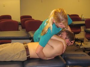
| | * [[File:Thoracic manip.JPG|right|frameless]]Thoracic discogenic pain syndrome (TDPS) is rare making it challenging for the healthcare team to diagnose and treat the condition. |
| | * The rarity of TDPS is attributed to the particular orientation, structure, and function of the thoracic spine in the vertebral column. |
| | * Despite this rarity, physiotherapists, physician assistants, and physicians should be familiar with its diagnosis and treatment and work as an interprofessional team to provide treatment<ref name=":3" /> |
| | * Physical therapy should be focussed on increasing the range of motion and pain relief, using a multiple-exercise based approach to strengthen muscles and postural support.<ref name="p0" /> <ref name=":7" /><br> |
|
| |
|
| The term ‘thoracic syndrome’ refers to all pathological clinical manifestations due to functional (physiopathological) disturbances and degenerative changes of the thoracic motion segments.<ref name="1" /> Due to the rarity of this subject, little importance is attached to it in the literature. <ref name="25" /><br>Several articles suggest an incidence between 0.2% and 5.0% of all intervertebral disc herniations. It should also be more common in men. <ref name="22" /><br>Mostly these disc disorders do not cause any symptoms. If, nevertheless, symptoms are extant, pain is the most common.(25) Other symptoms are sensory disturbances, referred pain, weakness in the abdominal and intercostal muscles, paresthesias, weakness of the lower extremities and bladder symptoms. <ref name="19" /><ref name="20" />(<ref name="21" /><ref name="22" />(<ref name="25" /><br>One of the main problems in the treatment of thoracic disc herniation has been the lack of accuracy of diagnostic tests. Disc herniation can be revealed by MRI but there aren’t really specific physical test to include this disease. Literature show that musculoskeletal, reflexes, sensory aspects, strength and provocative maneuvers should be tested.(<ref name="19" /><ref name="20" /><ref name="21" /><br>From medical research we can conclude, based on the study of Coppes. et.al , that the posterior transdural approach seems like a promising procedure to remove centrally herniated discs in patients with symptomatic throacic disc herniation. <ref name="16">Maarten H. Coppes; Posterior transdural discectomy: a new approach for the removal of central thoracic disc herniation; European Spine Journal; 2012; 21: 623-628; LoE: 2B</ref><br>Thoracic disc protrusions can be reduced by manipulation in 3 to 5 sessions.<br>Physical therapy should involve hyperextension strengthening <ref name="40">40Brown CW, Deffer PA Jr, Akmakjian J, Donaldson DH, Brugman JL. The naturalfckLRhistory of thoracic disc herniation. Spine (Phila Pa 1976). 1992 Jun;17(6fckLRSuppl):S97-102 (level 2a)</ref>, postural training <ref name="40" /> body mechanics education <ref name="40" />, McKenzie technique <ref name="42">Athletic Training and Sports Medicine; Schrijver: Robert C. Schenck; Editor: Jones &amp; Bartlett Learning, 1999 (LoE: 5)</ref> and Maitland technique <ref name="42" />.<br>
| | == References == |
| | | <references /> |
| <br>
| | [[Category:Thoracic Spine]] |
| | | [[Category:Musculoskeletal/Orthopaedics]] |
| == Recent Related Research ==
| | [[Category:Primary Contact]] |
| | | [[Category:Syndromes]] |
| Case Studies<br>In the case study of Bin Yue et.al they present two adult patients with thoracic intervertebral disc calcification and herniation. The purpose of this study was to report two cases of patients with acute paraplegia caused by a calcified thoracic disc prolapse and to discuss the clinical diagnosis and surgical treatment of this problem. <ref name="17">Bin Yue et.al; Thoracic intervertebral disc calcification and herniation in adults: a report of two cases; European Spine Journal; 2015; DOI 10.1007/s00586-015-4214-5; LoE: 4</ref><br>Case 1<br>In the first case, the patient was a woman, 57-years old with back pain already for 40 days. She experienced numbness in her lower extremity, bilateral and weakness for 20 days, bowel or bladder incontinence for 5 days. The findings of physical examination were: a lesser sensation below T11 level, bilateral lower extremity paraplegia with muscle force level zero, diffuse hyperreflexia(patellar jerk and Achilles tendon reflex for +++) and positive Babinski signs. CT imaging showed calcification of the nucleus pulposus with posterior central herniation within the disc space at level T11-T12. MRI showed a serious level of compression of the spinal cord at the disc of the segment T11-T12. After the diagnosis: calcified thoracic intervertebral disc with herniation, the patient underwent discectomy done with a posterior transfacet approach, which was followed by interbody fusion with instrumentation. The surgeon removed the calcified parts of the disc.
| | [[Category:Conditions]] |
| | | [[Category:Thoracic Spine - Conditions]] |
| <br>The results of the pathological examination of the removed parts was: degenerative fibrocartilage and calcium deposition. After a 2-year follow-up the patient did not show any important functional change in the postoperative period (such as muscle force, hyperreflexia, Babinksi signs). <ref name="17">Bin Yue et.al; Thoracic intervertebral disc calcification and herniation in adults: a report of two cases; European Spine Journal; 2015; DOI 10.1007/s00586-015-4214-5; LoE: 4</ref><br>Case 2<br>The second patient of this study was a 53-year old man with back pain for 2 months, paraplegia of the lower extremity bilateral and bowel or bladder incontinence for 15 days. The main complaint of the patient was back pain. The findings of physical examination were: paresis of the lower extremity bilateral below the level T10, paraplegia of the lower extremity bilateral with muscle force level zero and diffuse hyperreflexia(patellar jerk and Achilles tendon reflex for +++) with positive Babinski signs. CT imaging showed some high-density mass at the disc of the level T10-T11. MRI results showed a serious degree of spinal cord compression with a hyperintense central core and surrounding hypointense area at the intervertebral disc at level T10-T11. These findings led to the diagnosis: intervertebral disc calcification and protrusion. The surgical method of getting rid of the calcification was in this case also disectomy, through transversoarthropediculecttomy via bilateral posterolateral approach and interbody fusion with instrumentation. The researchers have found similar results as they have found in case 1: calcium deposition in the disc which was like ‘semisolid toothpaste’. This patient was followed-up 6 months and they have established full recovery (sensation, muscle force, hyperreflexia, and Babinski signs). <ref name="17">Bin Yue et.al; Thoracic intervertebral disc calcification and herniation in adults: a report of two cases; European Spine Journal; 2015; DOI 10.1007/s00586-015-4214-5; LoE: 4</ref><br>Researchers of this study found that if decompression surgery would not have been performed, spinal cord compression would have progressed further in these two patients which would have inevitably caused spinal cord injury. The research team therefore suggests to carry out decompression surgery as soon as possible for patients with early spinal myelopathy or paraplegia caused by a calcified protruded disc. <ref name="17">Bin Yue et.al; Thoracic intervertebral disc calcification and herniation in adults: a report of two cases; European Spine Journal; 2015; DOI 10.1007/s00586-015-4214-5; LoE: 4</ref><br><br>
| |