Tarsal Tunnel Syndrome: Difference between revisions
(replace wrong ampersand with single one) |
mNo edit summary |
||
| Line 6: | Line 6: | ||
== Definition/Description == | == Definition/Description == | ||
Tarsal tunnel syndrome is a compressive neuropathy of the posterior tibial nerve.<ref name=" | Tarsal tunnel syndrome is a compressive neuropathy of the posterior tibial nerve.<ref name="p1">McSweeney SC, Cichero M. Tarsal tunnel sydrome-A narrative literature review. The Foot 2015;25:244-50. (Level of Evidence 5)</ref>The tunnel lies posterior to the medial malleolus of the ankle, beneath the flexor retinaculum. Symptoms include pain radiating into the foot, usually this pain is worsened by walking (or weight-bearing acitvities). Examination may reveal Tinel’s sign over the tibial nerve at the ankle, weakness and atrophy of the small foot muscles or loss of sensation in the foot.<ref name="p2">https://www.ncbi.nlm.nih.gov/mesh/?term=tarsal+tunnel+syndrome</ref><br> | ||
== Clinically Relevant Anatomy == | == Clinically Relevant Anatomy == | ||
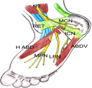
The tarsal tunnel is a fibrous osseus tunnel, it has a proximal floor and a distal floor. The proximal floor is formed by the flexor retinaculum (RET, Fig 1), it contains several flexor tendons (Tibialis posterior, Flexor digitorum longus, Flexor halluces longus), the posterior tibial nerve, the posterior tibial artery and the tibial veins. The distal floor is formed between the abductor hallucis muscle (H ABD, Fig. 1) and the deep quadratus plantae. This contains branches of the plantar vessels and the plantar nerves.<ref name=" | The tarsal tunnel is a fibrous osseus tunnel, it has a proximal floor and a distal floor. The proximal floor is formed by the flexor retinaculum (RET, Fig 1), it contains several flexor tendons (Tibialis posterior, Flexor digitorum longus, Flexor halluces longus), the posterior tibial nerve, the posterior tibial artery and the tibial veins. The distal floor is formed between the abductor hallucis muscle (H ABD, Fig. 1) and the deep quadratus plantae. This contains branches of the plantar vessels and the plantar nerves.<ref name="p3">Fantino O. Role of ultrasound in posteromedial tarsal tunnel syndrome: 81 cases. J Ultrasound 2014;17(2):99–112. (Level of Evidence 4)</ref><br> | ||
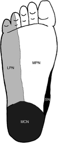
[[Image:Figuur 2.png|thumb|left|Fig. 2 Cutaneus innervation of the sole of the right foot (2)]] | [[Image:Figuur 2.png|thumb|left|Fig. 2 Cutaneus innervation of the sole of the right foot (2)]] | ||
| Line 38: | Line 38: | ||
Compression of the Tibial Nerve at the tarsal tunnel leads to plantar heel pain, this condition is called Tarsal Tunnel Syndrome. Other nerves that run in the foot also have risk of compression due to their anatomical situation. | Compression of the Tibial Nerve at the tarsal tunnel leads to plantar heel pain, this condition is called Tarsal Tunnel Syndrome. Other nerves that run in the foot also have risk of compression due to their anatomical situation. | ||
The LPN supplies most of the muscles of the foot, the lateral part of the skin (Fig. 2) and the fourth and fifth toe. This nerve runs between the abductor hallucis and the quadratus plantae, entrapment between these muscles is possible and also the most common. The first branch of the LPN runs close to the calcaneal tuberosity (ICN, Fig. 1), which is also a possible site of entrapment. The MCN supplies sensory innervation to most of the heel (heel fat pad, superficial tissues) (Fig. 2), this nerve is less likely to be compressed between its anatomical sturctures. But can be traumatised and irritated as a result of atrophy of the heel fat pad. The MPN innervates the abductor hallucis, flexor hallucis brevis, flexor digitorum brevis, first lumbrical and also the medial side of the plantar part (Fig. 2) of the foot.<ref name=" | The LPN supplies most of the muscles of the foot, the lateral part of the skin (Fig. 2) and the fourth and fifth toe. This nerve runs between the abductor hallucis and the quadratus plantae, entrapment between these muscles is possible and also the most common. The first branch of the LPN runs close to the calcaneal tuberosity (ICN, Fig. 1), which is also a possible site of entrapment. The MCN supplies sensory innervation to most of the heel (heel fat pad, superficial tissues) (Fig. 2), this nerve is less likely to be compressed between its anatomical sturctures. But can be traumatised and irritated as a result of atrophy of the heel fat pad. The MPN innervates the abductor hallucis, flexor hallucis brevis, flexor digitorum brevis, first lumbrical and also the medial side of the plantar part (Fig. 2) of the foot.<ref name="p4">Alshami AM, Souvlis T, Coppieters MW. A review of plantar heel pain of neural origin: Differntial diagnosis and management. Manual Therapy 2008;13:103-11. (Level of Evidence 5)</ref><br> | ||
== Epidemiology / Etiology == | == Epidemiology / Etiology == | ||
Tarsal Tunnel Syndrome is caused by any kind of entrapment or compression of the tibial nerve or its plantar branches. In many cases the cause is idiopathic or posttraumatic. Lau, M.D. et al. estimated that 20-40 % of cases were idiopathic.<ref name=" | Tarsal Tunnel Syndrome is caused by any kind of entrapment or compression of the tibial nerve or its plantar branches. In many cases the cause is idiopathic or posttraumatic. Lau, M.D. et al. estimated that 20-40 % of cases were idiopathic.<ref name="p1">Antoniadis G, Scheglmann K. Posterio Tarsal Tunnel Syndrome. Dtsch Arztebl Int. 2008;105(45):776-81. (Level of Evidence 5)</ref><ref name="p4">Lau JTC, et al. Tarsal Tunnel Syndrome: A Review of the Literature. Foot & Ankle Int. 1999;20(3):201-9. (Level of Evidence 5)</ref> | ||
Up to 10% of all cases are the result of the following diseases: arthrosis, tenosynovitis and [[Rheumatoid Arthritis|Rheumatoid Arthritis]]. Nearly twice as many cases have convoluted vessels as the origin.<ref name=" | Up to 10% of all cases are the result of the following diseases: arthrosis, tenosynovitis and [[Rheumatoid Arthritis|Rheumatoid Arthritis]]. Nearly twice as many cases have convoluted vessels as the origin.<ref name="p2">Oh S, Meyer R. Entrapment neuropathies of the tibial nerve. Neurologic Clinic 1999;17:593-615. (Level of Evidence 5)</ref> | ||
Some other rare causes are [[Diabetes|Diabetes Mellitus]], [[Hypothyroidism|Hypothyroidism]], [[Gout|Gout]], mucopolysaccharidoses, and (very rarely) hyperlipidemia.<ref name=" | Some other rare causes are [[Diabetes|Diabetes Mellitus]], [[Hypothyroidism|Hypothyroidism]], [[Gout|Gout]], mucopolysaccharidoses, and (very rarely) hyperlipidemia.<ref name="p1" /> | ||
Some muscles or tendons, medial of the talus bone, can entrap the tibial nerve due to hypertrophy or being accessory. As mentioned in the ‘anatomy’-section the tendon of the flexor hallucis longus muscle passes the tarsal tunnel along with blood vessels, the tibial nerve and other muscles. When enough hypertrophy occurs in one of these muscles the pressure within the tarsal tunnel increases. Sometimes this can even lead up to the muscle belly of the flexor hallucis longus entering the tarsal tunnel. This can cause an overstimulation of the tibial nerve or its branches. Depending on which nerve is being impinged the patient can get different uncomfortable sensations in its foot.<ref name=" | Some muscles or tendons, medial of the talus bone, can entrap the tibial nerve due to hypertrophy or being accessory. As mentioned in the ‘anatomy’-section the tendon of the flexor hallucis longus muscle passes the tarsal tunnel along with blood vessels, the tibial nerve and other muscles. When enough hypertrophy occurs in one of these muscles the pressure within the tarsal tunnel increases. Sometimes this can even lead up to the muscle belly of the flexor hallucis longus entering the tarsal tunnel. This can cause an overstimulation of the tibial nerve or its branches. Depending on which nerve is being impinged the patient can get different uncomfortable sensations in its foot.<ref name="p3">Rodriguez DI, Devos Bevernage B, et al. Tarsal tunnel syndrome and flexor hallucis longus tendon hypertrophy. Orthop Traumatol Surg Res. 2010;96(7):829-31. (Level of Evidence 4)</ref> | ||
Some persons are born with accessory muscles. These variations from the norm can cause more harm than good. Those muscles are not necessarily helpful, but it is a given that they do occupy space within the foot. Similar to hypertrophy of the muscles in the medial ankle region this can compress the tibial nerve possibly resulting in chronic pain.<ref name=" | Some persons are born with accessory muscles. These variations from the norm can cause more harm than good. Those muscles are not necessarily helpful, but it is a given that they do occupy space within the foot. Similar to hypertrophy of the muscles in the medial ankle region this can compress the tibial nerve possibly resulting in chronic pain.<ref name="p5">Cheung Y. Normal Variants: Accessory Muscles About the Ankle. Magn Reson Imaging Clin N Am. 2017;25(1):11-26. (Level of Evidence 5)</ref> | ||
Surgery or an overload on the ankle region can cause local inflammation and swelling, yet again causing pressure on the tibial nerve. Sports where sprinting and jumping play a significant role have been proven to be provocative for TTS. People with flat feet, talocalcaneal coalition or bony fragments around the tarsal tunnel are more vulnerable to develop the syndrome.<ref name=" | Surgery or an overload on the ankle region can cause local inflammation and swelling, yet again causing pressure on the tibial nerve. Sports where sprinting and jumping play a significant role have been proven to be provocative for TTS. People with flat feet, talocalcaneal coalition or bony fragments around the tarsal tunnel are more vulnerable to develop the syndrome.<ref name="p1" /><ref name="p4">Kinoshita M, Okuda R, Yasuda T, Abe M. Tarsal Tunnel Syndrome in Athletes. Am J Sport Med. 2006;34:1307-12. (Level of Evidence 4)</ref> | ||
People with flat feet, talocalcaneal coalition or bony fragments around the tarsal tunnel are more vulnerable to develop TTS.<ref name=" | People with flat feet, talocalcaneal coalition or bony fragments around the tarsal tunnel are more vulnerable to develop TTS.<ref name="p4" /> | ||
It might be important to mention that because tarsal tunnel syndrome is a relatively uncommon clinical entity, it can often be misdiagnosed in both children and adults due to the clinician’s low index of suspicion.<ref name=" | It might be important to mention that because tarsal tunnel syndrome is a relatively uncommon clinical entity, it can often be misdiagnosed in both children and adults due to the clinician’s low index of suspicion.<ref name="p1" /> McSweeney & Cichero (2015) also state in their review that the incidence of TTS is not known but the prevalence would be greater in females than males, predominately in adults.<ref name="p1" /> TTS also tends to be more common in athletes and individuals whom are subjected to prolonged weight-baring periods inclusive of standing, walking or intense physical activity.<ref name="p1" /><ref name="p5">Ahmad M ,Tsang K, Mackenney PJ, Adedapo AO. Tarsal tunnel syndrome: A literature review. Elsevier 2012;3:149-52. (Level of Evidence 5)</ref><ref name="p7">Tu P, Bytomski JR. Diagnosis of Heel Pain. American Family Physician 2011;84(8):909-16. (Level of Evidence 5)</ref> Pes planus deformity/hyperpronation may compromise the anatomical structures within the tarsal tunnel and thus lead to a physical decrease of space and an increase in tension of the nerve.<ref name="p1" /><ref name="p7" /> It would be one of the most common extrinsic factors to cause TTS.<ref name="p0">Lin D, Williams C, Zaw H. A rare case of an accessory flexor hallucis longus causing tarsal tunnel syndrome. Foot Ankle Surg. 2014;20:e37-39. (Level of Evidence 4)</ref> Alleviation of pain/complaints could be obtained with rest<ref name="p7" /> or neutral immobilization of the foot and ankle, and loose-fitting footwear.<ref name="p7" /> External compression resulting from footwear or tight plaster casts is said to be a most common cause.<ref name="p9">Kotnis N, Harish S, Popowich T. Medial Ankle and Heel: Ultrasound Evaluation and Sonographic Appearances of Conditions Causing Symptoms. Seminars in Ultrasound, CT and MRI 2011;32:125-41. (Level of Evidence 4)</ref><br> | ||
== Clinical Presentation == | == Clinical Presentation == | ||
| Line 62: | Line 62: | ||
TTS has the following symptoms: tingling or burning pain (paresthesia), hyperesthesia and sensory impairment (dysesthesia). The symptoms diffuse in the sole and/ or the heel or digits of the forefoot. The Valleix phenomenon, in which the symptoms extend midway to the lower leg due to percussion of the entrapped nerve, may also occur. | TTS has the following symptoms: tingling or burning pain (paresthesia), hyperesthesia and sensory impairment (dysesthesia). The symptoms diffuse in the sole and/ or the heel or digits of the forefoot. The Valleix phenomenon, in which the symptoms extend midway to the lower leg due to percussion of the entrapped nerve, may also occur. | ||
Symptoms are often unilaterally presented and that they may worsen as the day progresses.<ref name=" | Symptoms are often unilaterally presented and that they may worsen as the day progresses.<ref name="p1" /> This can also be accompanied by nocturnal awakening.<ref name="p2">Abouelela AA, Zohiery AK. The triple compression test for diagnosis of tarsal tunel syndrome. Elsevier The Foot 2012;3:146-49. (Level of Evidence 2b)</ref> Furthermore, in some cases muscle the patients may also present weakened, atrophied or paralyzed flexor and digital abductor muscles.<ref name="p2" /><ref name="p6">Moore KL, Dalley AF, Agur AMR. Clinically Oriented Anatomy. 6th ed. Philadelphia, Pa: Lippincott Williams and Wilkins;2010:617-18, 666-67. (Level of Evidence B)</ref><ref name="p7">Netter, FH. Atlas of Human Anatomy. 5th ed. Philadelphia, Pa: Saunders Elsevier 2011:529. (Level of Evidence B)</ref> Patients may be unable to move the hallux as a result of local tenderness and swelling. These movements include flexion, extension, abduction and adduction.<ref name="p2" /><ref name="p1" /><ref name="p6" /><ref name="p7" /> | ||
Pain becomes worse after or during weight-bearing activities and improves with rest.<ref name=" | Pain becomes worse after or during weight-bearing activities and improves with rest.<ref name="p1" /><ref name="p8">Kavlak Y, Uygur F. Effects of nerve mobilization exercise as an adjunct to the conservative treatment for patients with tarsal tunnel syndrome. Journal of manipulative and physiological therapeutics 2011;34(7):441-48. (Level of evidence 1b)</ref> Pain is the most prominent symptom, which is localized directly over the medial malleolus with radiation to the longitudinal arch and plantar aspect of the foot including the heel.<ref name="p1" /><ref name="p7" /> Pain begins in the plantar part of the forefoot and extends to the toes. It is usually aggravated in the night due to the modification of foot posture that causes the posterior tibial nerve to be restrained or venous congestion. There is rarely motor weakness or atrophy of intrinsic foot muscles.<ref name="p1" /><ref name="p6" /> Common manifestations of tarsal tunnel syndrome is a positive Tinel’s sign and pain felt on provocation using passively maximally dorsiflexion and eversion of the ankle while all the metatarsophalangeal joints are performing dorsiflexion. Most common and objective symptom is a diminished sensation.<ref name="p1" /><ref name="p8" /><br> | ||
== Differential Diagnosis<br> == | == Differential Diagnosis<br> == | ||
| Line 70: | Line 70: | ||
[[Posterior Tibial Tendon Dysfunction|'''Posterior Tibialis Tendon Dysfunction''']] | [[Posterior Tibial Tendon Dysfunction|'''Posterior Tibialis Tendon Dysfunction''']] | ||
'''Neuropathy''' - Most persons have once had sensation of so-called sleeping limbs, usually referred to as paresthesia. This feeling can also rise due to a pathogenic condition such as polyneuropathy. That’s when this numb, tingling feeling can no longer be put under control by the patient. It’s a condition often arises at the hands or feet. Considering one has paresthesia at the foot, the symptoms are very similar to those of the TTS.<ref name=" | '''Neuropathy''' - Most persons have once had sensation of so-called sleeping limbs, usually referred to as paresthesia. This feeling can also rise due to a pathogenic condition such as polyneuropathy. That’s when this numb, tingling feeling can no longer be put under control by the patient. It’s a condition often arises at the hands or feet. Considering one has paresthesia at the foot, the symptoms are very similar to those of the TTS.<ref name="p1" /> | ||
'''Ischaemia''' - A shortage of oxygen supply to tissue is called ischemia. Permanent damage can occur when this supply is put on hold for extensive time. As nerves start to lack oxygen their functionality slowly decreases. The result of extended ischemia can be devastating. However if a lack of blood flow is the cause, and it is normalized in time, damage can be near to none. Concerning nerves, in some cases permanent ischemic paresthesia can arise. This makes the neurons fire at random which gives the same sensation as the symptoms of TTS.<ref name=" | '''Ischaemia''' - A shortage of oxygen supply to tissue is called ischemia. Permanent damage can occur when this supply is put on hold for extensive time. As nerves start to lack oxygen their functionality slowly decreases. The result of extended ischemia can be devastating. However if a lack of blood flow is the cause, and it is normalized in time, damage can be near to none. Concerning nerves, in some cases permanent ischemic paresthesia can arise. This makes the neurons fire at random which gives the same sensation as the symptoms of TTS.<ref name="p1" /> | ||
'''Compartment Syndrome''' - A compartment syndrome can mostly be found in the upper arm and lower leg. It can show up after a pressure build-up on or within a muscle compartment with an overload of certain muscle groups or fluid build-up being possible origins of this syndrome. The tibial nerve runs along the deep calf muscles with a compartment. Excessive pressure may trigger this nerve to fire uncontrollably. The TTS can be misdiagnosed for this compartment syndrome if the compression of the nerve mostly takes place near the medial malleolus.<ref name=" | '''Compartment Syndrome''' - A compartment syndrome can mostly be found in the upper arm and lower leg. It can show up after a pressure build-up on or within a muscle compartment with an overload of certain muscle groups or fluid build-up being possible origins of this syndrome. The tibial nerve runs along the deep calf muscles with a compartment. Excessive pressure may trigger this nerve to fire uncontrollably. The TTS can be misdiagnosed for this compartment syndrome if the compression of the nerve mostly takes place near the medial malleolus.<ref name="p1" /> The former has also proven to be able to produce a distal tibial nerve lesion. A lesion is any damage or abnormal change in tissue. A lesion in or near a nerve can compromise its function. Therefore it can simulate the same symptoms as the TTS when a nerve near the medial malleolus is involved.<ref name="p1" /> | ||
'''Tumor''' - As a tumour is a group of cells which grow uncontrollably and can be benign, precancerous or malignant. Despite these three distinctions they do have in common that they take up space unnecessarily. Pressure caused by tumours rarely induces trouble to its surround tissues. It can however be a differential diagnosis for TTS if it overexcites a nerve at the medial malleolus.<ref name=" | '''Tumor''' - As a tumour is a group of cells which grow uncontrollably and can be benign, precancerous or malignant. Despite these three distinctions they do have in common that they take up space unnecessarily. Pressure caused by tumours rarely induces trouble to its surround tissues. It can however be a differential diagnosis for TTS if it overexcites a nerve at the medial malleolus.<ref name="p1" /><br> | ||
{| width="500" border="1" cellpadding="1" cellspacing="1" | {| width="500" border="1" cellpadding="1" cellspacing="1" | ||
|- | |- | ||
| '''A sum-up of possible differential diagnoses'''<ref name=" | | '''A sum-up of possible differential diagnoses'''<ref name="p1" /> | ||
|- | |- | ||
| | | | ||
| Line 89: | Line 89: | ||
*[[Calcaneal Spurs|Calcaneal Spurs]] , arthrosis, inflammatory changes of the fasciae and ligaments | *[[Calcaneal Spurs|Calcaneal Spurs]] , arthrosis, inflammatory changes of the fasciae and ligaments | ||
*Ischemia | *Ischemia | ||
*Infection<ref name=" | *Infection<ref name="p8">Baghla DP, Shariff S, Dega R. Calcaneal osteomyelitis presenting with acute tarsal tunnel syndrome: a case report. J Med Case Rep. 2010 Feb 23;4:66. doi: 10.1186/1752-1947-4-66 (Level of Evidence 4)</ref> | ||
*Lesion | *Lesion | ||
*Tumour | *Tumour | ||
| Line 100: | Line 100: | ||
== Diagnostic Procedures == | == Diagnostic Procedures == | ||
The diagnosis of TTS is mainly a clinical diagnosis based on detailed medical history and clinical examination. The clinical tests performed by physiotherapists are mostly provocative tests.<ref name=" | The diagnosis of TTS is mainly a clinical diagnosis based on detailed medical history and clinical examination. The clinical tests performed by physiotherapists are mostly provocative tests.<ref name="p2" /> These will be further described in the topic “Examination” (underneath). Adjunctive medical imaging and electrophysiological studies may assist in diagnosing<ref name="p6">Meadows JR, Finnoff JT. Lower extremity nerve entrapments in athletes. Current Sports Medicine Reports 2014;13(5):299-306. (Level of Evidence 5)</ref> and could provide additional information useful to plan management.<ref name="p5" />This can include electrodiagnostic studies, radiographs, ultrasound, MRI and computed tomography.<ref name="p1" /><ref name="p5" /><ref name="p6" /><ref name="p1">Omoumi P, de Gheldere A, Leemrijse T, Galant C, Van den Bergh P, Malghem J, Simoni P, Vande Berg BC, Lecouvet FE. Value of computed tomography arthrography with delayed acquisitions in the work-up of ganglion cysts of the tarsal tunnel: report of three cases. Skeletal Radiology 2010;39:381-86. (Level of Evidence 4)</ref><ref name="p6">Coraci D, Ioppolo F, Di Sante L, Santilli V, Padua L. Ultrasound in tarsal tunnel syndrome: correct diagnosis for appropriate treatment. Muscle & Nerve 2016;54(6):1148-49. (Level of Evidence 2c)</ref><ref name="p7" /><ref name="p9" /> It is, for example, also possible for the digital abductor and flexor muscles of the symptomatic foot to weaken, atrophy or even paralyse in some chronic circumstances.<ref name="p1" /> This is often difficult to detect clinically and may therefore require subsequent referral for medical imaging or nerve conduction studies.<ref name="p1" /> We should keep in mind though that these kinds of examinations are not substitutes for the clinical examination but they can play a key role in confirming or excluding the physician’s suspicion.<ref name="p6" /> | ||
As already mentioned, diagnosing the cause of TTS, and actually a lot of other medial ankle and heel pain, by physical examination alone can be challenging for the clinician.<ref name=" | As already mentioned, diagnosing the cause of TTS, and actually a lot of other medial ankle and heel pain, by physical examination alone can be challenging for the clinician.<ref name="p9" /> This is due to the complex anatomy of the medial aspect of the ankle and hindfoot, which makes localizing symptoms to a specific structure difficult.<ref name="p9" /> For example, plain X-rays of the ankle are useful in demonstrating structural abnormalities such as hind foot varus/valgus. Magnetic resonance imaging (MRI) adds further detail and is highly accurate (83%) when investigating space-occupying lesions.<ref name="p5" /> | ||
MRI is considered the gold standard in identifying suspected compression of the tarsal tunnel caused by the presence of obstructive foreign masses, lesions or tumours.<ref name=" | MRI is considered the gold standard in identifying suspected compression of the tarsal tunnel caused by the presence of obstructive foreign masses, lesions or tumours.<ref name="p1" /><ref name="p9" /> This type of imaging not only confirms the presence of a suspected lesion but also defines the depth, extent and margins of the lesion for accurate characterization.<ref name="p1" /> In a study undertaken by Frey (reviewed by McSweeney & Cichero, 2015), MRI was deemed to have shown significant findings in 88% of symptomatic tarsal tunnel candidates, thus assisting with etiological reasoning and surgical planning if required.<ref name="p1" /> MRI and high-resolution ultrasound have the diagnostic capability to detect and demonstrate the thickness of the flexor retinaculum, overall depth and contents within the tarsal tunnel, including the posterior tibial nerve cross-sectional area and its terminal branch derivatives.<ref name="p1" /> | ||
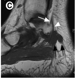
[[Image:Figuur 3A.png|thumb|left|Fig. 3A Kotnis, Harish & Popowich (2011). MRI show ganglion (black arrows) compressing the lateral plantar nerve (white arrowheads).]]<br> | [[Image:Figuur 3A.png|thumb|left|Fig. 3A Kotnis, Harish & Popowich (2011). MRI show ganglion (black arrows) compressing the lateral plantar nerve (white arrowheads).]]<br> | ||
| Line 142: | Line 142: | ||
<br> | <br> | ||
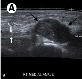
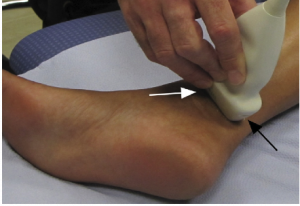
Ultrasound represents an accessible, portable and relatively inexpensive (less expensive than MRI) imaging tool for the assessment of medial ankle and heel pain.<ref name=" | Ultrasound represents an accessible, portable and relatively inexpensive (less expensive than MRI) imaging tool for the assessment of medial ankle and heel pain.<ref name="p9" /><ref name="p0">Rawool NM, Nazarian LN. Ultrasound of the Ankle and Foot. Seminars in Ultrasound, CT and MRI 2000;21(3):275-84. (Level of Evidence 5)</ref> In addition, it offers the advantage of comparison with the contralateral side.<ref name="p9" /> Although MRI (above) is considered the gold standard, ultrasound is effective in the diagnosis of pathologic conditions affecting the medial ankle and heel and correlates well with MRI.<ref name="p9" /> Ultrasound is able to demonstrate the complex anatomy of the tarsal tunnel and show the entire course of the tibial nerve and its branches at the medial ankle. It is also effective in the identification of space occupying lesions. Even small changes in the cross sectional area of the nerve can be detected on ultrasound in symptomatic patients.<ref name="p9" /><br> | ||
[[Image:Figuur US4.png|thumb|left|Fig. 4 Kotnis, Harish & Popowich (2011). Sonographic transducer position for examination of the tarsal tunnel structures. One end of the probe is placed on the medial malleolus (white arrow), and the other end is placed as much as possible towards the anterior edge of the Achiles tendon (black arrow).]] | [[Image:Figuur US4.png|thumb|left|Fig. 4 Kotnis, Harish & Popowich (2011). Sonographic transducer position for examination of the tarsal tunnel structures. One end of the probe is placed on the medial malleolus (white arrow), and the other end is placed as much as possible towards the anterior edge of the Achiles tendon (black arrow).]] | ||
| Line 158: | Line 158: | ||
<br> | <br> | ||
Electrodiagnostic testing can also assist in the diagnosis of tarsal tunnel syndrome.<ref name=" | Electrodiagnostic testing can also assist in the diagnosis of tarsal tunnel syndrome.<ref name="p1" /> These tests include nerve conduction studies that assess sensory conduction velocities of the tibial nerve or one of its branches, as well as the amplitude and duration of motor-evoked potentials.<ref name="p1" /> There is limited quality evidence based research that demonstrates high sensitivity and specificity of the electrophysiological techniques in TTS. Reduced amplitude and increased duration of the motor response are the more sensitive indicators of the presence of pathology.<ref name="p1" /> Unfortunately these investigations often yield an unacceptable level of false negative results, and should be utilized as an adjunctive assessment to confirm physical examination findings.<ref name="p1" /><ref name="p2" /> | ||
Saeed (reviewed by McSweeney & Cichero, 2015) discusses evidence of false positive readings in his study of 70 asymptomatic subjects<ref name=" | Saeed (reviewed by McSweeney & Cichero, 2015) discusses evidence of false positive readings in his study of 70 asymptomatic subjects<ref name="p1" /> and Ahmad et al (2012) report that false negative tests are not uncommon and therefore do not rule out the diagnosis.<ref name="p5" /> Thus, Abouelela & Zohiery (2012) state that provocative tests remain important in the diagnosis of TTS due to the unaccepted range of false negative results in electrodiagnostic testing.<ref name="p2" /> | ||
Last, plain weight-bearing radiographs and/or computed tomography of the foot and ankle should be acquired if suspecting morphological influences or structural anomalies from bony abnormalities, according to McSweeney & Cichero (2012).<ref name=" | Last, plain weight-bearing radiographs and/or computed tomography of the foot and ankle should be acquired if suspecting morphological influences or structural anomalies from bony abnormalities, according to McSweeney & Cichero (2012).<ref name="p1" /> Omoumi et al (2010) state that in practice, the visualization of articular communication with MRI or ultrasonography can be challenging.<ref name="p1" /> Computed tomography (arthrography) with delayed acquisitions has been shown to be a valuable technique for the detection of articular communication between structures and a joint.<ref name="p1" /> Closing, it is recommended that all tests should ideally be performed bilaterally for adequate observation and comparative study of the contralateral joint.<ref name="p1" /><br> | ||
== Examination<br> == | == Examination<br> == | ||
Tarsal tunnel syndrome may lead to a broad range of symptoms affecting the posteromedial ankle and plantar aspects of the foot.<ref name=" | Tarsal tunnel syndrome may lead to a broad range of symptoms affecting the posteromedial ankle and plantar aspects of the foot.<ref name="p1" /> This is due to the proximal TTS affecting the tibial nerve in the retromalleolar region, whereas distal TTS tends to affect its branches.<ref name="p9" /> According to Zheng et al (2016), paresthesia and/or pain in the sole of the foot is the widely accepted primary symptom of tarsal tunnel syndrome.<ref name="p5">Zheng C, Zhu Y, Jiang J, Ma X, Lu F, Jin X, Weber R. The prevalence of tarsal tunnel syndrome in patiens with lumbosacral radiculopathy. European Spine Journal 2016;25:895-905. (Level of Evidence 2b)</ref> Ahmad et al (2012) says that the predominant symptom is indeed pain, directly over the tarsal tunnel behind the medial malleolus with radiation to the longitudinal arch and plantar aspect of the foot including the heel.<ref name="p5" /> Tu & Bytomski (2011) talk about medial midfoot heel pain<ref name="p7" /> and Kavlak & Uygur (2011) about the symptom triad of pain, paresthesia and numbness. Other (sensory) symptoms may include dysesthesia and, as already stated above, paraesthesia (e.g. burning, tingling and numbness) at all or varied sites of the tarsal tunnel including: the posterior compartment, retromalleolar flexor retinaculum coverage, and both the anterior and posterior fibro-osseus tunnels branching to the plantar margins of the foot.<ref name="p1" /><ref name="p5" /><ref name="p2" /><ref name="p7" /> A varied and specific involvement of the differing nerve branches accounts for the diverse presentation and extent level of symptoms observed with the condition of tarsal tunnel syndrome.<ref name="p1" /> | ||
A very common used clinical test for the diagnosis of tarsal tunnel syndrome is the [[Tinel’s Test|Hoffmann-Tinel Sign]].<ref name=" | A very common used clinical test for the diagnosis of tarsal tunnel syndrome is the [[Tinel’s Test|Hoffmann-Tinel Sign]].<ref name="p1" /><ref name="p7" /> Originally used as a specific test for the carpal tunnel syndrome, it can also be used to examine tibial nerve compression in the ankle.<ref name="p3">http://www.physio-pedia.com/Tinel%E2%80%99s_Test</ref> During this test, the nerve is percussed or tapped at the suspected site of compression, the area below the medial malleolus.<ref name="p1" /> A positive diagnosis will cause paresthesia either locally or radiating along the course of the nerve.<ref name="p1" /> This positive result may be due to the entrapment of the nerve by surrounding tissues.<ref name="p7">Harries M, Williams C, Stanish WD, Micheli LJ. Oxford textbook of sports medicine. Great Britain: Butler &amp; Tanner ltd., frame, 2000, pp. 699 – 700. (Level of evidence 2B)</ref><ref name="p9">Nagaoka M, Satou K. Tarsal tunnel syndrome caused by ganglia. The journal of bone &amp; joins surgery 1998:607-10. (Level of Evidence 2b)</ref> When the test delivers a negative result, the patient feels no pain.<ref name="p3" /> It is furthermore postulated that greater than 50% of patients with compressive neuropathy of the tarsal tunnel will portray a positive Tinel’s sign of the posterior tibial nerve.<ref name="p8">Division of Physiotherapy, School of Health and Rehabilitation Sciences, The University of Queensland, QLD 4072 St. Lucia, Australia. Strain and excursion of the sciatic, tibial, and plantar nerves during a modified straight leg raising test. Pubmed. [Online] [Citaat van: 16 november 2010.] http://www.ncbi.nlm.nih.gov/pubmed/16838375 (Level of Evidence 1a)</ref> Alongside Tinel’s sign, the physiotherapist could also use the SLR (Straight Leg Raise) test to provoke symptoms similar to a nerve problem.<ref name="p8" /> | ||
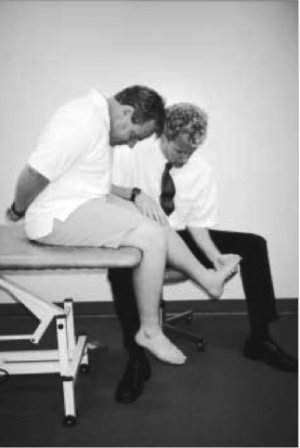
A second test that could be used (in addition to the Tinel’s sign) for making the symptoms become diagnostically apparent is the dorsiflexion-eversion test.<ref name=" | A second test that could be used (in addition to the Tinel’s sign) for making the symptoms become diagnostically apparent is the dorsiflexion-eversion test.<ref name="p1" /><ref name="p4" /><ref name="p7" /><ref name="p8">Kinoshita M, Okuda R, Morikawa J, Jotoku T, Abe M. The dorsiflexion-eversion test for diagnosis of tarsal tunnel syndrome. The Journal of Joint &amp; Bone Surgery 2001;83(12):1835-39. (Level of Evidence 2b)</ref> To perform this test the clinician both passively maximally everts and dorsiflexes the ankle whilst maximally dorsiflexing the metatarsophalangeal joints.<ref name="p1" /> This position is held for 5 – 10 s and will display further intensification of the symptoms if positive.<ref name="p1" /> On occasions pain may also ascend to the thigh following this means of testing (1; LOE: 5), although this would happen rather rarely.<ref name="p5" /> Kinoshita et al (2006) have reported that the dorsiflexion-eversion test reproduced or aggravated symptoms for 82% in symptomatic feet, with no replication evident in the healthy control group.<ref name="p4" /> In brief, what happens during the dorsiflexion-eversion test is that the distal posterior tibial nerve is stretched and compressed.<ref name="p4" /> | ||
[[Image:Figuur DE5.png|thumb|left|Fig. 5 McSweeney & Cichero (2015). The dorsiflexion-eversion test – the testing clinician passively maximally everts and dorsiflexes the ankle whilst maximally dorsiflexing the metatarsophalangeal joints.]] | [[Image:Figuur DE5.png|thumb|left|Fig. 5 McSweeney & Cichero (2015). The dorsiflexion-eversion test – the testing clinician passively maximally everts and dorsiflexes the ankle whilst maximally dorsiflexing the metatarsophalangeal joints.]] | ||
| Line 184: | Line 184: | ||
<br> | <br> | ||
Further intensification of symptoms can also be obtained by using the Trepman test or the plantar flexion-inversion test.<ref name=" | Further intensification of symptoms can also be obtained by using the Trepman test or the plantar flexion-inversion test.<ref name="p1" /><ref name="p7" /> This maneuver also increases pressure on the tibial nerve within the tarsal tunnel confines.<ref name="p1" /> Through intra-operative observation, Hendrix et al (reviewed by McSweeney & Cichero, 2015) acknowledged this combined movement not only reduced the overall width of the tarsal tunnel, but also compressed the lateral-planter nerve.<ref name="p1" /> So either dorsiflexion-eversion or plantar flexion-inversion can reproduce pain or increase the symptoms of tarsal tunnel syndrome.<ref name="p4" /> | ||
Abouelala & Zohiery (2012) investigated a fourth clinical test, called the triple compression stress test (TCST). This provocative test was supposed to be more specific and therefore sensitivity and specificity for diagnosing TTS was investigated. The TCST showed 85,9% sensitivity and 100% specificity. The TCST combines the Tinel’s sign test with the Trepman test<ref name=" | Abouelala & Zohiery (2012) investigated a fourth clinical test, called the triple compression stress test (TCST). This provocative test was supposed to be more specific and therefore sensitivity and specificity for diagnosing TTS was investigated. The TCST showed 85,9% sensitivity and 100% specificity. The TCST combines the Tinel’s sign test with the Trepman test<ref name="p1" /> by bringing the foot passively in full plantar flexion, inversion and applying an even and constant digital pressure over the posterior tibial nerve for 30s.<ref name="p2" /> A double compression on the nerve occurs from the plantar flexion and inversion, and along with a simultaneous third compression maneuver by direct digital pressure, the test was hence named triple compression stress test.<ref name="p2" /> The study found that clinical signs and symptoms of tarsal tunnel syndrome were apparent within a matter of seconds for 93,8% of symptomatic feet.<ref name="p2" /> Pain usually developed within 10 s and numbness within 30 s of the test. All control feet had negative clinical TCST. The researchers also state that the TCST achieves a simple, fast and very reliable provocative maneuver to increase sensitivity of TSS diagnosis, both clinically and electrophysiologically. | ||
[[Image:Figuur AB6.png|thumb|left|Fig.6 Abouelela & Zohiery (2012). TCST (A) ankle plantar flexion. (B) Heel and foot inversion. (C) Digital compression of the tibial nerve posterior to the medial malleolus.]]<br><br> | [[Image:Figuur AB6.png|thumb|left|Fig.6 Abouelela & Zohiery (2012). TCST (A) ankle plantar flexion. (B) Heel and foot inversion. (C) Digital compression of the tibial nerve posterior to the medial malleolus.]]<br><br> | ||
| Line 194: | Line 194: | ||
== Outcome Measures == | == Outcome Measures == | ||
There exists a wide variety in clinical outcomes that can be used to evaluate foot conditions. In 2013 Kenneth J. et al. concluded in their 10-year research that most of them were inconsistently used. Out of 139 clinical outcomes the American Orthopaedic Foot & Ankle Society (AOFAS), the visual analog scale (VAS) for pain, the Short Form-36 (SF-36) Health Survey, the Foot Function Index (FFI) and the American Academy of Orthopaedic Surgeons (AAOS) outcomes instruments were the most popular. The study underlined the need for consistent use of a responsive, valid, reliable and clinically meaningful outcome measurement tool.<ref name=" | There exists a wide variety in clinical outcomes that can be used to evaluate foot conditions. In 2013 Kenneth J. et al. concluded in their 10-year research that most of them were inconsistently used. Out of 139 clinical outcomes the American Orthopaedic Foot & Ankle Society (AOFAS), the visual analog scale (VAS) for pain, the Short Form-36 (SF-36) Health Survey, the Foot Function Index (FFI) and the American Academy of Orthopaedic Surgeons (AAOS) outcomes instruments were the most popular. The study underlined the need for consistent use of a responsive, valid, reliable and clinically meaningful outcome measurement tool.<ref name="p2">Hunt KJ, Hurwit D. Use of patient-reported outcome measures in foot and ankle research. J Bone Joint Surg Am. 2013;95(16):e118. (Level of Evidence 1a)</ref><br>One measurement tool that meets the requirements is the [[Foot and Ankle Ability Measure|Foot and Ankle Abitlity Measure]]. It was made in 2005 by RobRoy et al. It covers a wide variety of disorders in the lower extremity, namely the lower leg, foot and ankle. Examples are plantar fasciitis<ref name="p9">Digiovanni BF, Nawoczenski DA, Malay DP, Graci PA, Williams TT, Wilding GE, Baumhauer JF. Plantar fascia-specific stretching exercise improves outcomes in patients with chronic plantar fasciitis. J Bone Joint Surg Am. 2006;88(8):1775-81. (Level of Evidence 2b)</ref>, ankle instability<ref name="p1">Eechaute et al. The clinimetric qualities of patient-assessed instruments for measuring chronic ankle instability: A systematic review. BMC Musculoskeletal Disorders 2007, 8:6 doi:10.1186/1471-2474-8-6 (Level of evidence: 1a)</ref>, etc. Its clinical relevance was researched by Martin et al. and in the table underneath the validity, reliability and responsiveness of the tool is summerised.<ref name="p0">Martin R L, Irrgang JJ. A survey of self-reported outcome instruments for the foot and ankle. Journal of Orthopaedic & Sports Physical Therapy 2007;37(2):72-84. (Level of Evidence 5)</ref><br><br> | ||
{| width="600" border="1" cellpadding="1" cellspacing="1" | {| width="600" border="1" cellpadding="1" cellspacing="1" | ||
| Line 229: | Line 229: | ||
== Medical Management<br> == | == Medical Management<br> == | ||
The management goes into two directions: a conservative or non-operative management and surgery. The former is used prior to the latter approach, unless signs of muscle atrophy or motor involvement are obvious, according to McSweeney & Cichero. The latter is used when patients haven’t benefitted from the former approach.<ref name=" | The management goes into two directions: a conservative or non-operative management and surgery. The former is used prior to the latter approach, unless signs of muscle atrophy or motor involvement are obvious, according to McSweeney & Cichero. The latter is used when patients haven’t benefitted from the former approach.<ref name="p1" /><ref name="p1" /><ref name="p7">Dr. Karen Hudes, BCs, BS, DC: Conservative management of a case of tarsal tunnel syndrome. (Level of Evidence 4)</ref><ref name="p9">Bailie DS, Kelikian AS. Tarsal tunnel syndrome: diagnosis, surgical technique, and functional outcome. Foot & ankle international 1998;19(2):65-72. (Level of Evidence 4)</ref> (Levels of evidence: 5, 2A, 4, 4) | ||
<u>'''Conservative approach'''</u><br>In this approach the following can be included: “physical therapy, non-steroidal anti-inflammatory, analgesic, opioid or GABA analog medications, tri-cyclic antidepressants and vitamin B-complex supplements and corticosteroid injections”. In the literature there is unfortunately an absence of RCTs, and in existing case series the efficacy of treatment methods can’t be quantified.<ref name=" | <u>'''Conservative approach'''</u><br>In this approach the following can be included: “physical therapy, non-steroidal anti-inflammatory, analgesic, opioid or GABA analog medications, tri-cyclic antidepressants and vitamin B-complex supplements and corticosteroid injections”. In the literature there is unfortunately an absence of RCTs, and in existing case series the efficacy of treatment methods can’t be quantified.<ref name="p1" /><ref name="p1" /><ref name="p7" /><ref name="p9" /> (Levels of evidence: 5, 2A, 4, 4) | ||
<u>'''Surgical approach'''</u><br>When the non-operative management doesn’t work, surgical management is recommended.<ref name=" | <u>'''Surgical approach'''</u><br>When the non-operative management doesn’t work, surgical management is recommended.<ref name="p1" /><ref name="p1" /><ref name="p7" /><ref name="p9" /> (Levels of evidence: 5, 2A, 4, 4) This approach can include: “surgical decompression of the tibial nerve and its branches with division of the medial flexor retinaculum; release of the deep fascia of the abductor hallucis muscle and removal of impinging or pathological lesions”.<ref name="p1" /> (Level of evidence: 5) It is stated by McSweeney & Cichero that surgery may improve symptoms of the syndrome but that evidence of the efficacy of the surgery in the literature can conflict itself.<ref name="p1" /><ref name="p1" /><ref name="p7" /><ref name="p9" /> (Levels of evidence: 5, 2A, 4, 4) Another surgical approach is cryosurgery. McSweeney & Cichero state that there are different advantages of using cryosurgery for this syndrome, but as there is a lack of clinical trials the guideline for the use of this method is still undergoing change.<ref name="p1" /> (Level of evidence: 5)<br> | ||
== Physical Therapy Management<br> == | == Physical Therapy Management<br> == | ||
There is a lack of evidence in literature on treatment approaches.<ref name=" | There is a lack of evidence in literature on treatment approaches.<ref name="p4" /> (Level of evidence: 5) Small RCT’s would help to find succesfull rehabilitation excersices or other treatments for patients with tarsal tunnel syndrome.<ref name="p1" /> (Level of evidence: 5) Published papers have reported case studies, but empirical evidence of their efficacy is lacking.<ref name="p1" /> (Level of evidence: 5)<br>At the time patients who do not respond to physical therapy or other conservative treatment are reffered to a clinician for a surgical approach (e.g. decompression of the tarsal tunnel).<br> | ||
'''<u>Conservative treatment</u>''' | '''<u>Conservative treatment</u>''' | ||
There are three stages<ref name=" | There are three stages<ref name="p3">Plyler B, Martinez M, Melde C, Gieringer M. Tarsal Tunnel Syndrome: A Clinical Management Guideline. Texas State University, Department of Physical Therapy. August 2012. http://ptcoop.org/texas-state-ebp-tarsal-tunnel-syndrome/ (Level of Evidence 5)</ref><ref name="p4">Godges J, Loma L. Ankle Nerve Disorder: Tarsal Tunnel Syndrome. KPSoCal Orthopedic PT Residency. http://xnet.kp.org/socal_rehabspecialists/ptr_library/09FootRegion/07Ankle&amp;amp;amp;amp;amp;amp;amp;amp;amp;amp;amp;amp;amp;amp;Foot-RadiatingPain.pdf. (Level of Evidence 5)</ref> (Levels of evidence: 5, 5) in the development of TTS, in every stage there are different aspect that may be adressed in the management of the symptoms.<br><br> | ||
{| border="1" cellpadding="1" cellspacing="1" | {| border="1" cellpadding="1" cellspacing="1" | ||
| Line 312: | Line 312: | ||
<br> | <br> | ||
<u>Orthotics and taping<ref name=" | <u>Orthotics and taping<ref name="p1" /><ref name="p4" /><ref name="p8" /> (Levels of evidence: 5, 5, 1B)</u> | ||
#UCBL orthosis: A University of California Berkeley Laboratory orthosis can be used to improve hind foot alignment | #UCBL orthosis: A University of California Berkeley Laboratory orthosis can be used to improve hind foot alignment | ||
#CAM walker: Controlled Ankle Motion walker, with this boot the ROM of the patients ankle can be altered. | #CAM walker: Controlled Ankle Motion walker, with this boot the ROM of the patients ankle can be altered. | ||
#Plantar arch taping<ref name=" | #Plantar arch taping<ref name="p5" /> (Level of evidence: 4): <br>{{#ev:youtube|5Z2XlqsuQSY}}<br> | ||
#Patient education on footwear: the therapist should educate the patient on wearing appropriate footwear. Thight fitting shoes should be avoided. When dealing with athletes one must pay attention tot heir running mechanics and/or motions in their technique (sport specific) that may cause the symptoms.<ref name=" | #Patient education on footwear: the therapist should educate the patient on wearing appropriate footwear. Thight fitting shoes should be avoided. When dealing with athletes one must pay attention tot heir running mechanics and/or motions in their technique (sport specific) that may cause the symptoms.<ref name="p6" /> (Level of evidence: 5) | ||
<br><u>Therapeutic excersices</u><br>1) Stretching<ref name=" | <br><u>Therapeutic excersices</u><br>1) Stretching<ref name="p1" /><ref name="p4" /> (Level of evidence: 5, 5) | ||
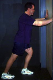
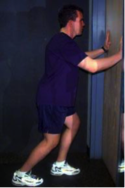
*Calf muscle: there are many ways to stretcht the calf muscle, one of the most frequently used is the calf stretch using a wall. The first picture focusses on stretching the gastrocnemius muscle, the second picture focusses on stretching the soleus muscle | *Calf muscle: there are many ways to stretcht the calf muscle, one of the most frequently used is the calf stretch using a wall. The first picture focusses on stretching the gastrocnemius muscle, the second picture focusses on stretching the soleus muscle | ||
| Line 413: | Line 413: | ||
<br> | <br> | ||
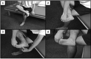
<br>2) Nerve mobility<ref name=" | <br>2) Nerve mobility<ref name="p8" /> (Level of evidence: 1B)<br>Nerve mobilization exercises have been used to treat carpal tunnel syndrome (nerve entrapment in the wrist) with contradicting results. In the present literature, only a limited number of case studies where nerve mobilization exercises have been used to treat plantar heel pain of neural origin.<br>Kavlak Y. and Uygur F. conducted a RCT where they used nerve mobilization techniques as an adjunct to conservative treatment. The studie reported a positive outcome for R.OM., muscle strenght and pain in both groups. The study group showed a significant improvement for 2-point discrimination, light touch and tinel’s sign. | ||
Nerve mobilization as described by Meyer et al.<ref name=" | Nerve mobilization as described by Meyer et al.<ref name="p5" /> (Level of evidence: 4) | ||
[[Image:Figuur 10.png|thumb|left|Fig 10 Nerve mobilization in slump position (image taken from Meyer et al.)]]<br> | [[Image:Figuur 10.png|thumb|left|Fig 10 Nerve mobilization in slump position (image taken from Meyer et al.)]]<br> | ||
| Line 447: | Line 447: | ||
<br> | <br> | ||
3) Tibialis posterior strenghtening<ref name=" | 3) Tibialis posterior strenghtening<ref name="p1" /><ref name="p5" /> (Level of evidence: 5, 4) | ||
<br>The function of the tibialis posterior muscle is to stabilize the ankle, it is also used for inversion of the ankle. Exercises can be classified into two categories: weight bearing and non-weigth bearing. | <br>The function of the tibialis posterior muscle is to stabilize the ankle, it is also used for inversion of the ankle. Exercises can be classified into two categories: weight bearing and non-weigth bearing. | ||
| Line 457: | Line 457: | ||
*Example of a weight bearing exercise:<br> {{#ev:youtube|aDyr2wAOIhM}}<br> | *Example of a weight bearing exercise:<br> {{#ev:youtube|aDyr2wAOIhM}}<br> | ||
<br><u>Other types of conservative treatment</u> may include<ref name=" | <br><u>Other types of conservative treatment</u> may include<ref name="p4" /> (Level of evidence: 5):<br>• Rest<br>• NSAID’s<br>• Corticosteroid injections<br>• Extracorporeal shockwave therapy<br>• Laser<br>• Local anaesthetic injections<br>• Heel pads and heel cups<br>• Night splints<br>• Medial longitudinal arch supports<br>• Strapping<br>• Soft-soled shoes<br>• Casting | ||
It is recommended that patients are treated conservatively prior to the surgical treatment. When patients do not respond to the conservative treatment or if there are signs of atrophy or motor involvment they should be reffered to a clinician. | It is recommended that patients are treated conservatively prior to the surgical treatment. When patients do not respond to the conservative treatment or if there are signs of atrophy or motor involvment they should be reffered to a clinician. | ||
<br>'''<u>Postoperative rehabilitation<ref name=" | <br>'''<u>Postoperative rehabilitation<ref name="p6">Joe Godges PT, Robert Klingman PT: Tarsal Tunnel Release. Loma Linda University DPT Program KPSoCal Ortho PT Residency. (Level of Evidence 5)</ref> (Level of evidence: 5)</u>'''<br> | ||
{| width="500" border="1" cellpadding="1" cellspacing="1" | {| width="500" border="1" cellpadding="1" cellspacing="1" | ||
| Line 550: | Line 550: | ||
== Clinical Bottom Line == | == Clinical Bottom Line == | ||
Tarsal tunnel syndrome is a rare condition and often underdiagnosed.<ref name=" | Tarsal tunnel syndrome is a rare condition and often underdiagnosed.<ref name="p1" /> A variety of symptoms are possible, such as: tingling or burning pain (paresthesia), hyperesthesia and sensory impairment (dysesthesia). These are felt on the plantar face of the ankel and foot.<br>There are a few test to identify tarsal tunnel syndrome or rule out other possibilities, these tests include: MRI, Ultrasound, Hoffman-tinels test, dorsiflexion-eversion test, trepman test and the triple compression stress test.<ref name="p4" /><br>Treatment of a tarsal tunnel syndrom should be attemted conservatively at first (see “Physical therapy management”). If conservative treatment fails a surgical aproach can be taken.<br> | ||
== References == | == References == | ||
Revision as of 12:46, 6 June 2017
Original Editor - Staci Burns, Jonathan Blondeel
Top Contributors - Admin, Blondeel Jonathan, Dieter Schuddinck, Rani Sileghem, Staci Burns, Kim Jackson, Rachael Lowe, Jesse Lemmens, Vidya Acharya, 127.0.0.1, Tarina van der Stockt, Jenny Wong, Naomi O'Reilly, Kai A. Sigel and WikiSysop
Definition/Description[edit | edit source]
Tarsal tunnel syndrome is a compressive neuropathy of the posterior tibial nerve.[1]The tunnel lies posterior to the medial malleolus of the ankle, beneath the flexor retinaculum. Symptoms include pain radiating into the foot, usually this pain is worsened by walking (or weight-bearing acitvities). Examination may reveal Tinel’s sign over the tibial nerve at the ankle, weakness and atrophy of the small foot muscles or loss of sensation in the foot.[2]
Clinically Relevant Anatomy[edit | edit source]
The tarsal tunnel is a fibrous osseus tunnel, it has a proximal floor and a distal floor. The proximal floor is formed by the flexor retinaculum (RET, Fig 1), it contains several flexor tendons (Tibialis posterior, Flexor digitorum longus, Flexor halluces longus), the posterior tibial nerve, the posterior tibial artery and the tibial veins. The distal floor is formed between the abductor hallucis muscle (H ABD, Fig. 1) and the deep quadratus plantae. This contains branches of the plantar vessels and the plantar nerves.[3]
The posterior tibial nerve (NT, Fig. 1) runs along the soleus in the posterior compartment. The nerve exits the compartment and passes deep behind the RET between the calcaneus and the medial malleolus. Before passing behind the flexor retinaculum the nerve branches, one part moves on behind the RET, the other part forms the Medial calcaneal nerve (MCN, Fig 1). At the distal floor the tibial nerve divides into two terminal branches: Medial plantar nerve (MPN, Fig. 1) and Lateral plantar nerve (LPN, Fig. 1), sometimes this happens at the proximal tunnel. The inferior calcaneal nerve runs branches off from the lateral plantar nerve and is also referred to as the motor nerve for the abductor muscle for the fifth toe (ABD V, Fig. 1)
Compression of the Tibial Nerve at the tarsal tunnel leads to plantar heel pain, this condition is called Tarsal Tunnel Syndrome. Other nerves that run in the foot also have risk of compression due to their anatomical situation.
The LPN supplies most of the muscles of the foot, the lateral part of the skin (Fig. 2) and the fourth and fifth toe. This nerve runs between the abductor hallucis and the quadratus plantae, entrapment between these muscles is possible and also the most common. The first branch of the LPN runs close to the calcaneal tuberosity (ICN, Fig. 1), which is also a possible site of entrapment. The MCN supplies sensory innervation to most of the heel (heel fat pad, superficial tissues) (Fig. 2), this nerve is less likely to be compressed between its anatomical sturctures. But can be traumatised and irritated as a result of atrophy of the heel fat pad. The MPN innervates the abductor hallucis, flexor hallucis brevis, flexor digitorum brevis, first lumbrical and also the medial side of the plantar part (Fig. 2) of the foot.[4]
Epidemiology / Etiology[edit | edit source]
Tarsal Tunnel Syndrome is caused by any kind of entrapment or compression of the tibial nerve or its plantar branches. In many cases the cause is idiopathic or posttraumatic. Lau, M.D. et al. estimated that 20-40 % of cases were idiopathic.[1][4]
Up to 10% of all cases are the result of the following diseases: arthrosis, tenosynovitis and Rheumatoid Arthritis. Nearly twice as many cases have convoluted vessels as the origin.[2]
Some other rare causes are Diabetes Mellitus, Hypothyroidism, Gout, mucopolysaccharidoses, and (very rarely) hyperlipidemia.[1]
Some muscles or tendons, medial of the talus bone, can entrap the tibial nerve due to hypertrophy or being accessory. As mentioned in the ‘anatomy’-section the tendon of the flexor hallucis longus muscle passes the tarsal tunnel along with blood vessels, the tibial nerve and other muscles. When enough hypertrophy occurs in one of these muscles the pressure within the tarsal tunnel increases. Sometimes this can even lead up to the muscle belly of the flexor hallucis longus entering the tarsal tunnel. This can cause an overstimulation of the tibial nerve or its branches. Depending on which nerve is being impinged the patient can get different uncomfortable sensations in its foot.[3]
Some persons are born with accessory muscles. These variations from the norm can cause more harm than good. Those muscles are not necessarily helpful, but it is a given that they do occupy space within the foot. Similar to hypertrophy of the muscles in the medial ankle region this can compress the tibial nerve possibly resulting in chronic pain.[5]
Surgery or an overload on the ankle region can cause local inflammation and swelling, yet again causing pressure on the tibial nerve. Sports where sprinting and jumping play a significant role have been proven to be provocative for TTS. People with flat feet, talocalcaneal coalition or bony fragments around the tarsal tunnel are more vulnerable to develop the syndrome.[1][4]
People with flat feet, talocalcaneal coalition or bony fragments around the tarsal tunnel are more vulnerable to develop TTS.[4]
It might be important to mention that because tarsal tunnel syndrome is a relatively uncommon clinical entity, it can often be misdiagnosed in both children and adults due to the clinician’s low index of suspicion.[1] McSweeney & Cichero (2015) also state in their review that the incidence of TTS is not known but the prevalence would be greater in females than males, predominately in adults.[1] TTS also tends to be more common in athletes and individuals whom are subjected to prolonged weight-baring periods inclusive of standing, walking or intense physical activity.[1][5][6] Pes planus deformity/hyperpronation may compromise the anatomical structures within the tarsal tunnel and thus lead to a physical decrease of space and an increase in tension of the nerve.[1][6] It would be one of the most common extrinsic factors to cause TTS.[7] Alleviation of pain/complaints could be obtained with rest[6] or neutral immobilization of the foot and ankle, and loose-fitting footwear.[6] External compression resulting from footwear or tight plaster casts is said to be a most common cause.[8]
Clinical Presentation[edit | edit source]
TTS has the following symptoms: tingling or burning pain (paresthesia), hyperesthesia and sensory impairment (dysesthesia). The symptoms diffuse in the sole and/ or the heel or digits of the forefoot. The Valleix phenomenon, in which the symptoms extend midway to the lower leg due to percussion of the entrapped nerve, may also occur.
Symptoms are often unilaterally presented and that they may worsen as the day progresses.[1] This can also be accompanied by nocturnal awakening.[2] Furthermore, in some cases muscle the patients may also present weakened, atrophied or paralyzed flexor and digital abductor muscles.[2][9][6] Patients may be unable to move the hallux as a result of local tenderness and swelling. These movements include flexion, extension, abduction and adduction.[2][1][9][6]
Pain becomes worse after or during weight-bearing activities and improves with rest.[1][10] Pain is the most prominent symptom, which is localized directly over the medial malleolus with radiation to the longitudinal arch and plantar aspect of the foot including the heel.[1][6] Pain begins in the plantar part of the forefoot and extends to the toes. It is usually aggravated in the night due to the modification of foot posture that causes the posterior tibial nerve to be restrained or venous congestion. There is rarely motor weakness or atrophy of intrinsic foot muscles.[1][9] Common manifestations of tarsal tunnel syndrome is a positive Tinel’s sign and pain felt on provocation using passively maximally dorsiflexion and eversion of the ankle while all the metatarsophalangeal joints are performing dorsiflexion. Most common and objective symptom is a diminished sensation.[1][10]
Differential Diagnosis
[edit | edit source]
Posterior Tibialis Tendon Dysfunction
Neuropathy - Most persons have once had sensation of so-called sleeping limbs, usually referred to as paresthesia. This feeling can also rise due to a pathogenic condition such as polyneuropathy. That’s when this numb, tingling feeling can no longer be put under control by the patient. It’s a condition often arises at the hands or feet. Considering one has paresthesia at the foot, the symptoms are very similar to those of the TTS.[1]
Ischaemia - A shortage of oxygen supply to tissue is called ischemia. Permanent damage can occur when this supply is put on hold for extensive time. As nerves start to lack oxygen their functionality slowly decreases. The result of extended ischemia can be devastating. However if a lack of blood flow is the cause, and it is normalized in time, damage can be near to none. Concerning nerves, in some cases permanent ischemic paresthesia can arise. This makes the neurons fire at random which gives the same sensation as the symptoms of TTS.[1]
Compartment Syndrome - A compartment syndrome can mostly be found in the upper arm and lower leg. It can show up after a pressure build-up on or within a muscle compartment with an overload of certain muscle groups or fluid build-up being possible origins of this syndrome. The tibial nerve runs along the deep calf muscles with a compartment. Excessive pressure may trigger this nerve to fire uncontrollably. The TTS can be misdiagnosed for this compartment syndrome if the compression of the nerve mostly takes place near the medial malleolus.[1] The former has also proven to be able to produce a distal tibial nerve lesion. A lesion is any damage or abnormal change in tissue. A lesion in or near a nerve can compromise its function. Therefore it can simulate the same symptoms as the TTS when a nerve near the medial malleolus is involved.[1]
Tumor - As a tumour is a group of cells which grow uncontrollably and can be benign, precancerous or malignant. Despite these three distinctions they do have in common that they take up space unnecessarily. Pressure caused by tumours rarely induces trouble to its surround tissues. It can however be a differential diagnosis for TTS if it overexcites a nerve at the medial malleolus.[1]
| A sum-up of possible differential diagnoses[1] |
|
Diagnostic Procedures[edit | edit source]
The diagnosis of TTS is mainly a clinical diagnosis based on detailed medical history and clinical examination. The clinical tests performed by physiotherapists are mostly provocative tests.[2] These will be further described in the topic “Examination” (underneath). Adjunctive medical imaging and electrophysiological studies may assist in diagnosing[9] and could provide additional information useful to plan management.[5]This can include electrodiagnostic studies, radiographs, ultrasound, MRI and computed tomography.[1][5][9][1][9][6][8] It is, for example, also possible for the digital abductor and flexor muscles of the symptomatic foot to weaken, atrophy or even paralyse in some chronic circumstances.[1] This is often difficult to detect clinically and may therefore require subsequent referral for medical imaging or nerve conduction studies.[1] We should keep in mind though that these kinds of examinations are not substitutes for the clinical examination but they can play a key role in confirming or excluding the physician’s suspicion.[9]
As already mentioned, diagnosing the cause of TTS, and actually a lot of other medial ankle and heel pain, by physical examination alone can be challenging for the clinician.[8] This is due to the complex anatomy of the medial aspect of the ankle and hindfoot, which makes localizing symptoms to a specific structure difficult.[8] For example, plain X-rays of the ankle are useful in demonstrating structural abnormalities such as hind foot varus/valgus. Magnetic resonance imaging (MRI) adds further detail and is highly accurate (83%) when investigating space-occupying lesions.[5]
MRI is considered the gold standard in identifying suspected compression of the tarsal tunnel caused by the presence of obstructive foreign masses, lesions or tumours.[1][8] This type of imaging not only confirms the presence of a suspected lesion but also defines the depth, extent and margins of the lesion for accurate characterization.[1] In a study undertaken by Frey (reviewed by McSweeney & Cichero, 2015), MRI was deemed to have shown significant findings in 88% of symptomatic tarsal tunnel candidates, thus assisting with etiological reasoning and surgical planning if required.[1] MRI and high-resolution ultrasound have the diagnostic capability to detect and demonstrate the thickness of the flexor retinaculum, overall depth and contents within the tarsal tunnel, including the posterior tibial nerve cross-sectional area and its terminal branch derivatives.[1]
Ultrasound represents an accessible, portable and relatively inexpensive (less expensive than MRI) imaging tool for the assessment of medial ankle and heel pain.[8][7] In addition, it offers the advantage of comparison with the contralateral side.[8] Although MRI (above) is considered the gold standard, ultrasound is effective in the diagnosis of pathologic conditions affecting the medial ankle and heel and correlates well with MRI.[8] Ultrasound is able to demonstrate the complex anatomy of the tarsal tunnel and show the entire course of the tibial nerve and its branches at the medial ankle. It is also effective in the identification of space occupying lesions. Even small changes in the cross sectional area of the nerve can be detected on ultrasound in symptomatic patients.[8]
Electrodiagnostic testing can also assist in the diagnosis of tarsal tunnel syndrome.[1] These tests include nerve conduction studies that assess sensory conduction velocities of the tibial nerve or one of its branches, as well as the amplitude and duration of motor-evoked potentials.[1] There is limited quality evidence based research that demonstrates high sensitivity and specificity of the electrophysiological techniques in TTS. Reduced amplitude and increased duration of the motor response are the more sensitive indicators of the presence of pathology.[1] Unfortunately these investigations often yield an unacceptable level of false negative results, and should be utilized as an adjunctive assessment to confirm physical examination findings.[1][2]
Saeed (reviewed by McSweeney & Cichero, 2015) discusses evidence of false positive readings in his study of 70 asymptomatic subjects[1] and Ahmad et al (2012) report that false negative tests are not uncommon and therefore do not rule out the diagnosis.[5] Thus, Abouelela & Zohiery (2012) state that provocative tests remain important in the diagnosis of TTS due to the unaccepted range of false negative results in electrodiagnostic testing.[2]
Last, plain weight-bearing radiographs and/or computed tomography of the foot and ankle should be acquired if suspecting morphological influences or structural anomalies from bony abnormalities, according to McSweeney & Cichero (2012).[1] Omoumi et al (2010) state that in practice, the visualization of articular communication with MRI or ultrasonography can be challenging.[1] Computed tomography (arthrography) with delayed acquisitions has been shown to be a valuable technique for the detection of articular communication between structures and a joint.[1] Closing, it is recommended that all tests should ideally be performed bilaterally for adequate observation and comparative study of the contralateral joint.[1]
Examination
[edit | edit source]
Tarsal tunnel syndrome may lead to a broad range of symptoms affecting the posteromedial ankle and plantar aspects of the foot.[1] This is due to the proximal TTS affecting the tibial nerve in the retromalleolar region, whereas distal TTS tends to affect its branches.[8] According to Zheng et al (2016), paresthesia and/or pain in the sole of the foot is the widely accepted primary symptom of tarsal tunnel syndrome.[5] Ahmad et al (2012) says that the predominant symptom is indeed pain, directly over the tarsal tunnel behind the medial malleolus with radiation to the longitudinal arch and plantar aspect of the foot including the heel.[5] Tu & Bytomski (2011) talk about medial midfoot heel pain[6] and Kavlak & Uygur (2011) about the symptom triad of pain, paresthesia and numbness. Other (sensory) symptoms may include dysesthesia and, as already stated above, paraesthesia (e.g. burning, tingling and numbness) at all or varied sites of the tarsal tunnel including: the posterior compartment, retromalleolar flexor retinaculum coverage, and both the anterior and posterior fibro-osseus tunnels branching to the plantar margins of the foot.[1][5][2][6] A varied and specific involvement of the differing nerve branches accounts for the diverse presentation and extent level of symptoms observed with the condition of tarsal tunnel syndrome.[1]
A very common used clinical test for the diagnosis of tarsal tunnel syndrome is the Hoffmann-Tinel Sign.[1][6] Originally used as a specific test for the carpal tunnel syndrome, it can also be used to examine tibial nerve compression in the ankle.[3] During this test, the nerve is percussed or tapped at the suspected site of compression, the area below the medial malleolus.[1] A positive diagnosis will cause paresthesia either locally or radiating along the course of the nerve.[1] This positive result may be due to the entrapment of the nerve by surrounding tissues.[6][8] When the test delivers a negative result, the patient feels no pain.[3] It is furthermore postulated that greater than 50% of patients with compressive neuropathy of the tarsal tunnel will portray a positive Tinel’s sign of the posterior tibial nerve.[10] Alongside Tinel’s sign, the physiotherapist could also use the SLR (Straight Leg Raise) test to provoke symptoms similar to a nerve problem.[10]
A second test that could be used (in addition to the Tinel’s sign) for making the symptoms become diagnostically apparent is the dorsiflexion-eversion test.[1][4][6][10] To perform this test the clinician both passively maximally everts and dorsiflexes the ankle whilst maximally dorsiflexing the metatarsophalangeal joints.[1] This position is held for 5 – 10 s and will display further intensification of the symptoms if positive.[1] On occasions pain may also ascend to the thigh following this means of testing (1; LOE: 5), although this would happen rather rarely.[5] Kinoshita et al (2006) have reported that the dorsiflexion-eversion test reproduced or aggravated symptoms for 82% in symptomatic feet, with no replication evident in the healthy control group.[4] In brief, what happens during the dorsiflexion-eversion test is that the distal posterior tibial nerve is stretched and compressed.[4]
Further intensification of symptoms can also be obtained by using the Trepman test or the plantar flexion-inversion test.[1][6] This maneuver also increases pressure on the tibial nerve within the tarsal tunnel confines.[1] Through intra-operative observation, Hendrix et al (reviewed by McSweeney & Cichero, 2015) acknowledged this combined movement not only reduced the overall width of the tarsal tunnel, but also compressed the lateral-planter nerve.[1] So either dorsiflexion-eversion or plantar flexion-inversion can reproduce pain or increase the symptoms of tarsal tunnel syndrome.[4]
Abouelala & Zohiery (2012) investigated a fourth clinical test, called the triple compression stress test (TCST). This provocative test was supposed to be more specific and therefore sensitivity and specificity for diagnosing TTS was investigated. The TCST showed 85,9% sensitivity and 100% specificity. The TCST combines the Tinel’s sign test with the Trepman test[1] by bringing the foot passively in full plantar flexion, inversion and applying an even and constant digital pressure over the posterior tibial nerve for 30s.[2] A double compression on the nerve occurs from the plantar flexion and inversion, and along with a simultaneous third compression maneuver by direct digital pressure, the test was hence named triple compression stress test.[2] The study found that clinical signs and symptoms of tarsal tunnel syndrome were apparent within a matter of seconds for 93,8% of symptomatic feet.[2] Pain usually developed within 10 s and numbness within 30 s of the test. All control feet had negative clinical TCST. The researchers also state that the TCST achieves a simple, fast and very reliable provocative maneuver to increase sensitivity of TSS diagnosis, both clinically and electrophysiologically.
Outcome Measures[edit | edit source]
There exists a wide variety in clinical outcomes that can be used to evaluate foot conditions. In 2013 Kenneth J. et al. concluded in their 10-year research that most of them were inconsistently used. Out of 139 clinical outcomes the American Orthopaedic Foot & Ankle Society (AOFAS), the visual analog scale (VAS) for pain, the Short Form-36 (SF-36) Health Survey, the Foot Function Index (FFI) and the American Academy of Orthopaedic Surgeons (AAOS) outcomes instruments were the most popular. The study underlined the need for consistent use of a responsive, valid, reliable and clinically meaningful outcome measurement tool.[2]
One measurement tool that meets the requirements is the Foot and Ankle Abitlity Measure. It was made in 2005 by RobRoy et al. It covers a wide variety of disorders in the lower extremity, namely the lower leg, foot and ankle. Examples are plantar fasciitis[8], ankle instability[1], etc. Its clinical relevance was researched by Martin et al. and in the table underneath the validity, reliability and responsiveness of the tool is summerised.[7]
| Instrument | Content validity | Construct validity | Reliability | Responsiveness |
| FAAM | 1-factor structure and high interal consistency (alpha .96 and .98 respectively) |
- No correlation to SF-36 physical function subscale and physical component summary score (r, 0.78 and 0.84, respectively)* - Low correlation to SF-36 mental health subscale and mental component summary score* |
- ADL subscale - Sport subscale ICC, 0.89 (SEM 4,5) - MDC95 of 6 points on the ADL subscale and 12 points on the sport subscale during 9 wk |
- Minimum clinically important differences of 8 and 9 points for the ADL and sports subscales, respectively, distinguishing between those improved versus not improved after 4 wk of physical therapy* - Significantly different change in scores during 4 wk in the group expected to change (P<.001)* |
Medical Management
[edit | edit source]
The management goes into two directions: a conservative or non-operative management and surgery. The former is used prior to the latter approach, unless signs of muscle atrophy or motor involvement are obvious, according to McSweeney & Cichero. The latter is used when patients haven’t benefitted from the former approach.[1][1][6][8] (Levels of evidence: 5, 2A, 4, 4)
Conservative approach
In this approach the following can be included: “physical therapy, non-steroidal anti-inflammatory, analgesic, opioid or GABA analog medications, tri-cyclic antidepressants and vitamin B-complex supplements and corticosteroid injections”. In the literature there is unfortunately an absence of RCTs, and in existing case series the efficacy of treatment methods can’t be quantified.[1][1][6][8] (Levels of evidence: 5, 2A, 4, 4)
Surgical approach
When the non-operative management doesn’t work, surgical management is recommended.[1][1][6][8] (Levels of evidence: 5, 2A, 4, 4) This approach can include: “surgical decompression of the tibial nerve and its branches with division of the medial flexor retinaculum; release of the deep fascia of the abductor hallucis muscle and removal of impinging or pathological lesions”.[1] (Level of evidence: 5) It is stated by McSweeney & Cichero that surgery may improve symptoms of the syndrome but that evidence of the efficacy of the surgery in the literature can conflict itself.[1][1][6][8] (Levels of evidence: 5, 2A, 4, 4) Another surgical approach is cryosurgery. McSweeney & Cichero state that there are different advantages of using cryosurgery for this syndrome, but as there is a lack of clinical trials the guideline for the use of this method is still undergoing change.[1] (Level of evidence: 5)
Physical Therapy Management
[edit | edit source]
There is a lack of evidence in literature on treatment approaches.[4] (Level of evidence: 5) Small RCT’s would help to find succesfull rehabilitation excersices or other treatments for patients with tarsal tunnel syndrome.[1] (Level of evidence: 5) Published papers have reported case studies, but empirical evidence of their efficacy is lacking.[1] (Level of evidence: 5)
At the time patients who do not respond to physical therapy or other conservative treatment are reffered to a clinician for a surgical approach (e.g. decompression of the tarsal tunnel).
Conservative treatment
There are three stages[3][4] (Levels of evidence: 5, 5) in the development of TTS, in every stage there are different aspect that may be adressed in the management of the symptoms.
| Physical agents | Orthotics and taping | Therapeutic exercises | Manual therapy | |
|
Acute stage: reduce inflammation, tissue stress and pain |
- Ice - Contrast baths - Ultrasound - Lidocane ointment - Iontopheresis - Interferential current therapy |
- UCBL othosis - CAM walker - Plantar arch taping - Medial heel wedge - Patient education on footwear |
- Calf stretching - Nerve mobility |
- Soft tissue massage - Neural mobilization |
| Subacute stage: improve strength and flexibility | See above | See above |
- Tibialis posterior strengthening - Impairment based interventions |
See above |
| Settled stage: improve functional mobility, strength and flexibility bilaterally | See above | See above |
- Tibialis posterior strengthening in weight-bearing - Impairment based interventions |
See above |
Orthotics and taping[1][4][10] (Levels of evidence: 5, 5, 1B)
- UCBL orthosis: A University of California Berkeley Laboratory orthosis can be used to improve hind foot alignment
- CAM walker: Controlled Ankle Motion walker, with this boot the ROM of the patients ankle can be altered.
- Plantar arch taping[5] (Level of evidence: 4):
- Patient education on footwear: the therapist should educate the patient on wearing appropriate footwear. Thight fitting shoes should be avoided. When dealing with athletes one must pay attention tot heir running mechanics and/or motions in their technique (sport specific) that may cause the symptoms.[9] (Level of evidence: 5)
Therapeutic excersices
1) Stretching[1][4] (Level of evidence: 5, 5)
- Calf muscle: there are many ways to stretcht the calf muscle, one of the most frequently used is the calf stretch using a wall. The first picture focusses on stretching the gastrocnemius muscle, the second picture focusses on stretching the soleus muscle
- Achilles tendon:
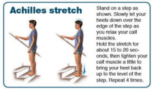
- Plantar fascia:
1) Cross you affected leg over your other leg.
2) Using the hand on your affected side, take hold of your affected foot and pull your toes back towards shin. This creates tension/stretch in the arch of the foot/plantar fascia.
3) Check for the appropriate stretch position by gently rubbing the thumb of your unaffected side left to right over the arch of the affected foot. The plantar fascia should feel firm, like a guitar string.
4) Hold the stretch for a count of 10. A set is 10 repetitions.
2) Nerve mobility[10] (Level of evidence: 1B)
Nerve mobilization exercises have been used to treat carpal tunnel syndrome (nerve entrapment in the wrist) with contradicting results. In the present literature, only a limited number of case studies where nerve mobilization exercises have been used to treat plantar heel pain of neural origin.
Kavlak Y. and Uygur F. conducted a RCT where they used nerve mobilization techniques as an adjunct to conservative treatment. The studie reported a positive outcome for R.OM., muscle strenght and pain in both groups. The study group showed a significant improvement for 2-point discrimination, light touch and tinel’s sign.
Nerve mobilization as described by Meyer et al.[5] (Level of evidence: 4)
The patient is seated at the egde of the table wit hand is asked to slump forward into a comfortable position with the hands behind the back (see image). The ankle is taken into end-range dorsiflexion and eversion to bring tension on the tibial nerve. The knee is extended to R1 (= the angle where the PT feels the first resistance) with the ankle in end-range dorsiflexion and eversion and back to the relaxed flexed state. Each extension-flexion takes about 4 seconds and is repeated 10 times
3) Tibialis posterior strenghtening[1][5] (Level of evidence: 5, 4)
The function of the tibialis posterior muscle is to stabilize the ankle, it is also used for inversion of the ankle. Exercises can be classified into two categories: weight bearing and non-weigth bearing.
- Example of a non-weight bearing exercise:
- Example of a weight bearing exercise:
Other types of conservative treatment may include[4] (Level of evidence: 5):
• Rest
• NSAID’s
• Corticosteroid injections
• Extracorporeal shockwave therapy
• Laser
• Local anaesthetic injections
• Heel pads and heel cups
• Night splints
• Medial longitudinal arch supports
• Strapping
• Soft-soled shoes
• Casting
It is recommended that patients are treated conservatively prior to the surgical treatment. When patients do not respond to the conservative treatment or if there are signs of atrophy or motor involvment they should be reffered to a clinician.
Postoperative rehabilitation[9] (Level of evidence: 5)
| Timeline | Goals | Intervention | |
| Phase I | Week 1 - 3 |
- Protect joint/nerve integrity - Control inflammation - Control pain/edema |
- Immobilization - Passive mobilization - Surgical site protection - Elevation and ice - Educate and monitor non-weight bearing crutch ambulation |
| Phase II | Week 3 - 6 |
- Prevent contraction, scar tissue - Soft tissue and joint mobility |
- Weight-bearing as tolerated - Gentle passive, active-assist, active ankle stretches - Genlte passive dorsiflexion - Tibial nerve gliding with anti-tension - Pain-free, weight-bearing dorsiflexion - Gait training wearing protective splint - Aqua therapy |
| Phase III | Week 6 - 12 |
- Normal gait mechanics - Symmetric ankle mobility - Single leg proprioception - Repeated single leg heel raises (pain free) - Sport/job - specific exercises
|
- Gatin training - Resistive ankle ex. (parin free) - Progress stretching ex. - Progress resistive ex. (to body weight) - Progress proprioceptive and balance ex. - Cardiovascular ex. (pain free) - Low level plyometric ex. |
Clinical Bottom Line[edit | edit source]
Tarsal tunnel syndrome is a rare condition and often underdiagnosed.[1] A variety of symptoms are possible, such as: tingling or burning pain (paresthesia), hyperesthesia and sensory impairment (dysesthesia). These are felt on the plantar face of the ankel and foot.
There are a few test to identify tarsal tunnel syndrome or rule out other possibilities, these tests include: MRI, Ultrasound, Hoffman-tinels test, dorsiflexion-eversion test, trepman test and the triple compression stress test.[4]
Treatment of a tarsal tunnel syndrom should be attemted conservatively at first (see “Physical therapy management”). If conservative treatment fails a surgical aproach can be taken.
References[edit | edit source]
- ↑ 1.00 1.01 1.02 1.03 1.04 1.05 1.06 1.07 1.08 1.09 1.10 1.11 1.12 1.13 1.14 1.15 1.16 1.17 1.18 1.19 1.20 1.21 1.22 1.23 1.24 1.25 1.26 1.27 1.28 1.29 1.30 1.31 1.32 1.33 1.34 1.35 1.36 1.37 1.38 1.39 1.40 1.41 1.42 1.43 1.44 1.45 1.46 1.47 1.48 1.49 1.50 1.51 1.52 1.53 1.54 1.55 1.56 1.57 1.58 1.59 1.60 1.61 1.62 1.63 1.64 1.65 1.66 McSweeney SC, Cichero M. Tarsal tunnel sydrome-A narrative literature review. The Foot 2015;25:244-50. (Level of Evidence 5) Cite error: Invalid
<ref>tag; name "p1" defined multiple times with different content Cite error: Invalid<ref>tag; name "p1" defined multiple times with different content Cite error: Invalid<ref>tag; name "p1" defined multiple times with different content - ↑ 2.00 2.01 2.02 2.03 2.04 2.05 2.06 2.07 2.08 2.09 2.10 2.11 2.12 https://www.ncbi.nlm.nih.gov/mesh/?term=tarsal+tunnel+syndrome Cite error: Invalid
<ref>tag; name "p2" defined multiple times with different content Cite error: Invalid<ref>tag; name "p2" defined multiple times with different content Cite error: Invalid<ref>tag; name "p2" defined multiple times with different content - ↑ 3.0 3.1 3.2 3.3 3.4 Fantino O. Role of ultrasound in posteromedial tarsal tunnel syndrome: 81 cases. J Ultrasound 2014;17(2):99–112. (Level of Evidence 4) Cite error: Invalid
<ref>tag; name "p3" defined multiple times with different content Cite error: Invalid<ref>tag; name "p3" defined multiple times with different content Cite error: Invalid<ref>tag; name "p3" defined multiple times with different content - ↑ 4.00 4.01 4.02 4.03 4.04 4.05 4.06 4.07 4.08 4.09 4.10 4.11 4.12 4.13 Alshami AM, Souvlis T, Coppieters MW. A review of plantar heel pain of neural origin: Differntial diagnosis and management. Manual Therapy 2008;13:103-11. (Level of Evidence 5) Cite error: Invalid
<ref>tag; name "p4" defined multiple times with different content Cite error: Invalid<ref>tag; name "p4" defined multiple times with different content Cite error: Invalid<ref>tag; name "p4" defined multiple times with different content - ↑ 5.00 5.01 5.02 5.03 5.04 5.05 5.06 5.07 5.08 5.09 5.10 5.11 5.12 Cheung Y. Normal Variants: Accessory Muscles About the Ankle. Magn Reson Imaging Clin N Am. 2017;25(1):11-26. (Level of Evidence 5) Cite error: Invalid
<ref>tag; name "p5" defined multiple times with different content Cite error: Invalid<ref>tag; name "p5" defined multiple times with different content - ↑ 6.00 6.01 6.02 6.03 6.04 6.05 6.06 6.07 6.08 6.09 6.10 6.11 6.12 6.13 6.14 6.15 6.16 6.17 Tu P, Bytomski JR. Diagnosis of Heel Pain. American Family Physician 2011;84(8):909-16. (Level of Evidence 5) Cite error: Invalid
<ref>tag; name "p7" defined multiple times with different content Cite error: Invalid<ref>tag; name "p7" defined multiple times with different content Cite error: Invalid<ref>tag; name "p7" defined multiple times with different content - ↑ 7.0 7.1 7.2 Lin D, Williams C, Zaw H. A rare case of an accessory flexor hallucis longus causing tarsal tunnel syndrome. Foot Ankle Surg. 2014;20:e37-39. (Level of Evidence 4) Cite error: Invalid
<ref>tag; name "p0" defined multiple times with different content Cite error: Invalid<ref>tag; name "p0" defined multiple times with different content - ↑ 8.00 8.01 8.02 8.03 8.04 8.05 8.06 8.07 8.08 8.09 8.10 8.11 8.12 8.13 8.14 8.15 Kotnis N, Harish S, Popowich T. Medial Ankle and Heel: Ultrasound Evaluation and Sonographic Appearances of Conditions Causing Symptoms. Seminars in Ultrasound, CT and MRI 2011;32:125-41. (Level of Evidence 4) Cite error: Invalid
<ref>tag; name "p9" defined multiple times with different content Cite error: Invalid<ref>tag; name "p9" defined multiple times with different content Cite error: Invalid<ref>tag; name "p9" defined multiple times with different content - ↑ 9.0 9.1 9.2 9.3 9.4 9.5 9.6 9.7 9.8 Moore KL, Dalley AF, Agur AMR. Clinically Oriented Anatomy. 6th ed. Philadelphia, Pa: Lippincott Williams and Wilkins;2010:617-18, 666-67. (Level of Evidence B) Cite error: Invalid
<ref>tag; name "p6" defined multiple times with different content Cite error: Invalid<ref>tag; name "p6" defined multiple times with different content Cite error: Invalid<ref>tag; name "p6" defined multiple times with different content - ↑ 10.0 10.1 10.2 10.3 10.4 10.5 10.6 10.7 Kavlak Y, Uygur F. Effects of nerve mobilization exercise as an adjunct to the conservative treatment for patients with tarsal tunnel syndrome. Journal of manipulative and physiological therapeutics 2011;34(7):441-48. (Level of evidence 1b) Cite error: Invalid
<ref>tag; name "p8" defined multiple times with different content Cite error: Invalid<ref>tag; name "p8" defined multiple times with different content Cite error: Invalid<ref>tag; name "p8" defined multiple times with different content
