Gastrocnemius: Difference between revisions
No edit summary |
Joao Costa (talk | contribs) No edit summary |
||
| (31 intermediate revisions by 8 users not shown) | |||
| Line 4: | Line 4: | ||
'''Top Contributors''' - {{Special:Contributors/{{FULLPAGENAME}}}} | '''Top Contributors''' - {{Special:Contributors/{{FULLPAGENAME}}}} | ||
</div> | </div> | ||
= Description | == Description == | ||
[[File:Gastrocnemius Primal.png|thumb|Gastrocnemius]] | |||
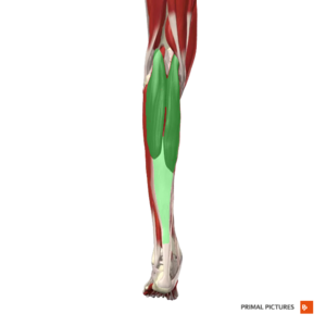
The gastrocnemius [[muscle]] is a complex muscle that is fundamental for [[Walking - Muscles Used|walking]] and [[posture]]<ref name=":0">Bordoni B, Varacallo M. [https://www.ncbi.nlm.nih.gov/books/NBK532946/ Anatomy, bony pelvis and lower limb, gastrocnemius muscle]. Available: https://www.ncbi.nlm.nih.gov/books/NBK532946/<nowiki/>(accessed 25.3.2022)</ref>. Gastrocnemius forms the major bulk at the back of lower leg and is a very powerful muscle. It is a two joint or biarticular muscle and has two heads and runs from back of [[knee]] to the heel. The definitive shape of the calf is as a result of the medial and lateral heads of the gastrocnemius, which are situated at the posterior, upper half of the lower leg. With the [[soleus]] and [[plantaris]], they form a composite muscle called the [[Triceps Surae|triceps surae]]. The two heads of the muscle form the lower boundaries of the [[Popliteal Fossa|popliteal fossa]].<ref name="pala">Palastanga N, Soames R. Anatomy and Human Movement: Structure and Function. 6th ed. London, United Kingdom: Churchill Livingstone; 2012.</ref>The gastrocnemius muscle is superficial, can be easily seen and can be touched on the back of your lower leg.<ref>Gastrocnemius Muscle: Anatomy, Function, and Conditions (verywellhealth.com)[https://www.verywellhealth.com/gastrocnemius-muscle-anatomy-4684083]</ref> | |||
== Anatomy == | |||
= | === Origin === | ||
[[File:Popliteal picture.jpg|thumb|250x250px|Popliteal Fossa]] | |||
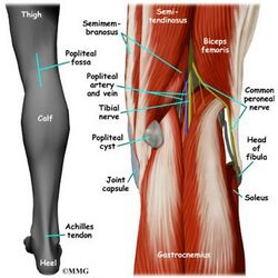
The two heads are located from the medial and lateral condyles of the [[femur]]. | |||
* The medial head from behind the medial supercondylar ridge and the adductor tubercle on the popliteal surface of the femur. | |||
* The lateral head from the outer aspect of the lateral condyle of the femur, just superior and posterior the lateral epicondyle.The fabella is an accessory ossicle most always found in the lateral head of the gastrocnemius.<ref>Fabella | Radiology Reference Article | Radiopaedia.org</ref> | |||
* Both heads have attachments from the knee joint capsule and from the oblique popliteal ligament.<ref name="pala" /> | |||
=== | === Insertion === | ||
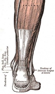
The | * [[File:Achilles tendon.jpg|thumb|300x300px|Achilles Tendon]]The bulk of the gastrocnemius muscle from each of the heads come together and insert into the posterior surface of a broad membranous tendon. | ||
* It then fuses with the soleus tendon to form the upper part of tendocalcaneus. | |||
* This broad tendon then narrows until it reaches the the calcaneous where it expands again for its insertion on the middle part of the posterior surface of the [[calcaneus]].<ref name="pala" /> | |||
=== Nerve supply === | |||
Both heads | * Both heads of the gastrocnemius is supplied by the [[Tibial Nerve|tibial nerve]] (S1 and 2). | ||
* Cutaneous supply is mainly provided by L4, 5 and S2.<ref name="pala" /> | |||
== | == Function == | ||
[[File:Gastrocnemius - Kenhub.png|alt=Gastrocnemius (highlighted in green) - posterior view|right|frameless|500x500px|Gastrocnemius (highlighted in green) - posterior view]] | |||
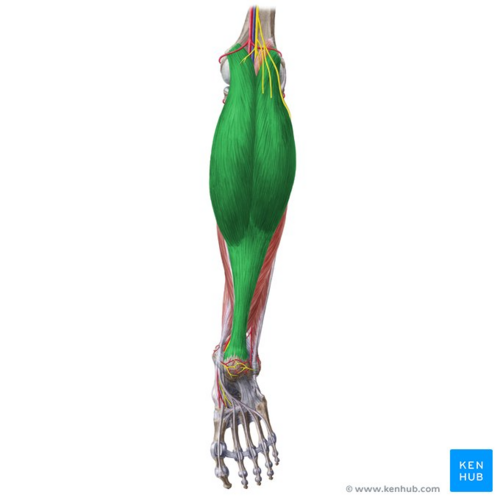
The gastrocnemius with the soleus, is the main plantarflexor of the [[Ankle Joint|ankle joint]]. The muscle is also a powerful [[Knee Flexors|knee flexor]]. It is not able to exert full power at both joints simultaneously, for example when the knee is flexed, gastrocnemius is unable to generate as much force at the ankle. The opposite is true when the ankle is flexed. | |||
When running, walking or jumping the gastrocnemius provides a significant amount of propulsive force. Consider the amount of force required to propel the body into the air, triceps surae can generate a lot of force. <ref name="pala" /> | |||
The gastrocnemius | The gastrocnemius muscle tension has many fascial connections, and this tension is transmitted not only to the foot but to the knee, hip, and lumbar area. A shortened gastrocnemius muscle could cause dysfunctions to the physiological movements of the hip, decreasing its anteversion (inward rotation of the femur). The fascial system plays a fundamental role in the transmission of the force produced by the contraction of the contractile component of the muscle.<ref name=":0" /> | ||
Image: Gastrocnemius (highlighted in green) - posterior view<ref >Gastrocnemius (highlighted in green) - posterior view image - © Kenhub https://www.kenhub.com/en/library/anatomy/gastrocnemius-muscle</ref> | |||
= Assessment | == Assessment == | ||
=== Palpation === | === Palpation === | ||
At the posterior aspect of the knee joint, the two large muscle bellies of gastrocnemius can be felt on either side of the upper portion of the calf. The medial head is projects higher and is lower than the lateral. Both can be felt joining the tendinous junction. | * At the posterior aspect of the [[knee]] joint, the two large muscle bellies of gastrocnemius can be felt on either side of the upper portion of the calf. The medial head is projects higher and is lower than the lateral. | ||
* Both can be felt joining the tendinous junction. | |||
* Further down the calf is the flattened tendocalcaneus which can be palpated to its insertional attachment at the posterior surface of the [[calcaneus]].<ref name="pala" /> | |||
=== Power === | |||
* Ankle plantarflexion in long-sitting (consider that gastrocnemius works against full body on a daily basis). | |||
*Double/single leg calf raise | |||
*Double/single leg calf raise | *Straight leg jump | ||
*Straight leg jump | *Functional tasks ([[Stair Gait|steps]], etc.) | ||
*Functional tasks (steps, etc.) | |||
=== Length === | === Length === | ||
| Line 52: | Line 59: | ||
*Body-weight lunge, measure the straight back leg. | *Body-weight lunge, measure the straight back leg. | ||
= Treatment | == Treatment == | ||
=== Weakness === | === Weakness === | ||
# | #Non-weight bearing and basic weight bearing exercises such as theraband exercises, double and single leg calf raises. | ||
#Weight-bearing exercises and gradually progresses stability exercises by (i) increasing load (ii) increasing the repetitions (iii) varying surface, for example introducing a wobble board | #Weight-bearing exercises and gradually progresses stability exercises by (i) increasing load (ii) increasing the repetitions (iii) varying surface, for example introducing a wobble board | ||
#Sport-specific movement patterns such as running, jumping, and bounding.< | #Sport-specific movement patterns such as running, jumping, and bounding. | ||
=== Stretching Exercises === | |||
In order to get effective gastrocnemius stretch, the [[knee]] must be in extension while the ankle is dorsiflexed. | |||
==== '''Patient position and procedure''' ==== | |||
* Long-sitting (knees extended) or with the knees partially flexed. | |||
* Instruct the patient to strongly dorsiflex the feet, attempting to keep the toes relaxed. | |||
==== '''Patient position and procedur'''e ==== | |||
* Long-sitting and with a towel or belt looped under the forefoot. | |||
* Instruct the patient to pull with equal force on both ends of the towel to move the [[Ankle and Foot|foot]] into dorsiflexion | |||
====Patient position and procedure ==== | |||
* Standing. | |||
* Instruct the patient to stride forward with one foot, keeping the heel of the back foot flat on the floor (the back foot is the one being stretched). | |||
* Have the patient brace his or her hands against a wall if necessary. To provide stability to the foot, the patient partially rotates the back leg inward so the foot assumes a supinated position and locks the joints. The patient then shifts body weight forward onto the front foot. | |||
* To stretch the gastrocnemius muscle, the knee of the back leg is kept extended. (To stretch the [[soleus]], the knee of the back leg is flexed.) | |||
{{#ev:youtube|EFnLllHNbQQ}}<ref>How to strech your Gastrocnemius. Available from:<nowiki>https://www.youtube.com/watch?v=EFnLllHNbQQ</nowiki></ref> | |||
====Patient position and procedure==== | |||
* Patient must be standing on an inclined board with feet pointing upward and heels downward. | |||
* Greater stretch occurs if the patient leans forward. Because the body weight is on the heels, there is little stretch on the long [[Arches of the Foot|arches of the feet]]. | |||
Precaution must be followed when a patient uses weight-bearing exercises to stretch the plantarflexor muscles, shoes with arch supports should be worn or a folded washcloth placed under the medial border of the foot to minimize the stress to the arches of the foot.<ref>Carolyn Kisner,Lynn Allen Colby,editors. Therapeutic Exercise : Foundations and Techniques. 6th Edition. Philadelphia.F. A. Davis Company,2012. p 883.</ref> | |||
=== Trigger Points === | === Trigger Points === | ||
[[Image:Gastrocnemius trigger points referred pain-1024x768.jpg|thumb|right|200px]]The gastrocnemius may contain up to four trigger points. | [[Image:Gastrocnemius trigger points referred pain-1024x768.jpg|thumb|right|200px]]The gastrocnemius may contain up to four [[Trigger Points|trigger points.]] | ||
*The two medial trigger points lie in the medial head of the gastrocnemius, with the upper trigger point found just below the crease of the knee, and the lower trigger point an inch or two below it. | *The two medial trigger points lie in the medial head of the gastrocnemius, with the upper trigger point found just below the crease of the knee, and the lower trigger point an inch or two below it. | ||
*The two lateral trigger points in the lateral head mirror the positioning of the medial trigger points, except that they lie slightly more distal (towards the foot) by about a half-inch.<ref>http://www.triggerpointtherapist.com/blog/gastrocnemius-pain/gastrocnemius-trigger-points-calf-cramp/</ref> | *The two lateral trigger points in the lateral head mirror the positioning of the medial trigger points, except that they lie slightly more distal (towards the foot) by about a half-inch.<ref>http://www.triggerpointtherapist.com/blog/gastrocnemius-pain/gastrocnemius-trigger-points-calf-cramp/</ref> | ||
= Resourses | == Resourses == | ||
{| width="100%" cellspacing="1" cellpadding="1" | {| width="100%" cellspacing="1" cellpadding="1" | ||
| Line 87: | Line 120: | ||
|} | |} | ||
= See also = | == See also == | ||
*[[Achilles Rupture|Achilles rupture]] | *[[Achilles Rupture|Achilles rupture]] | ||
*[[Achilles Tendinopathy|Achilles tendinopathy]] | *[[Achilles Tendinopathy|Achilles tendinopathy]] | ||
*[[Achilles | *[[Achilles Tendon|Achilles tendon]] | ||
*[[Baker's Cyst|Baker's cyst]] | *[[Baker's Cyst|Baker's cyst]] | ||
*[[Calcaneal Fractures|Calcaneal fracture]] | *[[Calcaneal Fractures|Calcaneal fracture]] | ||
| Line 98: | Line 131: | ||
*[[Fabella syndrome|Fabella syndrome]] | *[[Fabella syndrome|Fabella syndrome]] | ||
*[[Thompson Test|Thompson test]] | *[[Thompson Test|Thompson test]] | ||
*[[Soleus|Soleus]] | *[[Soleus|Soleus]]<div class="coursebox"></div> | ||
<div class="coursebox" | |||
= References = | == References == | ||
<references /> | <references /> | ||
<br> | |||
[[Category:Anatomy]] [[Category:Knee]] [[Category: | [[Category:Anatomy]] [[Category:Muscles]] [[Category:Knee]] [[Category:Ankle]] [[Category:Musculoskeletal/Orthopaedics]] | ||
[[Category:Knee - Anatomy]] [[Category:Ankle - Anatomy]] | |||
[[Category:Knee - Muscles]] [[Category:Ankle - Muscles]] | |||
Latest revision as of 02:47, 29 March 2022
Original Editor Aarti Sareen
Top Contributors - George Prudden, Aarti Sareen, Rishika Babburu, Admin, Andeela Hafeez, Kim Jackson, Evan Thomas, Venus Pagare, WikiSysop, Joao Costa, Lucinda hampton, Daphne Jackson and Samuel Adedigba
Description[edit | edit source]
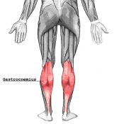
The gastrocnemius muscle is a complex muscle that is fundamental for walking and posture[1]. Gastrocnemius forms the major bulk at the back of lower leg and is a very powerful muscle. It is a two joint or biarticular muscle and has two heads and runs from back of knee to the heel. The definitive shape of the calf is as a result of the medial and lateral heads of the gastrocnemius, which are situated at the posterior, upper half of the lower leg. With the soleus and plantaris, they form a composite muscle called the triceps surae. The two heads of the muscle form the lower boundaries of the popliteal fossa.[2]The gastrocnemius muscle is superficial, can be easily seen and can be touched on the back of your lower leg.[3]
Anatomy[edit | edit source]
Origin[edit | edit source]
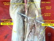
The two heads are located from the medial and lateral condyles of the femur.
- The medial head from behind the medial supercondylar ridge and the adductor tubercle on the popliteal surface of the femur.
- The lateral head from the outer aspect of the lateral condyle of the femur, just superior and posterior the lateral epicondyle.The fabella is an accessory ossicle most always found in the lateral head of the gastrocnemius.[4]
- Both heads have attachments from the knee joint capsule and from the oblique popliteal ligament.[2]
Insertion[edit | edit source]
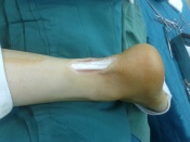
- The bulk of the gastrocnemius muscle from each of the heads come together and insert into the posterior surface of a broad membranous tendon.
- It then fuses with the soleus tendon to form the upper part of tendocalcaneus.
- This broad tendon then narrows until it reaches the the calcaneous where it expands again for its insertion on the middle part of the posterior surface of the calcaneus.[2]
Nerve supply[edit | edit source]
- Both heads of the gastrocnemius is supplied by the tibial nerve (S1 and 2).
- Cutaneous supply is mainly provided by L4, 5 and S2.[2]
Function[edit | edit source]
The gastrocnemius with the soleus, is the main plantarflexor of the ankle joint. The muscle is also a powerful knee flexor. It is not able to exert full power at both joints simultaneously, for example when the knee is flexed, gastrocnemius is unable to generate as much force at the ankle. The opposite is true when the ankle is flexed.
When running, walking or jumping the gastrocnemius provides a significant amount of propulsive force. Consider the amount of force required to propel the body into the air, triceps surae can generate a lot of force. [2]
The gastrocnemius muscle tension has many fascial connections, and this tension is transmitted not only to the foot but to the knee, hip, and lumbar area. A shortened gastrocnemius muscle could cause dysfunctions to the physiological movements of the hip, decreasing its anteversion (inward rotation of the femur). The fascial system plays a fundamental role in the transmission of the force produced by the contraction of the contractile component of the muscle.[1]
Image: Gastrocnemius (highlighted in green) - posterior view[5]
Assessment[edit | edit source]
Palpation[edit | edit source]
- At the posterior aspect of the knee joint, the two large muscle bellies of gastrocnemius can be felt on either side of the upper portion of the calf. The medial head is projects higher and is lower than the lateral.
- Both can be felt joining the tendinous junction.
- Further down the calf is the flattened tendocalcaneus which can be palpated to its insertional attachment at the posterior surface of the calcaneus.[2]
Power[edit | edit source]
- Ankle plantarflexion in long-sitting (consider that gastrocnemius works against full body on a daily basis).
- Double/single leg calf raise
- Straight leg jump
- Functional tasks (steps, etc.)
Length[edit | edit source]
- Passive dorsiflexion
- Body-weight lunge, measure the straight back leg.
Treatment[edit | edit source]
Weakness[edit | edit source]
- Non-weight bearing and basic weight bearing exercises such as theraband exercises, double and single leg calf raises.
- Weight-bearing exercises and gradually progresses stability exercises by (i) increasing load (ii) increasing the repetitions (iii) varying surface, for example introducing a wobble board
- Sport-specific movement patterns such as running, jumping, and bounding.
Stretching Exercises[edit | edit source]
In order to get effective gastrocnemius stretch, the knee must be in extension while the ankle is dorsiflexed.
Patient position and procedure[edit | edit source]
- Long-sitting (knees extended) or with the knees partially flexed.
- Instruct the patient to strongly dorsiflex the feet, attempting to keep the toes relaxed.
Patient position and procedure[edit | edit source]
- Long-sitting and with a towel or belt looped under the forefoot.
- Instruct the patient to pull with equal force on both ends of the towel to move the foot into dorsiflexion
Patient position and procedure[edit | edit source]
- Standing.
- Instruct the patient to stride forward with one foot, keeping the heel of the back foot flat on the floor (the back foot is the one being stretched).
- Have the patient brace his or her hands against a wall if necessary. To provide stability to the foot, the patient partially rotates the back leg inward so the foot assumes a supinated position and locks the joints. The patient then shifts body weight forward onto the front foot.
- To stretch the gastrocnemius muscle, the knee of the back leg is kept extended. (To stretch the soleus, the knee of the back leg is flexed.)
Patient position and procedure[edit | edit source]
- Patient must be standing on an inclined board with feet pointing upward and heels downward.
- Greater stretch occurs if the patient leans forward. Because the body weight is on the heels, there is little stretch on the long arches of the feet.
Precaution must be followed when a patient uses weight-bearing exercises to stretch the plantarflexor muscles, shoes with arch supports should be worn or a folded washcloth placed under the medial border of the foot to minimize the stress to the arches of the foot.[7]
Trigger Points[edit | edit source]
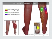
The gastrocnemius may contain up to four trigger points.
- The two medial trigger points lie in the medial head of the gastrocnemius, with the upper trigger point found just below the crease of the knee, and the lower trigger point an inch or two below it.
- The two lateral trigger points in the lateral head mirror the positioning of the medial trigger points, except that they lie slightly more distal (towards the foot) by about a half-inch.[8]
Resourses[edit | edit source]
|

|

|
|
|
See also[edit | edit source]
- Achilles rupture
- Achilles tendinopathy
- Achilles tendon
- Baker's cyst
- Calcaneal fracture
- Calf strain
- Deep vein thrombosis
- Fabella syndrome
- Thompson test
- Soleus
References[edit | edit source]
- ↑ 1.0 1.1 Bordoni B, Varacallo M. Anatomy, bony pelvis and lower limb, gastrocnemius muscle. Available: https://www.ncbi.nlm.nih.gov/books/NBK532946/(accessed 25.3.2022)
- ↑ 2.0 2.1 2.2 2.3 2.4 2.5 Palastanga N, Soames R. Anatomy and Human Movement: Structure and Function. 6th ed. London, United Kingdom: Churchill Livingstone; 2012.
- ↑ Gastrocnemius Muscle: Anatomy, Function, and Conditions (verywellhealth.com)[1]
- ↑ Fabella | Radiology Reference Article | Radiopaedia.org
- ↑ Gastrocnemius (highlighted in green) - posterior view image - © Kenhub https://www.kenhub.com/en/library/anatomy/gastrocnemius-muscle
- ↑ How to strech your Gastrocnemius. Available from:https://www.youtube.com/watch?v=EFnLllHNbQQ
- ↑ Carolyn Kisner,Lynn Allen Colby,editors. Therapeutic Exercise : Foundations and Techniques. 6th Edition. Philadelphia.F. A. Davis Company,2012. p 883.
- ↑ http://www.triggerpointtherapist.com/blog/gastrocnemius-pain/gastrocnemius-trigger-points-calf-cramp/
