Flexor Digitorum Longus: Difference between revisions
No edit summary |
No edit summary |
||
| Line 22: | Line 22: | ||
=== Artery === | === Artery === | ||
Posterior tibial artery | Posterior tibial artery | ||
== Function == | == Function == | ||
== Clinical relevance == | == Clinical relevance == | ||
== Assessment == | == Assessment == | ||
=== Palpation === | === Palpation === | ||
| Line 36: | Line 36: | ||
=== Length === | === Length === | ||
== Treatment == | == Treatment == | ||
=== Strengthening === | === Strengthening === | ||
| Line 49: | Line 49: | ||
|- | |- | ||
| {{#ev:youtube|WUS5BUB3fgM|270}} | | {{#ev:youtube|WUS5BUB3fgM|270}} | ||
| {{#ev:youtube|UHOExMbeJco|270}} | | {{#ev:youtube|UHOExMbeJco|270}} | ||
| {{#ev:youtube|LXIe209QYdI|270}} | | {{#ev:youtube|LXIe209QYdI|270}} | ||
|} | |} | ||
| Line 66: | Line 66: | ||
== See also == | == See also == | ||
*[[ | *[[Flexor_hallucis_longus|Flexor hallucis longus]] | ||
*[[ | *[[The Os Trigonum Syndrome|The Os Trigonum Syndrome]] | ||
*[[ | *[[Tarsal Tunnel syndrome|Tarsal Tunnel syndrome]] | ||
*[[ | *[[Posterior Tibial Tendon Dysfunction|Posterior Tibial Tendon Dysfunction]] | ||
*[[ | *[[Ankle & Foot|Ankle & Foot]] | ||
*[[ | *[[Compartment Syndrome of the Foot|Compartment Syndrome of the Foot]] | ||
*[[ | *[[Ankle Impingement|Ankle Impingement]] | ||
*[[ | *[[Hallux Valgus|Hallux Valgus]] | ||
*[[ | *[[Ankle Joint|Ankle Joint]] | ||
*[[Congenital talipes equinovarus (CTEV)|Congenital talipes equinovarus (CTEV)]] | |||
== Recent Related Research (from Pubmed) == | == Recent Related Research (from Pubmed) == | ||
<div class="researchbox"><rss>https://eutils.ncbi.nlm.nih.gov/entrez/eutils/erss.cgi?rss_guid=1hgsEQZ6hYlxDg0_4RJK6EmuRk1nkQQfv6dNixzAbdg-369MDj|charset=UTF-8|short|max=10</rss></div> | <div class="researchbox"><rss>https://eutils.ncbi.nlm.nih.gov/entrez/eutils/erss.cgi?rss_guid=1hgsEQZ6hYlxDg0_4RJK6EmuRk1nkQQfv6dNixzAbdg-369MDj|charset=UTF-8|short|max=10</rss></div> | ||
== References == | == References == | ||
Revision as of 21:13, 9 January 2017
Original Editor - George Prudden
Top Contributors - George Prudden, Kim Jackson, 127.0.0.1, Evan Thomas, WikiSysop, Abbey Wright, Pinar Kisacik and Patti Cavaleri;
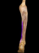
Description[edit | edit source]
Origin[edit | edit source]
Posterior surface of the body of the tibia.
Insertion[edit | edit source]
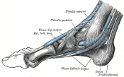
Plantar surface, base of the distal phalanges of the four lesser digits.
Nerve[edit | edit source]
Tibial nerve
Artery[edit | edit source]
Posterior tibial artery
Function[edit | edit source]
Clinical relevance[edit | edit source]
Assessment[edit | edit source]
Palpation[edit | edit source]
Power[edit | edit source]
Length[edit | edit source]
Treatment[edit | edit source]
Strengthening[edit | edit source]
Stretching[edit | edit source]
Manual techniques[edit | edit source]
Resources[edit | edit source]

|

|

|
File:FDL4.JPG | 
|
See also[edit | edit source]
- Flexor hallucis longus
- The Os Trigonum Syndrome
- Tarsal Tunnel syndrome
- Posterior Tibial Tendon Dysfunction
- Ankle & Foot
- Compartment Syndrome of the Foot
- Ankle Impingement
- Hallux Valgus
- Ankle Joint
- Congenital talipes equinovarus (CTEV)


