Deltoid
Original Editor - Wendy Walker
Lead Editors - Lucinda hampton, Sai Kripa, Wendy Walker, Naomi O'Reilly, Joao Costa, Chrysolite Jyothi Kommu, Andeela Hafeez, Kim Jackson, Vidya Acharya, WikiSysop, Aminat Abolade, Admin, Evan Thomas, George Prudden, Uchechukwu Chukwuemeka and Wanda van Niekerk
Description[edit | edit source]
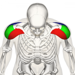
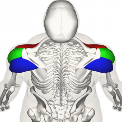
The Deltoid muscle is a large triangular shaped muscle which lies over the glenohumeral joint and which gives the shoulder its rounded contour. It is named after the Greek letter delta, which is shaped like an equilateral triangle. It comprises 3 distinct portions each of which produces a different movement of the glenohumeral joint, commonly named the anterior, mid (or lateral) and posterior heads.
Anatomy[edit | edit source]
Origin[edit | edit source]
Anterior Fibres/Head
Lateral third, Anterior Surface of the Clavicle (close to the lateral fibres of pectoralis major).
Mid/Lateral Head
Acromion Process, Superior Surface.
Posterior Head
Spine of the Scapula, Posterior Border.
Insertion[edit | edit source]
Fibres from all heads converge to insert into the deltoid tuberosity on the humerus.
The deltoid fascia is continuous with the brachial fascia and connects to the medial and lateral intermuscular septa[1].
Nerve Supply[edit | edit source]
Axillary Nerve, C5 & 6, posterior cord of the brachial plexus.
Blood Supply[edit | edit source]
Deltoid receives its blood supply from the posterior circumflex humeral artery.
Function[edit | edit source]
An important function of deltoid is the prevention of subluxation or even dislocation of the head of the humerus particularly when carrying a load. Deltoid is the prime mover of shoulder abduction.
Actions [edit | edit source]
All heads of deltoid work together to produce abduction of the Shoulder Joint. In addition, each individual head produces the following:
Anterior Fibres
- Flexes, abducts, medially rotates, and horizontally flexes the arm at the shoulder joint
Posterior Fibres
- Extends, abducts, laterally rotates, and horizontally extends the arm at the shoulder joint.
Clinical Relevance[edit | edit source]
In case of patients suffering from Subacromial bursitis, there occurs impingement of deltoid muscle below the acromion process when the arm is hanging by the side leading to pain. Moreover, there occurs no pain while performing abduction movement of the arm due to vanishing of bursa under the acromion process. Subacromial or Subdeltoid bursitis is commonly seen secondary to supraspinatus tendonitis.[3]
During dislocation of the shoulder or fracture of surgical neck of the humerus, there occurs damage to axillary nerve. Moreover, the damage to axillary nerve leads to paralysis of deltoid muscle, where the examiner ask the patient to abduct the arm to 90 degrees and the resistance will be provided. There occurs no contraction of the muscle during abduction and there will be complete loss of strength up to 90 degrees of shoulder abduction. Apart from that, there will be loss of sensation over the lower halves of the deltoid in a badge like area known as regimental badge.[3]
Intramuscular injections are often given to the deltoid muscle. It should be provided to the middle of the deltoid to avoid injury to the axillary nerve.[3]
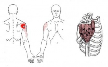
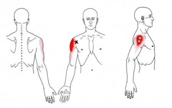
Trigger Point Referal Patterns[edit | edit source]
Techniques[edit | edit source]
Palpation[edit | edit source]
Flex elbow to 90 degrees and have patient abduct the shoulder against resistance.
Anterior Fibers
- Deltoid palpated with elbow extended, shoulder 90 degrees abduction and then resist horizontal adduction.
Posterior Fibers
- Position same as above and then resist horizontal abduction.
Length Tension Testing[edit | edit source]
Treatment[edit | edit source]
Exercises:[4]
Supine active assisted:
Lie down flat on your back, with a pillow supporting your head.
Bend your elbow as far as possible. Then raise your arm to 90 degrees vertical, using the stronger arm to assist if necessary. Once you have got to 90 degrees, you can straighten your elbow. Hold your arm in this upright position with its own strength.
Circles: Slowly with your fingers, wrist and elbow straight move the arm in small circular movements clockwise and counterclockwise. Gradually increase the circle as comfortable (this may take a few weeks to increase to bigger and bigger circles).
Progress to light weight:
As you get more confidence in controlling your shoulder movement, a lightweight e.g. a tin of beans or small paperweight, should be held in the affected hand.
Progress to sitting and standing:
Having more confidence in controlling your shoulder movement gradually go from lying down to sitting and eventually standing.
At this stage you may recline the head of your bed or put some pillows underneath your back to recline your position.
Repeat the same exercise again, this time against some gravity.
Start again from holding your arm in the upright position with its own strength.
Start first without any weights and progress to use the same lightweight you used before in the lying down position.
Resisted exercise:
For re-education of concentric contracture of the deltoid muscle.
Make a fist with the hand of the affected side. The flat hand of the opposite side is providing resistance. Push your affected side hand against resistance from the other hand. Whilst doing this, you will notice that you can fully elevate your arm (above your head).
Repeat these exercises in order to ‘learn’ and re-educate your Deltoid muscle to perform this ‘concentric contracture’ even without pushing against your other arm.
References[edit | edit source]
- ↑ Rispoli, Damian M.; Athwal, George S.; Sperling, John W.; Cofield, Robert H. (2009). "The anatomy of the deltoid insertion". J Shoulder Elbow Surg 18: 386–390
- ↑ Availble from:Kenhub-Learn Human Anatomy Help.https://youtu.be/FSSGkYrYwqM.Kenhub-Learn Human Anatomy.Deltoid Muscle:Origin,Insertion&Action-Human Anatomy|Kenhub{last accessed 29 April 2020}
- ↑ 3.0 3.1 3.2 Chaurasia BD. Human Anatomy Regional and Applied Dissection and Clinical. Vol 1. CBS Publishers and Distributors Pvt Ltd, 2010
- ↑ Shoulderdoc.co.uk. Available from:https://www.shoulderdoc.co.uk/article/1028 (accessed 10 Nov 2020).


