Deltoid
Original Editor - Wendy Walker
Lead Editors - Lucinda hampton, Sai Kripa, Wendy Walker, Naomi O'Reilly, Joao Costa, Chrysolite Jyothi Kommu, Andeela Hafeez, Kim Jackson, Vidya Acharya, WikiSysop, Aminat Abolade, Admin, Evan Thomas, George Prudden, Uchechukwu Chukwuemeka and Wanda van Niekerk
Description[edit | edit source]
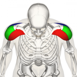
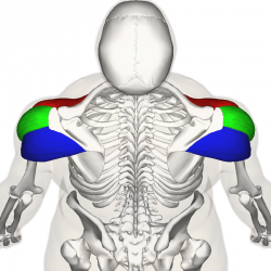
The Deltoid muscle is a large triangular shaped muscle which lies over the glenohumeral joint and which gives the shoulder its rounded contour. It is named after the Greek letter delta, which is shaped like an equilateral triangle. It comprises 3 distinct portions each of which produces a different movement of the glenohumeral joint, commonly named the anterior, mid (or lateral) and posterior heads.
Anatomy[edit | edit source]
Origin[edit | edit source]
Anterior Fibres/Head
Lateral third, Anterior Surface of the Clavicle (close to the lateral fibres of pectoralis major).
Mid/Lateral Head
Acromion Process, Superior Surface.
Posterior Head
Spine of the Scapula, Posterior Border.
Insertion[edit | edit source]
Fibres from all heads converge to insert into the deltoid tuberosity on the humerus.
The deltoid fascia is continuous with the brachial fascia and connects to the medial and lateral intermuscular septa[1].
Nerve Supply[edit | edit source]
Axillary Nerve, C5 & 6, posterior cord of the brachial plexus.
Blood Supply[edit | edit source]
Deltoid receives its blood supply from the posterior circumflex humeral artery.
Function[edit | edit source]
An important function of deltoid is the prevention of subluxation or even dislocation of the head of the humerus particularly when carrying a load. Deltoid is the prime mover of shoulder abduction.
Actions [edit | edit source]
All heads of deltoid work together to produce abduction of the Shoulder Joint. In addition, each individual head produces the following:
Anterior Fibres
- Flexes, abducts, medially rotates, and horizontally flexes the arm at the shoulder joint
Posterior Fibres
- Extends, abducts, laterally rotates, and horizontally extends the arm at the shoulder joint
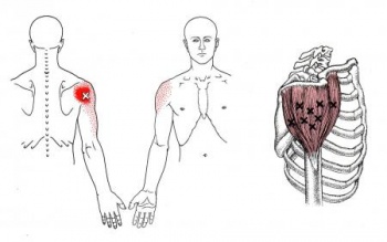
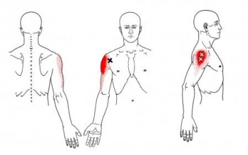
Trigger Point Referal Patterns[edit | edit source]
Techniques[edit | edit source]
Palpation[edit | edit source]
Flex elbow to 90 degrees and have patient abduct the shoulder against resistance.
Anterior Fibers
- Deltoid palpated with elbow extended, shoulder 90 degrees abduction and then resist horizontal adduction.
Posterior Fibers
- Position same as above and then resist horizontal abduction.
Length Tension Testing[edit | edit source]
Resources[edit | edit source]
Recent Related Research (from Pubmed)[edit | edit source]
Extension:RSS -- Error: Not a valid URL: Feed goes here!!|charset=UTF-8|short|max=10
References[edit | edit source]
References will automatically be added here, see adding references tutorial.
- ↑ Rispoli, Damian M.; Athwal, George S.; Sperling, John W.; Cofield, Robert H. (2009). "The anatomy of the deltoid insertion". J Shoulder Elbow Surg 18: 386–390
</div>


