|
|
| (24 intermediate revisions by 3 users not shown) |
| Line 1: |
Line 1: |
| <div class="editorbox"> | | <div class="editorbox"> |
| '''Original Editors ''' - [[User:Ward Willaert|Ward Willaert]] | | '''Original Editors ''' - [[User:Ward Willaert|Ward Willaert]] as part of the [[Vrije Universiteit Brussel Evidence-based Practice Project|Vrije Universiteit Brussel Evidence-Based Practice Project]] |
| '''Top Contributors''' - {{Special:Contributors/{{FULLPAGENAME}}}} | | '''Top Contributors''' - {{Special:Contributors/{{FULLPAGENAME}}}} |
| </div> | | </div> |
| == Description == | | == Introduction == |
| | [[File:Foot.jpg|right|frameless|493x493px]] |
| | Any joint in the [[Ankle and Foot|ankle]], [[Foot Anatomy|foot]] and toes can be affected by an arthropathy (arthropathy is a general term for any disease of the joints). |
| | * There are more than 100 forms of [[arthritis]], many of which affect the foot and ankle, causing [[Joint Classification|joint]] [[Pain Behaviours|pain]], swelling and stiffness. |
| | * Arthritis in the feet can make standing and walking painful and the feet and/or toes may change shape, making it harder to fit shoes and [[Activities of Daily Living]] may be affected. |
| | * Athropathies of the foot and ankle are an important public health challenge due to their increasing incidence combined with their substantial negative impact on patients’ [[Quality of Life|quality of life]]. |
| | * Although arthropathy is less common in the ankle than the [[Hip Anatomy|hip]] and [[knee]], it can be just as disabling.<ref name="Stauffer">Stauffer RN: Intra-articular ankle problems. In Evarts CM (ed): surgery of the musculoskeletal system, vol. 4. New York, Churchill-Livingstone, 1990.</ref> |
| | * Non-pharmacological treatments serve as the first line of treatment and are frequently used for patients with musculoskeletal conditions of the foot and ankle<ref name=":0">Rao S, Riskowski JL, Hannan MT. [https://www.ncbi.nlm.nih.gov/pmc/articles/PMC3414868/ Musculoskeletal conditions of the foot and ankle: assessments and treatment options.] Best Practice & Research Clinical Rheumatology. 2012 Jun 1;26(3):345-68. Available from:https://www.ncbi.nlm.nih.gov/pmc/articles/PMC3414868/ (last accessed 1.7.2020)</ref>. |
| | Arthropathy is a blanket or global term for a wide range of joint presentations. |
|
| |
|
| === Arthropathy ===
| | *Noninfectious arthritis eg [[Psoriatic Arthritis|Psoriatic arthritis]]; [[Ankylosing Spondylitis (Axial Spondyloarthritis)|Ankylosing spondylitis]]; [[Gout]]; [[Rheumatoid Arthritis]]; [[Osteoarthritis]]; Haemophilic Arthropathy (with severe haemophilia causes high levels of impairment) <ref name="Barg">Barg, A., et al. Haemophilic arthropathy of the ankle treated by total ankle replacement: a case series. Haemophilia 2010;16(4):647-655.</ref>; Post traumatic Arthritis. |
| | | *[[Reactive Arthritis|Reactive arthropathy]] occurs as a reaction against an infection site elsewhere in the body.<ref name="mayoclinic">Reactive Arthritis (Reiter’s Syndrome). www.mayoclinic.org. Retrieved May 16, 2011.(accessed 3 december 2016)</ref> |
| An arthropathy is a disease which affects a joint. Although the terms arthropathy and arthritis have very similar meanings, the former is traditionally used to describe the following conditions:
| | *[[Enteropathic Spondylitis|Enteropathic arthropathy]] Includes a group of rheumatic conditions such as arthritis caused by bacteria, parasitic infections and spondyloarthropathies. Other conditions that are included in this type of arthropathy are intestinal bypass arthritis, Whipple’s disease , collagenous colitis and [[Celiac Disease (Coeliac Disease)|celiac disease.]]<ref name="Björkengren">Björkengren A. G., Resnick D, Sartoris DJ. Enteropathic arthropathies. Radiologic Clinics of North America 1987: 189 </ref><ref name=":1" /> |
| | |
| *Reactive arthropathy occurs as a reaction against an infection site elsewhere in the body.<ref name="mayoclinic">Reactive Arthritis (Reiter’s Syndrome). www.mayoclinic.org. Retrieved May 16, 2011.(accessed 3 december 2016)</ref> | |
| *Enteropathic arthropathy is an arthropathy in association with, or as a reaction to, an enteric (usually colonic) inflammatory condition.<ref name="Björkengren">Björkengren A. G., Resnick D, Sartoris DJ. Enteropathic arthropathies. Radiologic Clinics of North America 1987: 189 </ref> | |
| *Crystal arthropathy is characterised by accumulation of tiny crystals in one or more joints.<ref name="McGill">McGill, Neil W. Gout and other crystal-associated arthropathies. Best Practice & Research Clinical Rheumatology 2000: 445-460 </ref> | | *Crystal arthropathy is characterised by accumulation of tiny crystals in one or more joints.<ref name="McGill">McGill, Neil W. Gout and other crystal-associated arthropathies. Best Practice & Research Clinical Rheumatology 2000: 445-460 </ref> |
| *Neuropathic arthropathy is gradual joint destruction when there is chronic damage of peripheral nerves and diminished proprioception. <ref name="Sanders (2013)"> Sanders, L.J., Edmonds, M.E. & Jeffcoate, W.J. Diabetologia (2013) 56: 1873. https://doi.org/10.1007/s00125-013-2961-6 </ref> | | *[[Charcot-Marie-Tooth Disease: A Case Study|Neuropathic arthropathy]] is gradual joint destruction when there is chronic damage of peripheral nerves and diminished proprioception (also called Charcot arthropathy and prominently affects patients with [[diabetes]])<ref name=":1">Scope heal [https://scopeheal.com/arthropathy/ Arthropathy] Available from:https://scopeheal.com/arthropathy/ (last accessed 1.7.2020)</ref> <ref name="Sanders (2013)">Sanders, L.J., Edmonds, M.E. & Jeffcoate, W.J. Diabetologia (2013) 56: 1873. https://doi.org/10.1007/s00125-013-2961-6 </ref> |
| *Diabetic arthropathy is a neuropathic arthropathy occurring in diabetic patients. <ref name="Medical dic">Medical dictionary. http://medical-dictionary.thefreedictionary.com/diabetic+arthropathy (Accessed 2 december 2016)</ref><br> | | *[[The Diabetic Foot|Diabetic arthropathy]] is a neuropathic arthropathy occurring in diabetic patients. <ref name="Medical dic">Medical dictionary. http://medical-dictionary.thefreedictionary.com/diabetic+arthropathy (Accessed 2 december 2016)</ref> |
| | | Be sure to look at all the links above for detailed information, this page is a general overview. |
| An arthropathy can be degenerative, such as osteoarthritis, or associated with inflammation, e.g. rheumatoid arthritis. A joint disease can also occur following trauma.
| |
| | |
| Although arthropathy is less common in the ankle than the hip and knee, it can be just as disabling.<ref name="Stauffer">Stauffer RN: Intra-articular ankle problems. In Evarts CM (ed): surgery of the musculoskeletal system, vol. 4. New York, Churchill-Livingstone, 1990.</ref>
| |
| | |
| Arthropathy is a blanket or global term for a wide range of joint presentations. Some specific arthropathies are detailed below:<br>
| |
| [[File:OA_ankle.jpg|right]]
| |
| | |
| === Osteoarthritis ===
| |
| | |
| Osteoarthritis (OA) results when mechanical and biological factors destabilise the normal coupling of degradation and synthesis of articular cartilage chondrocytes, extracellular matrix and subchondral bone. Destabilising factors include genetic, developmental, metabolic, and traumatic events. OA involves all the tissues of the diathrodial joint. <br>Ultimately, OA manifests as morphologic, biochemical, molecular and biomechanical change of tissues, cells and matrix, which soften and fibrillate articular cartilage. The sequelae then become ulceration and loss of articular cartilage, sclerosis and eburnation of subchondral bone, growth of osteophytes and development of subchondral cysts.<ref name="Huch">Huch, Klaus E. Kuettner, Dieppe P. Osteoarthritis in ankle and knee joints. Seminars in arthritis and rheumatism. 1997 Vol. 26. No. 4.</ref><br>
| |
| | |
| <br>
| |
| | |
| [[File:RA_ankle.jpg|right]]
| |
| | |
| === Rheumatoid arthritis ===
| |
| | |
| Rheumatoid arthritis (RA) in the foot is categorised anatomically, i.e. forefoot, midfoot and hindfoot pathologies.<ref name="Kerry">Kerry RM, Holt GM, Stockley I. The foot in chronic rheumatoid arthritis: a continuing problem. The Foot 1994: 201-203.</ref>Most of the studies have focused on forefoot and hindfoot pathologies; there are fewer studies on midfoot RA.<ref name="Chan">Chan, Pui‐Shan J, Kok Ooi Kong. Natural history and imaging of subtalar and midfoot joint disease in rheumatoid arthritis. International journal of rheumatic diseases 2013;16: 14-18.</ref> Rheumatoid arthritis is a multi-system, chronic, progressive, inflammatory disease. Joints become swollen, tender and painful which can lead to severe deformity and disability. <ref name="Chan" /><ref name="Alehata">Alehata, D., et al. Rheumatoid arthritis classification criteria: an American College of Rheumatology/European Union League Against Rheumatism collaborative initiative. Ann Rheum Dis 2010;69:1580-8.</ref><ref name="Lohkamp">Lohkamp M. et al. The prevalence of disabling foot pain in patients with early rheumatoid arthritis. The Foot 2006;16(4):201-207.</ref><ref name="Brenton">Brenton-Rule, Angela, et al. Foot and ankle characteristics associated with falls in adults with established rheumatoid arthritis: a cross-sectional study.BMC musculoskeletal disorders 2016;17(1):1.</ref><ref name="Baillet">Baillet, Athan, et al. Efficacy of resistance exercises in rheumatoid arthritis: meta-analysis of randomized controlled trials. Rheumatology 2012;51(3):519-527.(Level of evidence 1a)</ref>
| |
| | |
| <br>
| |
| | |
| [[File:HA_foot.jpg|right]]
| |
| | |
| === Haemophilic arthropathy ===
| |
| | |
| In patients with severe haemophilia, arthropathy causes high levels of impairment. The degenerative changes caused by recurrent haemarthrosis are progressive. The ankle is frequently affected in haemophilic patients. <ref name="Barg">Barg, A., et al. Haemophilic arthropathy of the ankle treated by total ankle replacement: a case series. Haemophilia 2010;16(4):647-655.</ref>
| |
| | |
| <br>
| |
| | |
| [[File:Diabetic_foot_arthropathy.jpg|right]]
| |
| | |
| === Diabetic foot arthropathy ===
| |
| | |
| Charcot neuropathic osteoarthropathy of the foot is a devastating neuropathic complication of diabetes and other metabolic illnesses. <ref name="Fauci">Fauzi, Aishah Ahmad, Chung Tze Yang.Bilateral diabetic Charcot foot. Australian family physician 2013;42:55.</ref><ref name="Jeffcoate">Jeffcoate WJ. Charcot foot syndrome. Diabetic Medicine 2015;32(6):760-770.</ref><ref name="Mabilleau">Mabilleau, Guillaume, et al. Number of circulating CD14-positive cells and the serum levels of TNF-α are raised in acute charcot foot. Diabetes Care 2011;34(3):e33-e33.</ref><ref name="Jeff">Jeffcoate, William J, Fran Game, Peter R. Cavanagh. The role of proinflammatory cytokines in the cause of neuropathic osteoarthropathy (acute Charcot foot) in diabetes. The Lancet 2005;366(9502):2058-2061.</ref><ref name="Smith">Smith, Caroline, Saravana Kumar, and Ryan Causby. The effectiveness of non‐surgical interventions in the treatment of Charcot foot. International Journal of Evidence‐Based Healthcare 2007;5(4):437-449.</ref><ref name="Kucera">Kucera, Tomas, Haroun Hassan Shaikh, and Pavel Sponer. Charcot Neuropathic Arthropathy of the Foot: A Literature Review and Single-Center Experience. Journal of Diabetes Research 2016</ref>
| |
| | |
| Charcot foot is a progressive and degenerative arthropathy of a single or multiple joints that ultimately leads to destruction of normal foot architecture and collapse of the arch. <ref name="Mabilleau" /> It also frequently leads to foot ulceration, gangrene and foot amputation. <ref name="Jeff" /><ref name="Smith" /><ref name="Kucera" />
| |
| | |
| The aetiology of Charcot neuropathic osteoarthropathy is thought to be unregulated inflammation that is triggered in people with peripheral neuropathy. The inflammatory process eventually stimulates the maturation of osteoclasts from osteoclast precursor cells. <ref name="Jeff" /><ref name="Kucera" />
| |
| | |
| <br>
| |
| | |
| [[File:Gout_foot.jpg|right]]
| |
| | |
| === Gout ===
| |
| | |
| Gout is a crystal-induced arthritis, in which intro-articular monosodiumurate (MSU) crystals precipitate, causing reactive inflammation in local soft tissues. A susceptibility to MSU crystal formation occurs in people with excessive blood levels of soluble urate, which is a product of purine nucleotide metabolism. Hyperuricemia is when serum urate levels rise above a saturation point for MSU, i.e. >6.8 mg/dL <ref name="Martillo">Martillo, Miguel A., Lama Nazzal, Daria B, Crittenden. The crystallization of monosodium urate. Current rheumatology reports 2014;16(2):1-8.</ref>. When levels are over 6.8 the risk of crystallisation increases.
| |
| | |
|
| |
| | |
| [[File:PsA_ankle.png|right]]
| |
| | |
| === Psoriatic arthritis ===
| |
| | |
| Psoriatic arthritis is a chronic inflammatory condition that affects the skin and musculoskeletal system. <ref name="Mahmood">Mahmood F, Helliwell P. Psoriatic Arthritis:a review.EMJ Rheumatol. 2016;3[1]:114-117.</ref><ref name="Coates">Coates L., Jaap F, Helliwell P. Defining minimal disease activity in psoriatic arthritis: a proposed objective target for treatment. Annals of the rheumatic diseases 2010;69(1):48-53.</ref> If not diagnosed early and treated effectively Psoriatic Arthritis can result in joint deformity and disability. <ref name="Mahmood" />
| |
| | |
| Psoriatic arthritis can cause considerable disability and pain, especially when not diagnosed and treated correctly. Approximately 15% of patients affected by psoriasis will develop associated joint disease. It was first recognised in 1964 and is now considered part of the spondyloarthropathy group of diseases. <ref name="Mahmood" /><br>
| |
| | |
| <br>
| |
| | |
| [[File:ReA_ankle.jpg|right]]
| |
| | |
| === Reactive arthritis ===
| |
| | |
| Reactive arthritis (ReA) is an infectious disease which may be initiated by several microbes in genetically susceptible hosts. <ref name="Toivanen">Toivanen A , Toivanen P. Reactive arthritis. Best Practice & Research Clinical Rheumatology 2004;18(5): 689-703.</ref> Reactive arthritis is one of the types known to primarily affect young men as it can be a complication of sexually transmitted infections.
| |
| | |
| ReA is classified as a type of spondyloarthritis. This group of joint diseases features mono- or oligoarthritis, often associated with extra-articular inflammatory manifestations involving the musculoskeletal, ophthalmologic, dermatologic, and genitourinary systems. These were previously referred to as seronegative spondyloarthritides because the rheumatoid factor is usually negative. <ref name="Kim">Kim PS., Klausmeier TL, Orr DP. Reactive arthritis: a review. Journal of Adolescent Health 2009;44(4):309-315.</ref>
| |
| | |
| <br>
| |
| | |
| == Clinically Relevant Anatomy ==
| |
| | |
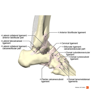
| More information about the anatomy of the ankle and foot can be found here: [http://www.physio-pedia.com/Biomechanics_of_Foot_and_Ankle Biomechanics of Foot and Ankle] and [http://www.physio-pedia.com/Ankle_Joint Ankle Joint].<br>
| |
| | |
| == Epidemiology /Etiology ==
| |
| | |
| === Osteoarthritis ===
| |
| | |
| Approximately 1% of the world’s adult population is affected by ankle OA, which results in pain, dysfunction, and impaired mobility. The mental and physical disability associated with end-stage ankle OA is at least as severe as that associated with end-stage hip OA. Numerous clinical and epidemiologic studies have identified previous trauma as the most common origin of ankle OA. 79.5% of patients with ankle OA had a verified history of 1 or more joint injuries. <ref name="Barg"/>
| |
| | |
| === Rheumatoid arthritis ===
| |
| | |
| The prevalence of foot pain in patients with RA has been reported in varying numbers within the published literature. The prevalence of foot pain depends on the stage of the disease. Ranging from 60 to 94 % at some stage of the disease. <ref name="Lohkamp" /><ref name="Brenton" /> In the early stages of RA the prevalence is lower: only 16 % in one study and 32 in another study. <ref name="Lohkamp" />
| |
| | |
| === Haemophilic arthropathy ===
| |
| | |
| Recurrent haemorrhages that occur with haemophilia result in severe joint destruction. Many authors have reported changes in the synovial membrane and cartilage in chronic haemarthrosis. Repeated episodes of intra-articular bleeding cause damage to the joint, leading to deformity. <br>Several joint disorders, degenerative ones, such as osteoarthritis, inflammation-mediated ones, such as rheumatoid arthritis, and blood-induced ones, such as haemophilic arthropathy, result in cartilage damage and changes in the synovial tissue. <ref name="Roosendal">Roosendaal, G., and Lafeber FP. Pathogenesis of haemophilic arthropathy. Haemophilia 2006;12:117-121.</ref>
| |
| | |
| === Diabetic foot arthropathy ===
| |
| | |
| Diabetes affects approximately 387 million people worldwide. Together with neuropathy, diabetes mellitus, is currently considered the main cause of Charcot neuropathic osteoarthropathy. The disease often goes undiagnosed amongst patients living with diabetes and its diagnosis greatly depends on the type of investigation: changes diagnosed by X-ray and corresponding to Charcot neuropathic osteoarthropathy are detected in up to 29% of diabetics, whereas under MRI the detection rate rises to 75%.<ref name="Kucera" />
| |
| | |
| === Gout ===
| |
| | |
| The global prevalence of gout is substantial and seems to be increasing in many parts of the world over the past 50 years. The distribution of gout is uneven across the globe, with prevalence being highest in Pacific countries. Developed countries tend to have a higher prevalence of gout than that of developing countries and incidence of the disease appears to be on the increase. <ref name="Kuo">Kuo CF, et al. Global epidemiology of gout: prevalence, incidence and risk factors. Nature Reviews Rheumatology 2015;11(11):649-662.</ref>
| |
| | |
| In various geographical regions, men are more likely to report gout than women <ref name="Kim2">Kim KY., et al. A literature review of the epidemiology and treatment of acute gout. Clinical therapeutics 2003;25(6):1593-1617.</ref>. Less than 10% of cases reported occur in women. Most women diagnosed with gout are reported to be 15 years or more beyond menopause. This type of arthropathy is rare in children. <ref name="Goodman">Goodman CC, Fuller KS. Pathology: Implications for the Physical Therapist. 3rd ed. Saint Louis, MO: Saunders; 2009.</ref>
| |
| | |
| The results of several studies suggest that environmental, racial, and hereditary differences may influence the development of gout. For example, Tokelauan migrants to New Zealand have a greater prevalence of gout than non-migrants and certain populations, such as the Maori, have a greater frequency of gout than other New Zealand populations. <ref name="Kim2" />
| |
| | |
| Some ethnic groups are particularly susceptible to gout, supporting the importance of genetic predisposition. <ref name="Kuo" />
| |
| | |
| Hyperuricemia is the most important risk factor for gout <ref name="Pascual">Pascual, Eliseo. "Hyperuricemia and gout." Current opinion in rheumatology 6.4 (1994): 454.</ref> and is affected by both genetic factors and environmental factors <ref name="Min">Min, Z., and M. Junwu. Research progress in the genetics of hyperuricaemia and gout. Yi chuan= Hereditas/Zhongguo yi chuan xue hui bian ji 2016;38(4):300-313.</ref>. Factors that increase serum urate levels include hypertension, thiazide diuretic intake, obesity, alcohol use, and a high animal protein diet <ref name="Reach">Reach G. Treatment adherence in patients with gout. Joint Bone Spine 2011;78(5):456-459.</ref>.
| |
| | |
| Socioeconomic and dietary factors, as well as comorbidities and medications that can influence uric acid levels and/or facilitate MSU crystal formation are also important in determining the risk of developing clinically evident gout. <ref name="Kuo" />
| |
| | |
| The risk of gout is lower in men who are more physically active, who maintain ideal body weight and who consume diets which are rich in fruit and limited in meat and alcohol. <ref name="Williams">Williams PT. Effects of diet, physical activity and performance, and body weight on incident gout in ostensibly healthy, vigorously active men. The American journal of clinical nutrition 2008;87(5):1480-1487.</ref>
| |
| | |
| === Psoriatic arthritis ===
| |
| | |
| In comparison to most other rheumatic disorders genetic predisposition plays a major role in the development of Psoriatic arthritis. <ref name="Amherd">Amherd‐Hoekstra A et al. Psoriatic arthritis: a review. JDDG: Journal der Deutschen Dermatologischen Gesellschaft 2010;8(5): 332-339.</ref> Psoriasis is known to affect approximately 2% to 3% of the general population, and the prevalence of Psoriatic arthritis in psoriasis patients is between 6% and 39%. It is possible that the condition remains generally underdiagnosed, related to lack of awareness by both the patient and physician. <ref name="Mease">Mease P. Psoriatic arthritis update." BULLETIN-HOSPITAL FOR JOINT DISEASES NEW YORK 64.1/2 (2006): 25.</ref>
| |
| | |
| It is still not clear what exact mechanism lies behind the development of psoriatic arthritis. It is thought to be multifactorial and secondary to environmental, genetic, and immunological factors. When compared with other inflammatory rheumatic conditions, psoriasis and psoriatic arthritis are strongly heritable. <ref name="Mahmood" />
| |
| | |
| === Reactive arthritis ===
| |
| | |
| Data indicates that approximately 50% of reactive arthritis and undifferentiated oligoarthritis cases can be attributed to a specific pathogen by a combination of culture and serology. The predominant organisms are Chlamydia, Salmonella, Shigella, Yersinia and Campylobactor species. The annual incidence of ReA was found to be 28/100.000 individuals in one study. This may exceed that of rheumatoid arthritis. <ref name="Klippel">Klippel, John H., John H. Stone, and Patience H. White. Primer on the rheumatic diseases. Springer Science & Business Media, 2008.</ref><br>
| |
| | |
| == Characteristics/Clinical Presentation ==
| |
| | |
| The characteristics and clinical presentation of ankle arthropathies such as different forms of arthritis can be described as followed:
| |
| | |
| *Ankle pain
| |
| *Stiffness
| |
| *Swelling
| |
| *Limited range of motion (ROM)
| |
| | |
| Pain usually increases on activities such as standing, walking or running.
| |
| | |
| “Start-up pain” is also a common complaint, where the patient experiences pain and stiffness in the ankle on moving after being asleep or sitting for a long period, which takes a few minutes of motion to settle. The ankle will tend to increasingly swell, however, as the day progresses, particularly with a higher level of activity. <br>
| |
| | |
| Pain is mostly felt in the entire ankle, although it may be more noticeable anteriorly if large boney spurs have formed. Arthritis often develops in an injured ankle joint. Degeneration of the overlying cartilage results in ankle arthropathy.
| |
| | |
| === Osteoarthritis ===
| |
| | |
| There are four degrees of severity with osteoarthritis:
| |
| | |
| #Degree I: normal joint with a minimal osteophyte.
| |
| #Degree II: osteophytose on two points with minimal subchondral sclerosis, normal joint space and no deformity.
| |
| #Degree III: moderate osteophytose, early deformity of the bone ends and a narrowing joint space.
| |
| #Degree IV: large osteophytes, deformity of bone ends, narrowing joint space, sclerosis and cysts <ref name="Kellgren">Kellgren J.H. 'Atlas of standard radiographs of arthritis'. Volume II of The Epidemiologic of Chronic Rheumatism. Oxford, Blackwell, 1963.</ref>
| |
| | |
| === Rheumatoid arthritis ===
| |
| | |
| Hallux valgus, splaying of the forefoot and pes planus are the most typically seen foot deformities in RA. <ref name="Chan" /><ref name="Brenton" /> The foot problems caused by RA can lead to mobility problems <ref name="Baillet" /><ref name="Carroll">Carroll M et al. Assessment of foot and ankle muscle strength using hand held dynamometry in patients with established rheumatoid arthritis. Journal of foot and ankle research 2013;6(1)</ref>, slower gait speed and a decrease in toe strength. <ref name="Lohkamp" />
| |
| | |
| A systematic review and meta-analysis by ''Carroll et al.'' <ref name="Carroll2">Carroll M et al. Gait characteristics associated with the foot and ankle in inflammatory arthritis: a systematic review and meta-analysis. BMC musculoskeletal disorders 2015;16(1):1.</ref> showed that the majority of the evaluated studies reported gait adaptations in patients with RA and that as a consequence they also have a greater risk of falling than other elderly individuals. <ref name="Brenton" />
| |
| | |
| Fatigue is a frequent symptom in RA, even among patients who display low and moderate levels of the disease with approximately 40% of RA patients with RA reporting severe levels of fatigue. <ref name="Rongen">Rongen‐van Dartel, S. A. A., et al. Effect of Aerobic Exercise Training on Fatigue in Rheumatoid Arthritis: A Meta‐Analysis.Arthritis care & research 2015;67(8): 1054-1062.</ref><br>
| |
| | |
| === Haemophilic arthropathy ===
| |
| | |
| Haemophilic arthropathy refers to permanent joint disease occurring in patients with haemophilia as a long-term consequence of repeated haemarthrosis. In an acute joint bleed, patients may report sensations of tingling and warmth, followed by pain, swelling and decreased motion. The pathogenesis of the progression from recurrent haemarthrosis to arthropathy is not fully understood, but it is characterised by inflammatory synovitis and cartilage destruction. <ref name="Leslie">Leslie, Raffini, and Manno Catherine. Modern management of haemophilic arthropathy. British journal of haematology 2007;136(6): 777-787. (level of evidence 5)</ref>
| |
| | |
| Haemophilic arthropathy, which shares some clinical and biological injury characteristics with rheumatoid arthritis, is characterised by 2 main features: chronic proliferative synovitis and cartilage destruction. It is the consequence of repeated extravasation of blood into joint cavities, but its exact pathogenesis, particularly with regard to early changes in the joint, is still not completely understood. <ref name="Lafeber">Lafeber FP., Miossec P, Valentino LA. Physiopathology of haemophilic arthropathy. Haemophilia 2008;14: 3-9</ref>
| |
| | |
| Over the long term, repeated episodes of haemarthrosis may cause irreversible damage to the joint, leading to haemophilic arthropathy, a polyarticular disease characterised by joint stiffness, chronic pain and a severely limited range of motion. In the most severe cases, the bones become fused, resulting in a completely disabled and misshapen joint. <ref name="Lafeber" /><br>
| |
| | |
| === Diabetic foot arthropathy ===
| |
| | |
| The Eichenholz classification describes the evolution of the condition over time. <ref name="Kucera" /><ref name="Shibata">Shibata, T. O. H. R. U., K. Tada, and C. Hashizume. The results of arthrodesis of the ankle for leprotic neuroarthropathy. J Bone Joint Surg Am 1990; 72: 749-756.</ref>
| |
| | |
| #Stage 0: Hot foot, usually painful, normal radiographic findings; MRI will show bone oedema and stress fractures
| |
| #Stage 1: Fragmentation, bone resorption, dislocations, fractures
| |
| #Stage 2: Coalescence, sclerosis, fracture healing, debris resorption
| |
| #Stage 3: Bone remodelling<br>
| |
| | |
| === Gout ===
| |
| | |
| There are four phases of gout. They include asymptomatic hyperuricemia, acute gout arthritis, intercritical gout and chronic tophaceous gout. <ref name="Harris">Harris, MARK D., LORI B. Siegel, and JEFFREY A. Alloway. Gout and hyperuricemia." American family physician 199;59(4):925-934.</ref>
| |
| # Asymptomatic hyperuricemia: Serum urate of more than 7 mg/dl but no symptoms are present. <ref name="Goodman" />
| |
| # Acute gout arthritis: joint pain, occurring suddenly at night, commonly in the first metatarsophalangeal joint. Localised and acute onset of intense pain, erythema, warmth, extreme tenderness and hypersensitivity are typically present. Chills, fever and tachycardia may accompany the joint complains. <ref name="Goodman" />
| |
| #Intercritical gout: Asymptomatic phase. <ref name="Goodman" />
| |
| #Tophaceous gout: sudden return of gout with increasing frequency and often in different joints. Characteristics are joint damage, functional loss, disability, deposits of monosodium urate crystals in soft tissue and bone abnormalities. <ref name="Goodman" /><br>
| |
| | |
| === Psoriatic arthritis ===
| |
| | |
| Typical symptoms include; inflammation in the Achilles tendon or the plantar fascia, and dactylitis (sausage-like swelling of the fingers or toes). <ref name="The">The Johns Hopkins University School of Medicine and the Johns Hopkins Arthritis Center.Psoriatic Arthritis. www.hopkinsmedicine.org/ (Accessed 27 november 2016</ref> Pain, swelling, or stiffness in one or more joints is also commonly present in psoriatic arthritis. <ref name="Amherd" /> Two features which are hallmarks of psoriatic arthritis are the presence of dactylitis and enthesitis (inflammation at the sites of insertion of tendons and ligaments into bone) <ref name="Mahmood" />
| |
| | |
| === Reactive arthritis ===
| |
| | |
| Reactive arthritis characteristically involves the joints of the lower extremities in an asymmetric, oligoarticular pattern. A dactylitis (‘sausage digit’) pattern in the feet is typical of reactive arthritis. Enthesopathy is often found in reactive arthritis. <ref name="Klippel" />
| |
| | |
| == Differential Diagnosis ==
| |
| | |
| Intra-articular pathologic lesions must be distinguished from surrounding joint tendinitis and [http://www.physio-pedia.com/Bursitis bursitis]. This can be achieved with diagnostic tests such as MRI or with injection of a local anesthetic.
| |
| | |
| Avascular necrosis must be considered in cases inwhich sclerosis of the talar dome is present. Patients may have a history of talar neck fracture, steroid or alcohol usage, or nonspecific injuries. Avascular necrosis of the talus can result in progressive segmental collapse and an increasing amount of matter into the joint.
| |
| | |
| === Osteoarthritis-Rheumatoid arthritis ===
| |
| | |
| Primary, osteoarthritis is a diagnosis of exclusion.
| |
| | |
| Post traumatic osteoarthritis<u></u>is is the most common form of ankle arthritis. Post traumatic disease can be present after intra-articular fractures or improper joint biomechanics after extra-articular fractures. Frequently, deformity is present in the joint. The extent of bone loss after trauma and joint space collapse can be assessed with weightbearing radiographs and CT scans.<br>Osteoarthritis is best differentiated from rheumatoid arthritis by a thorough history and examination. Three factors can distinguish the two disorders: the absence of systemic inflammatory signs and symptoms, onset in later life, and the pattern of joint involvement. <br>
| |
| | |
| Systematic inflammatory diseases such as rheumatoid arthritis should be excluded prior to considering operative intervention. Ankle arthritis can be effectively treated medically prior to considering surgical intervention, particularly during a flare up of the disease. The majority of patients with rheumatoid arthritis will test positive for rheumatoid factors and a rheumatoid arthritis diagnosis requires the presence of certain other symptoms: morning stiffness, multiple joint swelling, rheumatoid nodules, and evidence of joint erosion on radiographs.
| |
| | |
| Patients with an absence of rheumatoid factor in the serum, but who show signs of inflammatory arthritis are classified as having seronegative arthropathy. The four major disorders include ankylosis spondylitis, psoriatic arthritis, Reiter’s syndrome, and inflammatory bowel arthritides.
| |
| | |
| Metabolic and infectious causes of arthritis must also be considered. This can include gonococcal disease, Lyme disease, and gouty uricemia. A patient history should also include questions about any possible exposure to disease sources for sexually transmitted diseases and insect bites.
| |
| | |
| === Haemophilic arthropathy ===
| |
| | |
| Haemophilic arthropathy occurs in individuals who have haemophilia. Bleeds within a joint can cause multiple defects to the joint, as the result of a number of mechanisms affecting the synovial lining which becomes progressively fibrotic and the hyaline cartilage which becomes degenerative and is eventually lost. Mechanical and chemical processes cause degeneration of cells, but enzymatic processes appear to be primarily responsible for the degradation of the matrix of the articular cartilage.
| |
| | |
| Charcot osteoarthropathy or pedal neuropathic joint disease is a condition associated with peripheral neuropathy , it is a progressive deterioration of weight-bearing joints, usually in the foot or ankle, and is characterised in its early stages by acute inflammation that leads to bone and joint fracture, dislocation, instability and Gross deformaties and often seen in patients with longstanding diabetes,
| |
| | |
| In the early stages of Charcot osteoarthropathy, the patient presents with a warm, erythematous and oedematous foot with or without associated pain or reported previous injury and it can clinically mimic cellulitis or gout. This can lead to gross structural deformities of the foot and ankle, and subsequent skin ulceration and lower limb amputation from soft tissue or bony infection. The Charcot foot occurs most often in patients with diabetic neuropathy, but other predisposing conditions include alcoholic neuropathy, sensory loss caused by cerebral palsy or leprosy, and congenital insensitivity to pain. However, it is often goes unrecognised, with deleterious consequences..<br>
| |
| | |
| Differentiating between osteomyelitis and diabetic foot arthropathy is crucial, but can be difficult when both acute inflammation and bony changes are present. <ref name="Jeffcoate" /><span style="font-size: 13.28px;"><ref name="Kucera" /></span>
| |
| | |
| === Gout ===
| |
| | |
| Gout may masquerade as septic arthritis, rheumatoid arthritis or neoplasm. <ref name="Goodman" />
| |
| | |
| Pseudogout often resembles gout and, like gout, is caused by the formation of crystals in the joints. Instead of being composed of uric acid, as true gout crystals are, the crystals in pseudogout are composed of a salt called calcium pyrophosophate dehydrate (CPPD). The condition is also called CPPD disease. <ref name="www">http://www.arthritis.org/living-with-arthritis/tools-resources/expert-q-a/gout-questions/what-is-pseudogout.php (Accessed 3 december 16)</ref>
| |
| | |
| == Diagnostic Procedures ==
| |
| | |
| === Osteoarthritis ===
| |
| | |
| Diagnosing an osteoarthritic ankle joint starts with a clinical assessment and includes assessing alignment, stability and range of motion. Different radiographic modalities may help to recognise and analyse the underlying reasons for ankle OA. Radiographs of the foot and ankle should be taken on weight bearing only. Additional imaging modalities such as MRI and SPECT-CT may help to evaluate the extent of degenerative changes and their biological activities. <ref name="Barg" /><br>
| |
| | |
| === Rheumatoid arthritis ===
| |
| | |
| Different imaging techniques, e.g. MRI, CT and ultrasonography (US), can help clinicians to detect early onset or subclinical foot problems as the clinical signs of foot disease in RA are often subtle. <ref name="Chan" /><ref name="Wakefield">Wakefield, Richard J., et al. The value of sonography in the detection of bone erosions in patients with rheumatoid arthritis. Arthritis Rheum 2000;43(12):2762-70.</ref>
| |
| | |
| Ultrasonography and MRI have shown to be superior to clinical examination in detecting joint inflammation. <ref name="Lehtinen">Lehtinen, Ari, et al. Painful ankle region in rheumatoid arthritis Analysis of soft-tissue changes with ultrasonography and MR imaging. Acta Radiologica 1996;37(3):572-577.</ref> Sonography is increasingly being used and has been found to be effective for the detection of bony erosions in patients with RA. Ultrasonography detected 6.5 times more bony erosions in the early stages of the disease than radiography. <ref name="Wakefield" /> As US is more readily available and less expensive than an MRI it is recommended as the second diagnostic imaging method, after radiography. <ref name="Lehtinen" />
| |
| | |
| === Haemophilic arthropathy ===
| |
| | |
| Radiography remains the go to method in the diagnosis and follow-up of haemophilic arthropathy. Radiographical results of arthropathy often show an expected sequence of events and are found to be similar in different joints. An MRI, however, has advantages over radiography due to its ability to display soft tissue and cartilage changes in haemophilic joints. The recent development and standardisation of MRI scoring systems for measuring soft tissue and cartilage abnormalities may enable the comparison of pathological joint findings in clinical trials conducted at different institutions across the world <ref name="Doria">Doria, A. S. "State‐of‐the‐art imaging techniques for the evaluation of haemophilic arthropathy: present and future." Haemophilia 2010; 16:107-114.</ref>
| |
| | |
| === Diabetic foot arthropathy ===
| |
| | |
| Diagnosis is based on the patient’s history, clinical examination, and imaging results. Due a lowered perception of pain, patients are quite often not aware of any injury.<ref name="Kucera" /> Localised inflammation is the main symptom for diagnosing diabetic foot arthopathy. <ref name="Jeffcoate" />
| |
| | |
| In Charcot feet arthropathies it is very important that the disease is diagnosed quickly, because a delay can lead to worsening structural damage or even limb loss.<ref name="Jeffcoate" /><ref name="Wukich">Wukich, D. K., et al. The consequences of complacency: managing the effects of unrecognized Charcot feet. Diabetic Medicine 2011;28(2):195-198.</ref><ref name="KJ">Kalish J, Hamdan A. Management of diabetic foot problems. Journal of vascular surgery 2010; 51(2):476-486.</ref><ref name="Varma">Varma, Ajit Kumar. Charcot neuroarthropathy of the foot and ankle: a review. The journal of foot and ankle surgery 2013;52(6):740-749.</ref> Unfortunately the diagnosis is often missed at first presentation. A possible reason for the missed diagnosis is that Charcot feet are not emphasized in medical training. The result is that it is difficult to advocate the right choice of approach due to low evidence based information. <ref name="Jeffcoate" />
| |
| | |
| Acute Charcot activity can be diagnosed if the temperature of the affected foot is 2°C or more than the contralateral unaffected foot. <ref name="Varma" /><ref name="Kucera" />
| |
| | |
| === Gout ===
| |
| | |
| Gout is ideally diagnosed through identification of characteristic negatively birefringent crystals under polarized light microscopy in fluid aspirated from end-organ deposits, typically from a joint <ref name="Wall">Wallace, Stanley L., et al. Preliminary criteria for the classification of the acute arthritis of primary gout. Arthritis & Rheumatism 1977;20: 895-900.</ref>. However, fewer than 10% of patients with gout see a rheumatologist, and most cases of gout are diagnosed in the primary care setting based on signs, symptoms, and serum uric acid level <ref name="Owens">Owens D., Whelan B, McCarthy G. A survey of the management of gout in primary care. Irish medical journal 2008;101(5):147-149.</ref>.
| |
| | |
| === Psoriatic arthritis ===
| |
| | |
| A diagnostic test for psoriatic arthritis does not exist unlike in RA which is cyclic citrullinated peptide and rheumatoid factor positive. As in other inflammatory conditions, markers such as erythrocyte sedimentation rate and C-reactive protein can be raised in psoriatic arthritis. <ref name="Mahmood" />
| |
| | |
| Scoring systems have been developed to try and identify psoriatic arthritis at an early stage and criteria have been developed to aid in classification of the disease from the other SPAs and inflammatory arthritides. Not only are they useful for identifying psoriatic arthritis earlier, they can also help identify cases of psoriatic arthritis which do not present in the typical manner. Some criteria include psoriatic arthritis with the SPA group. The classification for psoriatic arthritis (CASPAR) criteria was developed specifically for psoriatic arthritis. It has good sensitivity and specificity for those presenting with disease of <2 years’ duration. Although primarily used for classification, it can be used for diagnostic purposes. <ref name="Mahmood" />
| |
| | |
| Further imaging such as MRI can help to identify soft tissue involvement in further detail, particularly when a patient is suffering from enthesitis. Ultrasound has also become a useful tool in the investigation of arthritis as it can help to identify bony erosions in those patients where synovitis or dactylitis is not always evident clinically. Studies have shown that ultrasound and MRI are more sensitive for detecting inflammation than plain radiographs in psoriatic arthritis. <ref name="Mahmood" />
| |
| | |
| == Outcome Measures ==
| |
| | |
| === Osteoarthritis ===
| |
| | |
| The Ankle Osteoarthritis Scale (two subscales: pain and disability) <ref name="Salk">Salk, Robert S., et al.Sodium hyaluronate in the treatment of osteoarthritis of the ankle: a controlled, randomized, double-blind pilot study. J Bone Joint Surg Am 2006;88(2):295-302.</ref> is a reliable and valid self-assessment instrument that specifically measures patient symptoms and disabilities related to ankle arthritis.<ref name="Domsic">Domsic RT, Saltzman CL. Ankle osteoarthritis scale.Foot & ankle international 1998;19(7):466-471.</ref>
| |
| | |
| More outcome measures of ankle osteoarthritis can be found on the page [http://www.physio-pedia.com/Ankle_Osteoarthritis#Outcome_Measures Ankle Osteoarthritis]
| |
| | |
| === Rheumatoid arthritis ===
| |
| | |
| American College of Rheumatology (ACR) response criteria for RA. <ref name="Gladman">Gladman, Dafna D., et al. Outcome measures in psoriatic arthritis. The Journal of Rheumatology 2007;34(5):1159-1166.</ref> The ACR20 response criteria requires a 20% improvement in both tender and swollen joint counts and a 20% improvement in 3 of 5 items:
| |
| * Patient global assessment (VAS),
| |
| * Physician global assessment (VAS),
| |
| * Patient pain score (VAS),
| |
| * Health Assessment Questionnaire (HAQ),
| |
| * Erythrocyte sedimentation rate or C-reactive protein (CRP). <ref name="Gladman" />
| |
| | |
| === Haemophilic arthropathy ===
| |
| | |
| MRI images of bone or cartilage damage in the index joints can be used as an outcome measure. Tentative haemophilic arthropathy scales based on MRI findings have been developed in the last decade. In 2005, the International Prophylaxis Study Group (IPSG) presented a preliminary comprehensive scoring scheme that combined the Denver and European MRI scores. The use of such scales should result in a more consistent assessment of haemophilic joints and should facilitate the development of more targeted treatment to prevent or delay further destructive osteoarticular changes.<ref name="Doria" />
| |
| | |
| === Diabetic foot arthropathy ===
| |
| *
| |
| | |
| === Gout ===
| |
| | |
| Numerous different instruments can be used to assess the acute gout core domains; VAS, 5 point Likert scales, 4 point Likert scales of index joint swelling and tenderness and 5 point PGART instruments meet the criteria for the Outcome Measures in Rheumatology (OMERACT) filter. <ref name="Dalbeth">Dalbeth, Nicola, et al.Outcome measures in acute gout: a systematic literature review. The Journal of rheumatology 2014;41(3):558-568.</ref>
| |
| | |
| === Psoriatic arthritis ===
| |
| | |
| The Psoriatic Arthritis Response Criteria (PsARC) is recommended in the assessment and monitoring of PsA. It consists of four components: assessment of joint tenderness and swelling utilizing 68/66 joint counts respectively, the patient’s opinion of their global health and the physician’s global assessment. <ref name="Gladman" /><ref name="Little">Littlewood S. et al. Evaluation of a Psoriatic Arthritis Response Criteria Standardization Training Session for Clinicians. Rheumatology 2014;53(1):152.</ref>
| |
| | |
| The ACR20 response criteria require a 20% improvement in both tender and swollen joint counts, and a 20% improvement in 3 of 5 items:
| |
| * Patient global assessment (visual analog scale, VAS),
| |
| * Physician global assessment (VAS),
| |
| * Patient pain score (VAS),
| |
| * Health Assessment Questionnaire (HAQ),
| |
| * Erythrocyte sedimentation rate or C-reactive protein (CRP).
| |
| For some PsAstudies the joint count was increased to 78 to include distal interphalangeal (DIP) joints of the feet. To achieve an ACR50 or ACR70 response, the same guidelines apply but the level of response is 50% or 70% improvement, respectively. <ref name="Gladman" />
| |
| | |
| === Reactive arthritis ===
| |
| | |
| The outcome measures of reactive arthritis can be found on the page [http://www.physio-pedia.com/Reactive_Arthritis#Outcome_Measures Reactive Arthritis]<br>
| |
| | |
| == Examination ==
| |
| | |
| === Osteoarthritis ===
| |
| | |
| The examination of osteoarthritis can be found on the page [http://www.physio-pedia.com/Ankle_Osteoarthritis Ankle Osteoarthritis]
| |
| | |
| === Rheumatoid arthritis ===
| |
| | |
| The examination of rheumatoid arthritis can be found on the page [http://www.physio-pedia.com/Rheumatoid_Arthritis Rheumatoid Arthritis]<br>
| |
| | |
| == Medical Management ==
| |
| | |
| === Osteoarthritis ===
| |
| | |
| There is no cure for osteoarthritis. There are several treatments that can be subdivided into pharmacological, non-pharmacological and surgical. The choice of treatment of ankle and foot OA depends on the severity of the disease. The main goals of OA management in the foot and ankle are, pain control, improvement in function and in quality of life. A number of different aspects like discomfort, comorbidity and radiologic damage also need to be considered. <ref name="DM">D.M. Reid, C.G. Miller (eds.), Clinical Trials in Rheumatoid Arthritis and Osteoarthritis, Springer-Verlag London Limited 2008, 325p.</ref>
| |
| | |
| '''Pharmacological treatments'''
| |
| | |
| {| width="600" border="1" cellspacing="1" cellpadding="1"
| |
| |-
| |
| | ''<u></u><sub></sub><sup></sup><strike></strike> ''Analgesics
| |
| |
| |
| Simple painkillers like paracetamol or acetaminophen are usually enough to have a significant relief of symptoms, for patients with mild to moderate pain<ref name="DM" />
| |
| | |
| |-
| |
| | NSAID
| |
| |
| |
| <u></u><sub></sub><sup></sup>NSAIDs are often used by patients with severe pain, where analgesics aren’t strong enough to relief symptoms <ref name="Witteveen">Witteveen, Angelique GH, Cheriel J. Hofstad, and Gino MMJ Kerkhoffs. "Hyaluronic acid and other conservative treatment options for osteoarthritis of the ankle." The Cochrane Library (2015)</ref>. Research shows improvements for short duration use. There is a lack of research in their long term use <ref name="DM" />.
| |
| | |
| |-
| |
| | Opioids <br> <br>
| |
| |
| |
| When NSAIDs do not offer enough relief, opioids are often used. Opioids are mostly taken by patients with moderate to severe pain. Some patients use simple analgesics in combination with a low dose opioid for a greater improvement in pain at rest as well as in motion<ref name="DM" />.
| |
| | |
| Opioids decrease pain in the long term increasing quality of sleep and enjoyment of life. Common opioid side effects are present, but decrease as therapy continues<ref name="Roth">Roth, Sanford H., et al. Around-the-clock, controlled-release oxycodone therapy for osteoarthritis-related pain: placebo-controlled trial and long-term evaluation. Archives of Internal Medicine 2000;160(6):853-860.</ref>.
| |
| | |
| |-
| |
| | <u></u><sub></sub><sup></sup> Intra-articular injections
| |
| |
| |
| When simple analgesics have failed, injections are used in a hospital environment. Most of the time, hyaluronic acid is recommended if there is inadequate response to simple analgesics. Hyaluronic acid Is a natural component of synovial fluid. These gel-like fluid injections help to lubricate the joint and to act as a shock absorber for joint loads<ref name="Witteveen" />.<u></u><sub></sub><sup></sup>Research indicates that there is an improvement in pain, but repeated injections in the same joint showed a decreased response<ref name="DM" />. It remains unclear, however, which dosage schedule should be used and which patients would benefit the most from it. Other research showed that there is a relief of pain, but more difficulty in walking on uneven surfaces, especially when compared with exercise therapy<ref name="Karatosun">Karatosun, V., et al. Intra-articular hyaluronic acid compared to exercise therapy in osteoarthritis of the ankle. A prospective randomized trial with long-term follow-up. Clinical and experimental rheumatology 2008; 26(2):288. (Level of evidence 1b)</ref>.
| |
| | |
| |}
| |
| | |
| '''''Surgical treatment'''''<br>
| |
| | |
| {| width="600" border="1" cellspacing="1" cellpadding="1"
| |
| |-
| |
| | Osteotomy
| |
| |
| |
| Low tibial osteotomy (a change of alignment of the bone) is indicated for osteoarthritis in the ankle, more specifically for varus type osteoarthritis in a moderate stage<ref name="Tanaka">Tanaka, Y., et al. "Low tibial osteotomy for varus-type osteoarthritis of the ankle." Bone & Joint Journal 88.7 (2006): 909-913.</ref>.
| |
| | |
| |-
| |
| | Arthroscopy
| |
| |
| |
| There was no significant improvement in instability. But there was significant improvement in pain, swelling, stiffness, limp, and activity in the patients postoperatively compared with their preoperative status, but not with all patients however. Approximately one third of patients experienced no benefit from arthroscopic intervention<ref name="Ogilvie">Ogilvie-Harris DJ, Sekyi-Otu A. Arthroscopic debridement for the osteoarthritic ankle. Arthroscopy: The Journal of Arthroscopic & Related Surgery 1995;11(4):433-436.</ref>.
| |
| | |
| |-
| |
| | Arthrodesis
| |
| |
| |
| The joint becomes fixed, so there no movement in the joint postoperatively. This method is used in end stage osteoarthritis, but research shows that arthroplasty offers better pain relief<ref name="Saltzman">Saltzman, Charles L., Robert G. Kadoko, and Jin Soo Suh. Treatment of isolated ankle osteoarthritis with arthrodesis or the total ankle replacement: a comparison of early outcomes.Clinics in orthopedic surgery 2010;2(1):1-7.</ref>.
| |
| | |
| |-
| |
| | Arthroplasty
| |
| |
| |
| This method is used, like arthrodesis, in end stage osteoarthritis. This is a total replacement of the affected joint and the most effective of all medical interventions. The pain and disability of end stage osteoarthritis can be eliminated and restores patients to near normal function<ref>D.T. Felson et al., Osteoarthritis : New inights, part 2: treatment approach, (level of evidence: 2A) (last consulted on 20/11/2016)</ref>.
| |
| | |
| |}
| |
| | |
|
| |
| | |
| === Rheumatoid arthritis ===
| |
| | |
| '''''Pharmacological treatments'''''
| |
| | |
| {| width="600" border="1" cellspacing="1" cellpadding="1"
| |
| |-
| |
| | DMARD’s<br><br><br>
| |
| |
| |
| <u></u><sub></sub><sup></sup>DMARD’s are Disease-modifying antirheumatic drugs. Frequency of use ha increased over the years and enhanced the success of RA management<ref name="Chan" />.
| |
| | |
| |-
| |
| | Anti-TNF treatment <br>
| |
| |
| |
| The use of anti-TNF therapy on patients suffering from RA has accelerated ulcer healing <ref name="Murosaki">Murosaki, Takamasa, et al. "Foot ulcers caused by rheumatoid vasculitis in a patient with rheumatoid arthritis undergoing etanercept treatment." Internal Medicine 51.22 (2012): 3181-3183.</ref>. <u></u><sub></sub><sup></sup>
| |
| | |
| The difference between DMARD combination treatments, including or excluding TNF inhibitors, is small however. Due to the huge cost differences, RA guidelines should recommend combination DMARD treatment before the initiation of TNF inhibitors<ref name="Graudal">Graudal, Niels, et al. "Combination Therapy With and Without Tumor Necrosis Factor Inhibitors in Rheumatoid Arthritis: A Meta‐Analysis of Randomized Trials." Arthritis care & research 67.11 (2015): 1487-1495.</ref>.
| |
| | |
| |-
| |
| | Steroids<u></u><sub></sub><sup></sup>
| |
| |
| |
| <u></u><sub></sub><sup></sup>''Choy et al.'' demonstrated that monthly injections of 120 mg intramuscular depomedrone in addition to a DMARD’s treatment showed only a transient clinical benefit. The value of steroids is still controversial. Previous studies found that steroids only provide short and medium term symptomatic improvements in RA. The level of steroid related adverse events such as diabetes, vertebral fractures and osteoporosis, exclude its use as a long term treatment strategy. Therefore, patients should not be given long term steroid treatment, but rather an alternative or additional DMARD’s treatment<ref>Choy, Ernest H., et al. "A two year randomised controlled trial of intramuscular depot steroids in patients with established rheumatoid arthritis who have shown an incomplete response to disease modifying antirheumatic drugs." Annals of the rheumatic diseases 64.9 (2005): 1288-1293.</ref>.
| |
| | |
| |}
| |
| | |
| === <br>'''Haemophilic arthropathy''' ===
| |
| | |
| '''Pharmacological treatments'''
| |
| | |
| {| width="600" border="1" cellspacing="1" cellpadding="1"
| |
| |-
| |
| | Narcotics
| |
| |
| |
| This is effective, but leads to dependence<ref name="Leslie" />.
| |
| | |
| |-
| |
| | NSAIDs
| |
| |
| |
| NSAIDS act by inhibiting cyclooxygenase enzymes, which result in an analgesic and anti-inflammatory effect.<br>3 types: Aspirin, Traditional NSAIDS and Cox-2 inhibitors<br>Further evaluation of the safety and efficacy of drugs in patients with haemophilia is needed.<ref name="Leslie" />.
| |
| | |
| |-
| |
| | Steroid-/ hyaluronic acid-injections
| |
| |
| |
| Both injections decrease pain <ref name="Leslie" />
| |
| | |
| |}
| |
| | |
| '''''Surgical treatments''''' <u></u><sub></sub><sup></sup><u></u><sub></sub><sup></sup>
| |
| | |
| {| width="600" border="1" cellspacing="1" cellpadding="1"
| |
| |-
| |
| | Synovectomy
| |
| |
| |
| Excision of the friable synovium. This approach is frequently used to reduce pain in patients who experience recurrent haemarthrosis. This can be achieved by direct surgical excision or arthroscopic synovectomy. Another way is to inject a radioactive or chemical agent which causes fibrosis of sclerosis of the synovium. If there are several advanced changes in the joint, a joint arthroplasty may be considered<ref name="Leslie" />.
| |
| | |
| |-
| |
| | Joint arthroplasty
| |
| |
| |
| <u></u><sub></sub><sup></sup>Patients with haemophilia who develop severe arthropathy may experience relentless pain, loss of motion and functional disability. If conservative management fails (analgesics, orthotics and physical therapy) these patients may benefit from total joint replacement<ref name="Leslie" />.<br>
| |
| | |
| |}
| |
| | |
| <br><u></u><sub></sub><sup></sup>
| |
| | |
| === Diabetic foot arthropathy ===
| |
| | |
| A multidisciplinary approach is recommended for the management of Charcot foot.<ref name="Smith" />
| |
| | |
| '''Pharmacological treatments'''''<u></u><sub></sub><sup></sup>''
| |
| | |
| {| width="600" border="1" cellspacing="1" cellpadding="1"
| |
| |-
| |
| | Biphosphonates
| |
| |
| |
| There is currently little evidence to support the use of bisphosphonates as part of the routine management of patients with diabetes complicated by acute Charcot neuropathic osteoarthropathy<ref>Richard, J-L., M. Almasri, and S. Schuldiner. "Treatment of acute Charcot foot with bisphosphonates: a systematic review of the literature." Diabetologia 55.5 (2012): 1258-1264.</ref><ref name="Kucera" /><ref name="Shibata" /> but some authors found that bisphosphonates may improve the healing of Charcot foot by reducing skin temperature and the disease activity of Charcot foot. Bisphosphonates should be used in addition to standard interventions to control the position and shape of the foot<ref name="Jeffcoate" />.
| |
| | |
| |-
| |
| | Anti-TNF treatment
| |
| |
| |
| It is plausible to suggest the use of anti-TNF biological therapies for controlling Charcot neuropathic osteoarthropathy based on the excessive bone resorption due to circulating osteoclast precursors and serum levels of TNF-a<ref name="Mabilleau" />.
| |
| | |
| |}
| |
| | |
| '''Surgical treatments'''
| |
| | |
| {| width="600" border="1" cellspacing="1" cellpadding="1"
| |
| |-
| |
| | Surgery
| |
| |
| |
| Surgery should be avoided during the active inflammatory stage because of the perceived risk of wound infection<ref name="Shibata" />.A patient only becomes a candidate for surgical treatment after the failure of conservative management<ref name="Kucera" />.<br>Specific methods for the surgical treatment of Charcot neuropathic osteoarthropathy to save the extremity or delay major amputation are still being developed. Below the knee amputation is the preferred option. <ref name="Kucera" />.
| |
| | |
| |-
| |
| | Magnetic therapy treatment
| |
| |
| |
| S<u></u><sub></sub><sup></sup>ignificant reduction in the amount of deformity and reduced healing time to consolidation after receiving magnetic therapy treatment<ref name="Smith" />.
| |
| | |
| |}
| |
| | |
| === Gout ===
| |
| '''<u></u><sub></sub><sup></sup>Pharmacotherapy'''
| |
| {| width="600" border="1" cellspacing="1" cellpadding="1"
| |
| |-
| |
| | Urate level
| |
| |
| |
| The goal of pharmacotherapy for gout is to maintain the serum urate level below 50–60 mg/L (300–360 moll/L). When urate levels fall below 50–60 mg/L, the urate crystals start to dissolve <ref name="Reach" />. Currently used urate lowering medications include xanthine-oxidase inhibitors such as allopurinol, febuxostat, as well as available uricosuric agents. However, evidence comparing these agents remains low. Based on the reported data, febuxostat can play a major role in the treatment of hyperuricaemia and gout. Febuxostat is a suitable pharmacological option for first line treatment of gout, given its established efficacy and safety, documented in a high number of clinical studies and in daily practice<ref>Borghi, C., and F. Perez-Ruiz. "Urate lowering therapies in the treatment of gout: a systematic review and meta-analysis." European review for medical and pharmacological sciences 20.5 (2016): 983-992.</ref><br>During the first few months of urate-lowering therapy (ULT), an anti-inflammatory drug must be taken to also prevent the occurrence of gouty attacks<ref name="Reach" />
| |
| | |
| |-
| |
| | NSAIDs
| |
| |
| |
| These are frequently used as first line therapies for acute gout. However, these agents may have serious side effects such as gastrointestinal toxicity, renal toxicity, or gastrointestinal bleeding<ref name="Cronstein">Cronstein, Bruce N., and Robert Terkeltaub. "The inflammatory process of gout and its treatment." Arthritis Research & Therapy 8.1 (2006): 1.</ref>
| |
| | |
| |-
| |
| | Systemic corticosteroids
| |
| |
| |
| Systemic corticosteroids have also exhibited significant efficacy in patients with acute gout; intra-articular corticosteroids are frequently used in patients with monoarticular gout, particularly in patients who cannot receive oral therapy<ref name="Cronstein" />.
| |
| | |
| |-
| |
| | ACTH (Adrenocorticotropic hormone)
| |
| |
| |
| This is also quite useful in treating acute gout, particularly in those patients with renal and/or gastrointestinal contraindications to other therapies. Synthetic ACTH is effective partly via induction of adrenal glucocorticosteroids, and partly via rapid peripheral suppression of leukocyte activation by melatonin receptor 3 signalling<ref name="Cronstein" />
| |
| | |
| |-
| |
| | Oral colchicine
| |
| |
| |
| In patients with contraindications or who cannot tolerate NSAIDs or systemic (either oral or parenteral) corticosteroids, oral colchicine is generally the next choice for primary therapy<ref name="Cronstein" /><br>As the acute attack resolves, it is appropriate to use low doses of oral colchicine as an adjunct to NSAID, glucocorticosteroid, or ACTH treatment, and continuation of low dose oral colchicine can help to suppress rebound flares after treatment<ref name="Cronstein" />
| |
| | |
| |}
| |
| | |
| === Psoriatic arthritis ===
| |
| | |
| Treatments such as oral disease modifying anti-rheumatic drugs and biologic therapy are effective, but have side effects which could limit their use in certain individuals<ref name="Mahmood" /><br>
| |
| | |
| ''Pharmacological treatments''
| |
| | |
| {| width="600" border="1" cellspacing="1" cellpadding="1"
| |
| |-
| |
| | Corticosteroids
| |
| |
| |
| Corticosteroids are used in psoriatic arthritis and in many other inflammatory arthropathies to provide immediate symptom relief. They can rapidly diminish the inflammatory response seen in these conditions, causing a reduction in swelling and stiffness in the joints. They can be given as an intramuscular injection for general widespread relief or directly injected into the affected joint for a more targeted response. Long term steroid use should be avoided due to the side effect profile which includes diabetes, hypertension, osteoporosis, and immunosuppression. There is also a risk of skin psoriasis flares after intramuscular injections. In most cases steroids are used for short term relief and occasionally as bridging therapy while establishing the patient on more long term immunosuppressants<ref name="Mahmood" />
| |
| | |
| |-
| |
| | Disease‐modifying anti‐rheumatic agents or immunosuppressive drugs
| |
| |
| |
| Disease modifying anti-rheumatic drugs (DMARDs) are used in psoriatic arthritis as in other inflammatory conditions. They are a group of drugs which can help to reduce inflammation as well as to slow disease progression to prevent joint damage, as opposed to nonsteroidal anti-inflammatory drugs and steroids which treat the inflammation, but not the underlying cause. However, evidence behind their use in psoriatic arthritis is not as robust as in RA. Use of DMARDs is mostly based on a clinician’s experience rather than evidence. Methotrexate can cause improvement in both the skin and peripheral joints. Sulfasalazine is useful for peripheral arthropathy although only with weak evidence. Cyclosporine has beneficial effects on the skin. Small studies have shown that leflunomide has similar efficacy for use in psoriatic arthritis as compared with RA. The main side effect with DMARDs is their immunosuppression, which can expose the patient to potentially serious infections such as neutropenic sepsis<ref name="Mahmood" />
| |
| | |
| |-
| |
| | Intra –articular injections: TNF-α blockers
| |
| | Biologic drugs that are aimed at blocking TNF-α have been effective therapies for such conditions<ref name="Mahmood" />
| |
| |}
| |
| | |
| === Reactive arthritis ===
| |
| | |
| The treatment of reactive arthritis comprises of mainly non-steroidal anti-inflammatory drugs, intra-articular steroid injections, and physical treatment. <ref name="Fry">Fryden, Aril, et al. "Early antibiotic treatment of reactive arthritis associated with enteric infections: clinical and serological study." BMJ 301.6764 (1990): 1299-1302.</ref><br>
| |
| | |
| '''Pharmacological treatments'''
| |
| | |
| {| width="600" border="1" cellspacing="1" cellpadding="1"
| |
| |-
| |
| | NSAIDs
| |
| |
| |
| Non‐steroidal anti‐inflammatory drugs (NSAIDs) form the basis of pharmacological therapy. They should be used in proper dosage and not be stopped too early. During recovery, insufficient analgesia may lead to limited joint movement and prolong the rehabilitation, but their use should be reduced as appropriate to recovery and not used unnecessarily<ref name="Toivanen" />.
| |
| | |
| |-
| |
| | <u></u><sub></sub><sup></sup> Corticosteroids
| |
| |
| |
| Corticosteroids are a potent group of drugs to be used in ReA. Intra‐articular application results most often in prompt relief of the joint inflammation. This is of great value for a young, active and employed person. The injection can be repeated a number of times, if necessary. However, this treatment is not applicable if a number of joints is involved. There is the risk of infectious complications and true septic arthritis as however. <ref name="Toivanen" /><br>In severe cases and when several joints are involved, peroral corticosteroids are useful. They should be started in high dosage, e.g. prednisolone 30–40 mg/day, and tapered rapidly down according to relief of symptoms. The drug should then be continued for a brief period in low dose, and stopped gradually. Corticosteroids are, in a similar manner, also useful in the treatment of acute relapses. If the patient later suffers from chronic arthralgias, corticosteroids should be avoided<ref name="Toivanen" />. Steroids are administered when inflammatory symptoms are resistant to NSAIDs<ref>Palazzi, Carlo, et al. "Management of reactive arthritis." Expert opinion on pharmacotherapy 5.1 (2004): 61-70.</ref>.
| |
| | |
| |-
| |
| | Antibiotics
| |
| |
| |
| Antibiotics seem to be ineffective in postenteric reactive arthritis. By the time the patient seeks help for joint inflammation, the initial infection has already passed and antibiotics are no longer effective<ref>van der Linden, Sjef, and Désirée van der Heijde. "Clinical aspects, outcome assessment, and management of ankylosing spondylitis and postenteric reactive arthritis." Current opinion in rheumatology 12.4 (2000): 263-268.</ref>.
| |
| | |
| |-
| |
| | <u></u><sub></sub><sup></sup>Disease modifying anti‐rheumatic agents or immunosuppressive drugs
| |
| |
| |
| The value of disease modifying anti‐rheumatic agents or immunosuppressive drugs has not been established, although ''Mielants and Veys''<ref>Mielants, H., et al. "HLA-B27 related arthritis and bowel inflammation. Part 2. Ileocolonoscopy and bowel histology in patients with HLA-B27 related arthritis." The Journal of rheumatology 12.2 (1985): 294-298.</ref> suggest that ileocolonoscopy should be performed for patients with ReA more often than is currently customary.<br>Experiences with other DMARDs such as azathioprine, methotrexate and cyclosporin, have been sporadically reported and they can be used in patients that are unresponsive to the more usual medicaments<ref name="Palazzi" />.
| |
| | |
| |-
| |
| | <u></u><sub></sub><sup></sup>Intra –articular injections
| |
| |
| |
| Intra-articular steroid injections can be used as treatment for reactive arthritis<ref name="Fry" />.
| |
| | |
| |-
| |
| | TNF-α blockers
| |
| |
| |
| In more aggressive cases, or when ReA evolves towards ankylosing spondylitis, TNF-α blockers could represent an effective choice<ref name="Palazzi">Palazzi, Carlo, et al. "Management of reactive arthritis." Expert opinion on pharmacotherapy 5.1 (2004): 61-70.</ref>.
| |
| | |
| |}
| |
| | |
| == Physical Therapy Management ==
| |
| <u></u><sub></sub><sup></sup>
| |
| === Osteoarthritis<br> ===
| |
| | |
| Non care management and care management have been tested. Non care management includes resting and relaxing, although rigorous evidence for its effectiveness has not proved conclusive. On the other hand, physical activity and/or exercise is generally adopted and recognised by health professionals and patients. Specific joints can be targeted to improve general mobility, function and the reduction of pain. More intensive exercise can strengthen muscles around the affected joint of the foot. A limited number of OA patients may have increased pain symptoms while exercising, but with an exercise program that is appropriately targeted and adjusted to the individual's pace, it should be possible for every OA patient to be active <ref name="Scott">Scott, A., Gidlow, C., Clinical exercise science, Taylor & Francis Ltd, 2016</ref>. (LOE: 1A)
| |
| | |
| '''Non-pharmacological treatments'''
| |
| | |
| {| width="600" border="1" cellspacing="1" cellpadding="1"
| |
| |-
| |
| | Aerobic exercise
| |
| | Guidelines were developed for training parameters in people with osteoarthritis pain.
| |
| *2-5 sessions a week
| |
| *20-30 minutes/session
| |
| *Holding each contraction 1-6 seconds
| |
| *Avoidance of high-impact exercises
| |
| *Targets should be achievable for the patient
| |
| *Other exercises (flexibility, postural balance, strength) may be incorporated to achieve specific goals based on the patient his individual assessments<ref name="Scott" /> (LOE: 1A)
| |
| | |
| |-
| |
| | Range of motion and flexibility exercise
| |
| | Joint motion and flexibility exercises of the periarticular tissues on a regular basis are important for cartilage nutrition and health, protection of joint structures from damaging impact loads and comfort in daily activities. Those exercises are routines of low-intensity, controlled movements that do not increase the pain in foot or ankle<ref name="DT">D.T. Felson et al., Osteoarthritis : New inights, part 2: treatment approach (level of evidence 1b)</ref>.(LOE: 2B)
| |
| |}
| |
| | |
| <br>
| |
| | |
| === Rheumatoid arthritis ===
| |
| | |
| '''Physical therapy'''
| |
| | |
| {| width="600" border="1" cellspacing="1" cellpadding="1"
| |
| |-
| |
| | Exercise therapy
| |
| | Education is key to patients adhering to their training program even when supervision is withdrawn, as the gains in muscle mass and strength dependent function diminish when the training stops. <ref name="Karatosun"/>.(LOE: 1A)<br>Consistency is also key to reduce the rate of damage in the foot joints<ref name="Karatosun" />. <br>Patients with RA who participate in long term high intensity weight bearing exercise classes develop less radiological joint damage in the feet in comparison with patients participating in the more standard physical therapy protocols<ref name="Karatosun" />.<br>Resistance exercise therapy is safe and offers a clinically significant amelioration of functional capacity and disability. Further RCT’s are required to determine whether supervised exercises are clinically relevant<ref name="Baillet" />.(LOE: 1A)<br>The maintenance of long-term positive effects gained from resistance exercise has not been extensively evaluated, and remains controversial, yet it is crucial for long-term benefits that after the intervention of a structured physical activity program is maintained, because the effects of detraining occur rapidly<ref name="Baillet" /><ref name="Baillet" />.<br>A 3 year follow up after completing a high-intensity progressive resistance training program <ref name="Lemmey">Lemmey, Andrew B., et al. "Are the benefits of a high‐intensity progressive resistance training program sustained in rheumatoid arthritis patients? A 3‐year followup study." Arthritis care & research 64.1 (2012): 71-75. (level of evidence 1b)</ref>. (LOE: 2B)<br>
| |
| |}
| |
| | |
| '''Custom made foot orthoses'''
| |
| | |
| {| width="600" border="1" cellspacing="1" cellpadding="1"
| |
| |-
| |
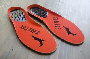
| | (Orthotic) Insoles and specialised shoes
| |
| | ''Cho et al'' suggest that the use of insoles combined with prescribed forefoot rocketed shoes with a wide toebox resulted in a significant reduction in foot pain and dysfunction in patients with rheumatoid foot problems, although custom made insoles showed no differences in terms of the anatomical location of foot pathology<ref name="Cho">Cho, Nam Soon, et al. "Randomized controlled trial for clinical effects of varying types of insoles combined with specialized shoes in patients with rheumatoid arthritis of the foot." Clinical rehabilitation 23.6 (2009/): 512-521. (level of evidence 1b)</ref>.(LOE: 1B)
| |
| <br>The use of orthotic insoles is common in patients with RA and often part of an early conservative treatment which is strongly recommended to protect the rheumatoid foot from potentially destructive processes<ref name="Wood">Woodburn, James, Sharon Barker, and Philip S. Helliwell. "A randomized controlled trial of foot orthoses in rheumatoid arthritis." The Journal of rheumatology 29.7 (2002): 1377-1383. (level of evidence 1b)</ref>(LOE: 3A)<ref name="Nagel">Nagel, Arne, Henrik Schmiegel Andreas Lars, and Dieter Rosenbaum. "Long-term effects of orthotic insoles in rheumatoid arthritis—A one-year year longitudinal follow-up." Gait & Posture 24 (2006): S178-S179.</ref>(LOE: 2B).<br>Orthotic insoles can decrease foot pain and reduce disability<ref name="Nagel" /><ref name="Mejjad">Mejjad, Othmane, et al. "Foot orthotics decrease pain but do not improve gait in rheumatoid arthritis patients." Joint Bone Spine 71.6 (2004): 542-545.</ref><span style="font-size: 13.28px;">,(LOE: 2B)</span> but are not sufficient to correct gait.
| |
| | |
| |-
| |
| | Foot Orthoses
| |
| | ''Conceição et al.'' found pain improvements due to foot orthoses, but no effect on disability<ref>Conceição, Cristiano Sena da, et al. "Systematic review and meta-analysis of effects of foot orthoses on pain and disability in rheumatoid arthritis patients." Disability and rehabilitation 37.14 (2015): 1209-1213.</ref>(LOE: 1B)
| |
| |}
| |
| | |
| <br>
| |
| | |
| === ''Haemophilic arthropathy'' ===
| |
| | |
| Physiotherapy is an integral component in haemophilic foot and ankle arthropathy. Physiotherapy can be used at the acute and chronic stages or after surgery. To prevent the progression of haemophilic arthropathy, regular exercise (30 min at least three time/week) are recommended<ref name="Leslie" />(LOE: 1B), to reduce bleeding, pain and improve articular mobility of the ankle<ref name="Cuesta">Cuesta-Barriuso, Rubén, Antonia Gómez-Conesa, and José-Antonio López-Pina. "Manual therapy in the treatment of ankle hemophilic arthropathy. A randomized pilot study." Physiotherapy theory and practice 30.8 (2014): 534-539.</ref>.(LOE: 2B)<br>
| |
| | |
| '''Physical therapy'''
| |
| | |
| {| width="600" border="1" cellspacing="1" cellpadding="1"
| |
| |-
| |
| | Therapy goals
| |
| | -restoration/maintenance of ROM<br>-muscle strengthening<br>-pain management<br>-improved balance/coordination/ proprioception<ref name="Leslie" />.(LOE: 1B)<br>
| |
| |-
| |
| | Varied therapy
| |
| | -passive mobilisations<br>-passive/active stretching (specifically: the anterior and posterior tibialis, peroneal, gastrocnemius and soleus muscles)<br>-proprioception exercises<br>-active exercises of strength with exercise bands<br>-joint stabilisation exercises<ref name="Cuesta" />.(LOE: 2B)<br>
| |
| |}
| |
| | |
| <br>
| |
| | |
| '''Custom made foot orthoses'''''<br>''
| |
| | |
| {| width="600" border="1" cellspacing="1" cellpadding="1"
| |
| |-
| |
| | Orthopaedic insoles(OI) and shoes(OS)
| |
| | ''Lobet et al.'' demonstrated that foot orthoses may have beneficial effects on ankle joints in hemophilic arthropathy. OI and OS provided significant pain relief, functional improvement and improved comfort in more than half of patients, with minimal side effects<ref name="Lobet">Lobet, Sébastien, et al. "Functional impact of custom‐made foot orthoses in patients with haemophilic ankle arthropathy." Haemophilia 18.3 (2012): e227-e235.</ref>.(LOE: 1B)
| |
| |-
| |
| | OI/OS combined with physiotherapy
| |
| | Combined physiotherapy and OI/OS results in excellent patient satisfaction scores and significant pain reduction<ref name="Lobet" />.
| |
| |}
| |
| | |
| === Diabetic foot arthropathy ===
| |
| | |
| The treatment of Charcot neuroarthropathy is mostly conservative<ref name="Kucera" />.(LOE: 1A) The first step in the management Charcot foot is to offload the affected extremity, prevent further collapse and deformity and protect the opposite foot. This can be done using a castor splint immobilisation to promote healing of the joint during the active stage of the disease<ref name="Kalish">Kalish, Jeffrey, and Allen Hamdan. "Management of diabetic foot problems." Journal of vascular surgery 51.2 (2010): 476-486.</ref>(LOE: 1B)<ref name="Kucera" /><ref name="Shibata" /><span style="font-size: 13.28px;">.(LOE: 1B)</span>
| |
| | |
| '''Non-pharmacological treatments'''
| |
| | |
| {| width="600" border="1" cellspacing="1" cellpadding="1"
| |
| |-
| |
| | Total contact cast
| |
| | The use of a total contact cast in an early stage of Charcot foot is also recommended<ref>R.T. Crews, J.S. Wrobel, Physical management of the Charcot foot, Clin Podiatr Med Surg, Volume 25, Issue 1; Pages 71-9, vii, 2008.</ref>(LOE: 2B) It should be changed 3 days after initial application and then every week. <br>The period of fixation depends on the reduction in oedema and a drop in skin temperature below 2°C compared to the contralateral leg. The usage of a wheelchair as a preventive means against overloading the other extremity can be considered<ref name="Kucera" />(LOE: 1A)<br>
| |
| |-
| |
| |
| |
| CROW
| |
| | |
| [[Image:CROW.jpg]]
| |
| | |
| Figure 10<br>
| |
| | |
| | Alternatively, it is also possible to use Charcot Restraint Orthotic Walker (CROW). <br>''Kalish et al''<ref name="Kalish" /> (LOE: 1B)suggest that the use of accommodative footwear is essential for the long term effective management of Charcot foot.<br>
| |
| |}
| |
| | |
| <br>
| |
| | |
| === Gout ===
| |
| | |
| A patient with a history of gout, hyperuricemia, and/or a septic joint presentation should be referred for medical evaluation before treatment. <ref name="Goodman" />(LOE: 1B)
| |
| | |
| During acute exacerbations, the focus should be on reinforcement of the management programme and splinting, orthotics, or other assistive devices to protect the affected joint(s). During the inter-critical phases, maintaining ROM, strength, and function is essential and a suitable exercise routine should be designed along with advice on keeping weight under control. <ref name="Goodman" /> (LOE: 1B)<br>
| |
| | |
| '''Non-pharmacological treatments'''<br>
| |
| | |
| {| width="600" border="1" cellspacing="1" cellpadding="1"
| |
| |-
| |
| | Cryotherapy
| |
| | Cryotherapy, may be a useful adjunct to treatment of acute gouty arthritis, reducing pain.<ref>Schlesinger, Naomi, et al. "Local ice therapy during bouts of acute gouty arthritis." The Journal of rheumatology 29.2 (2002): 331-334.</ref> (LOE: 1B)
| |
| |-
| |
| | Acupuncture
| |
| | The effectiveness of acupuncture as a complementary therapy may be effective for gouty arthritis patients, decreasing uric acid levels in some patients and the VAS score. <ref>Lee, Won Bock, et al. "Acupuncture for gouty arthritis: a concise report of a systematic and meta-analysis approach." Rheumatology 52.7 (2013): 1225-1232. (Level of evidence 1a)</ref> (LOE: 1A)
| |
| |}
| |
| | |
| <br>
| |
| | |
| === Psoriatic arthritis ===
| |
| | |
| The physical therapy management of psoriatic arthritis can be found on the page [https://www.physio-pedia.com/Psoriatic_Arthritis Psoriatic Arthritis]. <br>
| |
| === Reactive arthritis ===
| |
| | |
| An exercise regime that includes regular aerobic activity as well as exercises to promote joint ROM and muscle strengthening should be followed. Strengthening should target muscles surrounding the affected joints. A brief period of non-weight bearing may be advised to decrease inflammation and limit pressure on the inflamed joints.
| |
| | |
| <br>Immobilisation and inactivity are discouraged as they can lead to decreased range of motion, contractures, joint stiffness, decreased muscle strength and decreased flexibility, as well as overall decreased cardiovascular fitness, which can cause a cascade effect on other body systems. <ref name="phy" />(LOE: 1A)
| |
| | |
| <br>
| |
| | |
| '''''Physical therapy<br>'''''
| |
| | |
| {| width="600" border="1" cellspacing="1" cellpadding="1"
| |
| |-
| |
| | Therapy goals
| |
| | -pain relief
| |
| -improved activities of daily living
| |
| | |
| -reduce joint swelling
| |
| | |
| -prevention of joint damage and disability. <ref name="phy" />
| |
| |-
| |
| | Aerobic exercise
| |
| | Aerobic exercise should include low impact activities, such as swimming, walking or cycling, depending on the patient's cardiovascular level. <ref name="phy">http://www.physio-pedia.com/Reiter's_Syndrome#Physical_Therapy_Management_.28 current_best_evidence.29</ref>
| |
| |-
| |
| | Range of motion and flexibility exercise
| |
| | Stretching and ROM exercises should be completed to prevent muscle atrophy, especially if several joints are involved<ref name="Kim" /><ref name="Toivanen" />(LOE: 1A)to avoid stiffness and deformities and to promote mobility and strength<ref name="Verbruggen">Verbruggen L. Reumatologie, Reactieve arthritis. Vrije Universiteit Brussel, 3de Bachelor REVAKI 2011;48-51.(Level of evidence 5)</ref>(LOE: 4)<ref name="Koehler">Koehler L, Kuipers JG, Zeidler H. Managing seronegative spondarthritides. Rheumatol 2000;39:360-368.</ref>. (LOE: 1A)
| |
| |}
| |
| | |
| <br>
| |
| | |
| '''Custom made foot orthoses'''
| |
| | |
| {| width="600" border="1" cellspacing="1" cellpadding="1"
| |
| |-
| |
| | Orthopaedic insoles(OI) and shoes(OS)
| |
| | If enthesitis is present, heel support and orthosis can be considered to decrease pain and improve mobility<ref name="Toivanen" />(LOE: 1A)<ref name="Olivieri">Olivieri I, Barozzi L, Padula A. Enthesiopathy: clinical manifestations, imaging and treatment. Baillieres Clin Rheumatol 1998;12(4).</ref>(LOE: 1B)<ref name="Kim" />(LOE: 1A)
| |
| |}
| |
| | |
| == Key Research ==
| |
| | |
| === Osteoarthritis ===
| |
| | |
| D.M. Reid, C.G. Miller (eds.), Clinical Trials in Rheumatoid Arthritis and Osteoarthritis, Springer-Verlag London Limited 2008, 325p.
| |
| | |
| Witteveen, Angelique GH, Cheriel J. Hofstad, and Gino MMJ Kerkhoffs. "Hyaluronic acid and other conservative treatment options for osteoarthritis of the ankle." The Cochrane Library (2015).
| |
| | |
| Roth, Sanford H., et al. "Around-the-clock, controlled-release oxycodone therapy for osteoarthritis-related pain: placebo-controlled trial and long-term evaluation." Archives of Internal Medicine 160.6 (2000): 853-860.
| |
| | |
| Karatosun, V., et al. "Intra-articular hyaluronic acid compared to exercise therapy in osteoarthritis of the ankle. A prospective randomized trial with long-term follow-up." Clinical and experimental rheumatology 26.2 (2008): 288.
| |
| | |
| === Rheumatoid arthritis ===
| |
| | |
| Conceição, Cristiano Sena da, et al. "Systematic review and meta-analysis of effects of foot orthoses on pain and disability in rheumatoid arthritis patients." Disability and rehabilitation 37.14 (2015): 1209-1213.
| |
| | |
| Mejjad, Othmane, et al. "Foot orthotics decrease pain but do not improve gait in rheumatoid arthritis patients." Joint Bone Spine 71.6 (2004): 542-545.
| |
| | |
| Baillet, Athan, et al. "Efficacy of resistance exercises in rheumatoid arthritis: meta-analysis of randomized controlled trials." Rheumatology 51.3 (2012): 519-527.
| |
| | |
| Rongen‐van Dartel, S. A. A., et al. "Effect of Aerobic Exercise Training on Fatigue in Rheumatoid Arthritis: A Meta‐Analysis." Arthritis care & research67.8 (2015): 1054-1062. A Meta-Analysis, Arthritis Care & Research, Vol. 67, No. 8, August 2015, pp 1054–1062.
| |
| | |
| Carroll, Matthew, et al. "Gait characteristics associated with the foot and ankle in inflammatory arthritis: a systematic review and meta-analysis." BMC musculoskeletal disorders 16.1 (2015): 1.
| |
| | |
| Graudal, Niels, et al. "Combination Therapy With and Without Tumor Necrosis Factor Inhibitors in Rheumatoid Arthritis: A Meta‐Analysis of Randomized Trials." Arthritis care & research 67.11 (2015): 1487-1495.
| |
| | |
| === Diabetic foot arthropathy ===
| |
| | |
| Kalish, Jeffrey, and Allen Hamdan. "Management of diabetic foot problems." Journal of vascular surgery 51.2 (2010): 476-486.
| |
| | |
| Varma, Ajit Kumar. "Charcot neuroarthropathy of the foot and ankle: a review." The journal of foot and ankle surgery 52.6 (2013): 740-749.
| |
| | |
| Smith, Caroline, Saravana Kumar, and Ryan Causby. "The effectiveness of non‐surgical interventions in the treatment of Charcot foot." International Journal of Evidence‐Based Healthcare 5.4 (2007): 437-449.
| |
| | |
| Kucera, Tomas, Haroun Hassan Shaikh, and Pavel Sponer. "Charcot Neuropathic Arthropathy of the Foot: A Literature Review and Single-Center Experience." Journal of Diabetes Research 2016 (2016).
| |
| | |
| Richard, J-L., M. Almasri, and S. Schuldiner. "Treatment of acute Charcot foot with bisphosphonates: a systematic review of the literature." Diabetologia 55.5 (2012): 1258-1264.
| |
| | |
| === Gout ===
| |
| | |
| Min, Z., and M. Junwu. "Research progress in the genetics of hyperuricaemia and gout." Yi chuan= Hereditas/Zhongguo yi chuan xue hui bian ji 38.4 (2016): 300-313.
| |
| | |
| Harris, MARK D., LORI B. Siegel, and JEFFREY A. Alloway. "Gout and hyperuricemia." American family physician 59.4 (1999): 925-934.
| |
| | |
| Owens, D., B. Whelan, and G. McCarthy. "A survey of the management of gout in primary care." Irish medical journal 101.5 (2008): 147-149.
| |
|
| |
|
| Borghi, C., and F. Perez-Ruiz. "Urate lowering therapies in the treatment of gout: a systematic review and meta-analysis." European review for medical and pharmacological sciences 20.5 (2016): 983-992.
| | == Relevant Anatomy == |
| | [[File:Bones of the foot.png|right|frameless|485x485px]] |
| | Each foot has 28 bones and more than 30 joints. The most common foot joints that arthritis affects are: |
| | # The [[Ankle Joint|Ankle joint]] |
| | # The 3 joints of the foot that involve the calcaneus, the navicular , and the cuboid bone. |
| | # The !st MTP joint <ref>Cleveland clinic [https://my.clevelandclinic.org/health/diseases/13900-foot-and-ankle-arthritis Foot and ankle arthritis] Available from:https://my.clevelandclinic.org/health/diseases/13900-foot-and-ankle-arthritis (last accessed 1.7.2020)</ref> |
| | More information about the anatomy of the ankle and foot can be found here: [http://www.physio-pedia.com/Biomechanics_of_Foot_and_Ankle Biomechanics of Foot and Ankle] and [http://www.physio-pedia.com/Ankle_Joint Ankle Joint]. |
|
| |
|
| Cronstein, Bruce N., and Robert Terkeltaub. "The inflammatory process of gout and its treatment." Arthritis Research & Therapy 8.1 (2006): 1.
| | == Signs and Symptoms == |
| | In general the symptoms are pain in the joints, reduction of the functions of the structure of connection or support and inflammation in the tendons, ligaments, joints, muscles and bones. |
| | * Decreased range of motion, effusion, neumarthrosis, bone erosion. |
| | * The symptoms are similar to rheumatic conditions and include pain, swelling and stiffness. |
| | * Skeletal muscle is also affected by pain and inflammation of bones, structure, muscles, and is a type of joint disease<ref name=":1" />. |
| | * Pain usually increases on activities such as standing, walking or running. |
| | * “Start-up pain” is also a common complaint, where the patient experiences pain and stiffness in the ankle on moving after being asleep or sitting for a long period, which takes a few minutes of motion to settle. |
| | * The affected joints will tend to increasingly swell as the day progresses, particularly with a higher level of activity. |
| | == Assessment == |
| | [[File:OA_ankle.jpg|right|frameless|200x200px]]A clinical examination of the foot includes ‘traditional’ components such as history, palpation, and assessments of sensation, range of motion and strength, as well as special tests that provoke specific tissues. On observation, toe deformities and skin health (dryness, sweating, perfusion) should be noted.<ref name=":0" /> |
|
| |
|
| Schlesinger, Naomi, et al. "Local ice therapy during bouts of acute gouty arthritis." The Journal of rheumatology 29.2 (2002): 331-334.
| | Salient Points of Examination include: |
| | # Patients’ self-reported outcomes eg [[Visual Analogue Scale|VAS,]] [[Patient Specific Functional Scale|PSFS]] |
| | # Static Foot Structure and Alignment eg [[Foot Posture Index (FP1-6)|the Foot Posture Index]] |
| | # Joint Range of Motion - Quantified using a [[goniometer]], and note position tested in. |
| | #[[File:Walk.jpg|right|frameless]]Muscle strength - Decreased toe flexor strength and foot pain are independently associated with fall risk. Strength deficits have also been reported in individuals with foot pain secondary to tibialis posterior tendinopathy<ref name=":0" />. |
| | # Footwear assessment - Inspect for fit (length and width) and design features such as the presence of a heel cup, arch support, torsional and toe-break flexibility. Patterns of wear on the sole of the shoe and/or or scuffing should be noted. The Footwear Assessment Form is a simple and well-organized tool with established reliability and face-validity<ref name=":0" />. |
| | # Dynamic Assessment of Foot Motion (including Gait Analysis) - Involves an observational or quantitative assessment of foot and lower extremity mechanics during a weight-bearing task (e.g., walking, running, single limb squat, step down). Particularly relevant in a clinical foot exam because evidence indicates that there is only a weak relationship between static and dynamic measures of arch height and large between-person variability<ref name=":0" /> |
| | # Dynamic Assessment of Plantar Load Distribution - When performing a clinical assessment, the plantar aspect of the foot should be inspected for patterns of calluses and weight-bearing. |
| | # Provocational Tests - The final part of the clinical examination comprises provocational tests that provoke specific tissues. The [[Windlass Test]]<nowiki/>tstretcheshe plantar fascia and is considered positive if the patient reports pain when the 1st metatarso-phalanegeal joint is passively dorsiflexed. Limited extensibility in the gastrocnemius-soleus complex or the [[flexor hallucis longus]] can be assessed using passive muscle length testing. Symptoms related to the [[Sesamoiditis|sesamoids]] may manifest as plantar pain and localized tenderness to palpation.<ref name=":0" /> |
|
| |
|
| Lee, Won Bock, et al. "Acupuncture for gouty arthritis: a concise report of a systematic and meta-analysis approach." Rheumatology 52.7 (2013): 1225-1232.
| | == Medical Treatment == |
| | Management of ankle and foot arthropathies generally commences with conservative interventions, including analgesic or anti-inflammatory medications, therapeutic injections, physical therapy, footwear modifications and foot orthoses. If these treatments are ineffective, surgical options may be considered.<ref name=":2">Roddy E, Menz HB. [https://www.ncbi.nlm.nih.gov/pmc/articles/PMC5871064/ Foot osteoarthritis: latest evidence and developments.] Therapeutic advances in musculoskeletal disease. 2018 Apr;10(4):91-103. Available from:https://www.ncbi.nlm.nih.gov/pmc/articles/PMC5871064/ (last accessed 2.7.2020)</ref> |
|
| |
|
| === Psoriatic arthritis === | | The medications used to treat arthropathies vary depending on the diagnosis. Commonly used medications include: |
| | #[[File:Methotrexate.jpg|right|frameless]][[Pain Medications|Painkillers]] eg paracetamol, For more-severe pain, opioids might be prescribed, |
| | # Oral non-steroidal anti-inflammatory drugs (NSAIDs) are the most widely used first-line pharmacological treatment. Some NSAIDs are also available as creams or gels, which can be rubbed on joints.<ref name=":3">Mayo clinic [https://www.mayoclinic.org/diseases-conditions/arthritis/diagnosis-treatment/drc-20350777 Arthritis] Available from:https://www.mayoclinic.org/diseases-conditions/arthritis/diagnosis-treatment/drc-20350777 (last accessed 2.7.2020)</ref> |
| | # Counterirritants. Some varieties of creams and ointments contain menthol or capsaicin, the ingredient that makes hot peppers spicy. Rubbing these preparations on the skin over your aching joint may interfere with the transmission of pain signals from the joint itself. |
| | # Disease-modifying antirheumatic drugs ([[DMARDs in the Management of Rheumatoid Arthritis|DMARDs]]). Often used to treat rheumatoid arthritis, DMARDs slow or stop your immune system from attacking your joints. eg methotrexate (Trexall, Rasuvo, others) and hydroxychloroquine (Plaquenil). |
| | # [[BDMARDs in the Management of Rheumatoid Arthritis|Biologic response modifiers]]. Typically used in conjunction with DMARDs, biologic response modifiers are genetically engineered drugs that target various protein molecules that are involved in the immune response. eg Tumor necrosis factor (TNF) inhibitors are commonly prescribed. Other medications target other substances that play a role in inflammation eg interleukin-1 and certain types of white blood cells known as B cells and T cells. |
| | # [[Corticosteroids in the Management of Rheumatoid Arthritis|Corticosteroids]]. This class of drugs, which includes prednisone (Prednisone Intensol, Rayos) and cortisone (Cortef), reduces inflammation and suppresses the immune system. Corticosteroids can be taken orally or can be injected directly into the painful joint<ref name=":3" />. |
| | [[Therapeutic Corticosteroid Injection|Therapeutic Injections]]: Provide an effective alternative financially and some evidence exists that they are effective in pain alleviation (current evidence is limited and the benefit described from injection therapy has been short-lived in most cases)<ref>Urits I, Smoots D, Franscioni H, Patel A, Fackler N, Wiley S, Berger AA, Kassem H, Urman RD, Manchikanti L, Abd-Elsayed A. I[https://link.springer.com/article/10.1007/s40122-020-00157-5 njection Techniques for Common Chronic Pain Conditions of the Foot]: A Comprehensive Review. Pain and therapy. 2020 Feb 27:1-6. Available from:https://link.springer.com/article/10.1007/s40122-020-00157-5 (last accessed 2.7.2020)</ref>. |
| | * The most widely used are: corticosteroid injections and Viscosupplementation, the intra-articular injection of a lubricating fluid (hyaluronan) (aim of restoring the viscoelasticity of the synovial fluid).<ref name=":2" /> |
| | * Regenerative Injections are another option 1. Platelet-rich plasma (PRP) injections - these injections use your own blood and platelets to promote healing. Platelets contain growth factors and proteins that aid healing in soft tissues. Research shows PRP injections can alter the immune response to help reduce inflammation; 2. Placental tissue matrix (PTM) injections - injections of placental tissue (obtained after a healthy baby is delivered from a healthy mother), there is a large number of growth factors in placental tissue that promote healing<ref>Clevelandclinic [https://health.clevelandclinic.org/4-injections-that-can-banish-joint-pain-for-months/ 4 Therapeutic injections] Available from:https://health.clevelandclinic.org/4-injections-that-can-banish-joint-pain-for-months/ (last accessed 2.7.2020)</ref>; 3. Mesenchymal Stem Cells (MSC) - Mesenchymal stem cells are usually collected from the patient’s fat tissue, blood, or bone marrow. When administering stem cell injections, many physicians use medical imaging, such as ultrasound, in order to deliver cells precisely to the site of cartilage damage.When administering stem cell injections, many physicians use medical imaging, such as ultrasound, in order to deliver cells precisely to the site of damage<ref>arthritis health [https://www.arthritis-health.com/treatment/injections/stem-cell-therapy-arthritis SCT] Available from: https://www.arthritis-health.com/treatment/injections/stem-cell-therapy-arthritis (last accessed 2.7.2020)</ref>. |
|
| |
|
| Amherd‐Hoekstra, Anne, et al. "Psoriatic arthritis: a review." JDDG: Journal der Deutschen Dermatologischen Gesellschaft 8.5 (2010): 332-339.
| | == Physiotherapy == |
| | [[File:Foot massage.jpg|right|frameless]] |
| | The primary aim of treatment is to afford pain relief, restore mechanics (alignment, motion and/or load distribution) and return the patient to their desired level of activity participation. Individualised treatment is paramount taking into account clients diagnosis. |
|
| |
|
| === Reactive arthritis ===
| | The plan of care should be designed with the goal of targeting impairments noted during assessment. Options include (see links also): |
| | # [[File:ShoeCue insole.jpg|right|frameless]]Lifestyle Modifications |
| | * In the early stages of arthritathies, limiting the amount of force being placed on the foot and ankle may relieve pain. For example, low-impact activities such as swimming and yoga can provide cardiovascular benefits without putting stress on the foot or ankle. |
| | * Losing weight may decrease pressure on the joints. The foot and ankle support the entire body every time we stand and move, and being overweight increases this pressure and may cause arthritis to progress more quickly. |
| | 2. [[Introduction to Orthotics|Orthoses]] and [[Foot Orthoses|footwear]] |
|
| |
|
| Olivieri I, Barozzi L, Padula A. Enthesiopathy: clinical manifestations, imaging and treatment. Baillieres Clin Rheumatol 1998;12(4).
| | 3. [[Manual Therapy|Manual therapy]] examples include |
| | * [[File:Standing Heel Rise.jpg|right|frameless]][[Massage]] – with sufficient pressure through the superficial tissue to reach the deep lying structures (used to increase blood flow, decrease swelling, reduce muscle spasm and promote normal tissue repair). |
| | * Mobilisation – joint and soft tissues are gently moved to restore normal range, lubricate tissues and relieve pain. eg Talar glides, Maitland mobilisations |
| | 4. [[Stretching]] and [[Exercise -Therapeutic|therapeutic exercises]], eg Stretches that target hamstrings, calves, foot arch and heel are particularly effective and complete range of motion exercises. Strengthening exercises like "small foot exercise", attempting to pick up pegs with toes, walking bare feet in sand. |
|
| |
|
| Kim, Paul S., Thomas L. Klausmeier, and Donald P. Orr. "Reactive arthritis: a review." Journal of Adolescent Health 44.4 (2009): 309-315.
| | 5. [[Cryotherapy]] or [[Thermotherapy]] |
|
| |
|
| Silman, Alan J., and Marc C. Hochberg. Epidemiology of the rheumatic diseases. No. Ed. 2. Oxford University Press, 2001.
| | 6. [[Therapeutic Ultrasound|Ultrasonic Therapy]] – to reduce tissue spasm, accelerates healing and pain relief. |
|
| |
|
| == Clinical Bottom Line ==
| | 7. I[[Interferential Therapy]] – used for differing treatment effects. E.g. pain relief, muscle or nerve stimulation, promoting blood flow and reducing inflammation. |
|
| |
|
| Ankle and foot arthropathies covers all joint disease types of the foot and ankle. Physical therapy can be effective in all cases of ankle and foot arthropathies, but an accurate differentail diagnosis is important so that the most appropriate management protocol is implemented for the specific disease.
| | 8. [[Gait|Gait re-education]] – assess and treat appropriately with eg gait aides |
|
| |
|
| == References == | | 9. [[Taping]] |
| | == Epidemiology /Etiology == |
| | *[[File:Ligaments_of_the_ankle_lateral_aspect_Primal.png|right|frameless]]Osteoarthritis - Approximately 1% of the world’s adult population is affected by ankle OA <ref name="Barg" /> |
| | * Rheumatoid Arthritis - The prevalence of foot pain in patients with RA has been reported at ranges from 60 to 94 % at some stage of the disease. <ref name="Lohkamp">Lohkamp M. et al. The prevalence of disabling foot pain in patients with early rheumatoid arthritis. The Foot 2006;16(4):201-207.</ref><ref name="Brenton">Brenton-Rule, Angela, et al. Foot and ankle characteristics associated with falls in adults with established rheumatoid arthritis: a cross-sectional study.BMC musculoskeletal disorders 2016;17(1):1.</ref> |
| | * Diabetic Foot Arthropathy - Charcot neuropathic osteoarthropathy are detected in up to 29% of diabetics, whereas under MRI the detection rate rises to 75%.<ref name="Kucera">Kucera, Tomas, Haroun Hassan Shaikh, and Pavel Sponer. Charcot Neuropathic Arthropathy of the Foot: A Literature Review and Single-Center Experience. Journal of Diabetes Research 2016</ref> |
| | * Gout - The global prevalence of gout is substantial and is increasing in many parts of the world over the past 50 years. The distribution of gout is uneven across the globe, with prevalence being highest in Pacific countries. Developed countries tend to have a higher prevalence of gout than that of developing countries and incidence of the disease appears to be on the increase. <ref name="Kuo">Kuo CF, et al. Global epidemiology of gout: prevalence, incidence and risk factors. Nature Reviews Rheumatology 2015;11(11):649-662.</ref> |
| | * Psoriatic Arthritis - Prevalence of Psoriatic arthritis in psoriasis patients is between 6% and 39%. It is possible that the condition remains generally underdiagnosed, related to lack of awareness by both the patient and physician. <ref name="Mease">Mease P. Psoriatic arthritis update." BULLETIN-HOSPITAL FOR JOINT DISEASES NEW YORK 64.1/2 (2006): 25.</ref><u></u> |
| | == ''<u></u><sub></sub><sup></sup>''<u></u><sub></sub><sup></sup><u></u><sub></sub><sup></sup><u></u><sub></sub><sup></sup><u></u><sub></sub><sup></sup><u></u><sub></sub><sup></sup><u></u><sub></sub><sup></sup><u></u><sub></sub><sup></sup>Clinical Bottom Line == |
| | <u></u><sub></sub><sup></sup><u></u><sub></sub><sup></sup><u></u><sub></sub><sup></sup><u></u><sub></sub><sup></sup>''<u></u><sub></sub><sup></sup>''<u></u><sub></sub><sup></sup>'''<u></u><sub></sub><sup></sup>'''<u></u><sub></sub><sup></sup><u></u><sub></sub><sup></sup><u></u><sub></sub><sup></sup><u></u><sub></sub><sup></sup> |
| | *[[File:Ankle pumps.gif|right|frameless]]Ankle and foot arthropathies covers all joint disease types of the foot and ankle. |
| | * Physical therapy can be effective in all cases of ankle and foot arthropathies |
| | * Accurate differential diagnosis is important so that the most appropriate management protocol is implemented for the specific disease. |
| | * Recent studies show that individuals with foot and ankle pain have multiple co-existing impairments in alignment, motion, load distribution and muscle performance. |
| | * A comprehensive assessment of the foot and ankle should include patients’ self-reported outcomes and measures of alignment, motion, strength and provocative tests. |
| | * Increasing evidence highlights the importance of evaluating dynamic function and regional interdependence (advances in motion capture and plantar load distribution offer exciting opportunities to obtain precise, clinically relevant measures). |
| | Non-surgical interventions are important factors to ease foot pain and slow the disease-related progression of foot and ankle conditions often seen with the rheumatic diseases. Increasingly, studies are showing that orthoses, footwear and other rehabilitation interventions may play an important role in rheumatology-related foot treatment<ref name=":0" />. |
|
| |
|
| | == References == |
| <references /> | | <references /> |
|
| |
|
| <br> | | <br> |
|
| |
|
| [[Category:Foot]][[Category:Foot_and_Ankle_Conditions]][[Category:Ankle]] | | [[Category:Foot]] |
| | [[Category:Ankle]] |
| | [[Category:Foot - Conditions]] |
| | [[Category:Ankle - Conditions]] |
| | [[Category:Vrije Universiteit Brussel Project]] |
| | [[Category:Conditions]] |
| | [[Category:Osteoarthritis]] |