Alar ligaments: Difference between revisions
Evan Thomas (talk | contribs) mNo edit summary |
(citations added, references updated and added, visual edits, information added to page) |
||
| (10 intermediate revisions by 4 users not shown) | |||
| Line 6: | Line 6: | ||
</div> | </div> | ||
== Description == | == Description == | ||
[[File:Tectorial_membrane.png|alt=|right|frameless|400x400px]] | |||
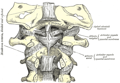
The alar ligaments are two strong rounded cords that attach the dens of C2 ([[Axis]]) to the occipital condyles<ref name=":0">Ishak B, Glinski AV, Dupont G, Lachkar S, Yilmaz E, Iwanaga J, Unterberg A, Oskouian R, Tubbs RS, Chapman JR. [https://journals.sagepub.com/doi/epub/10.1177/2192568220941452 Update on the biomechanics of the craniocervical junction, part II: alar ligament.] Global Spine Journal. 2021 Sep;11(7):1064-9.</ref>. The ligaments' orientation is often described as supernatural but they tend to be more horizontal<ref name=":0" /><ref name=":1">Osmotherly PG, Rivett DA, Mercer SR. [https://www.ncbi.nlm.nih.gov/pmc/articles/PMC3540300/ Revisiting the clinical anatomy of the alar ligaments]. European Spine Journal. 2013 Jan;22(1):60-4.</ref>.The alar and [[Transverse Ligament of the Atlas|transverse]] ligaments are the strongest ligaments that stabilise the cranocervical junction <ref name=":2">Rizvi A, Iwanaga J, Oskouian RJ, Loukas M, Tubbs RS. [https://www.ncbi.nlm.nih.gov/pmc/articles/PMC6116888/pdf/cureus-0010-00000002893.pdf Duplication of the Alar Ligaments: A Case Report.] Cureus. 2018 Jun 29;10(6).</ref> with the alar ligament only failing at a mean force of 394 ± 52 N<ref name=":0" /><ref name=":0" /> | |||
== Attachments == | |||
= | Origin: Arise from either side of the dens/odontoid process (varying attachment points)<ref name=":1" /> | ||
Insertion: Usually the medial aspect of the occipital condyles but some studies have found it to insert on the lateral walls of the foramen magnum<ref name=":1" />. | |||
== Function == | == Function == | ||
* Limits C0-C2 extension<ref name=":0" /> | |||
* Limits axial rotation<ref name=":0" /><ref name=":2" /><ref name=":1" /> | |||
Play a role in stabilizing C1 and C2, especially in rotation<ref name="Magee">Magee DJ (2007). Orthopedic Physical Assessment (5th ed). St Louis, MO: Saunders Elsevier.</ref>. | * Limits lateral bending<ref name=":0" /><ref name=":2" /> | ||
* Limits flexion secondarily<ref name=":2" /> | |||
* Play a role in stabilizing C1 and C2, especially in rotation<ref name="Magee">Magee DJ (2007). Orthopedic Physical Assessment (5th ed). St Louis, MO: Saunders Elsevier.</ref>. | |||
== Pathology == | == Pathology == | ||
Isolated alar ligament ruptures are rare<ref name=":3">Unal TC, Dolas I, Unal OF. [https://www.sciencedirect.com/science/article/abs/pii/S187887501932114X Unilateral alar ligament injury: diagnostic, clinical, and biomechanical features]. World Neurosurgery. 2019 Dec 1;132:e878-84.</ref> but are hypothesised to occur when the head is subjected to sudden rotation and hyperflexion<ref name=":0" />. Usually alar ligment ruptures occur together with fractures and other ligament injuries after trauma to the craniovertebral junction<ref name=":3" />. | |||
== Examination | == Examination == | ||
[http://www.physio-pedia.com/Alar_Ligament_Test Alar ligament test] | [http://www.physio-pedia.com/Alar_Ligament_Test Alar ligament test] | ||
== References == | == References == | ||
<references /> | <references /> | ||
[[Category: | [[Category:Cervical Spine - Anatomy]] [[Category:Ligaments]] [[Category:Cervical Spine - Ligaments]] [[Category:Cervical Spine]] | ||
Latest revision as of 17:12, 16 December 2022
Original Editor - Rachael Lowe
Top Contributors - Rachael Lowe, Kim Jackson, Evan Thomas, WikiSysop, Alistair James, George Prudden and Wendy Snyders
Description[edit | edit source]
The alar ligaments are two strong rounded cords that attach the dens of C2 (Axis) to the occipital condyles[1]. The ligaments' orientation is often described as supernatural but they tend to be more horizontal[1][2].The alar and transverse ligaments are the strongest ligaments that stabilise the cranocervical junction [3] with the alar ligament only failing at a mean force of 394 ± 52 N[1][1]
Attachments[edit | edit source]
Origin: Arise from either side of the dens/odontoid process (varying attachment points)[2]
Insertion: Usually the medial aspect of the occipital condyles but some studies have found it to insert on the lateral walls of the foramen magnum[2].
Function[edit | edit source]
- Limits C0-C2 extension[1]
- Limits axial rotation[1][3][2]
- Limits lateral bending[1][3]
- Limits flexion secondarily[3]
- Play a role in stabilizing C1 and C2, especially in rotation[4].
Pathology[edit | edit source]
Isolated alar ligament ruptures are rare[5] but are hypothesised to occur when the head is subjected to sudden rotation and hyperflexion[1]. Usually alar ligment ruptures occur together with fractures and other ligament injuries after trauma to the craniovertebral junction[5].
Examination[edit | edit source]
References[edit | edit source]
- ↑ 1.0 1.1 1.2 1.3 1.4 1.5 1.6 1.7 Ishak B, Glinski AV, Dupont G, Lachkar S, Yilmaz E, Iwanaga J, Unterberg A, Oskouian R, Tubbs RS, Chapman JR. Update on the biomechanics of the craniocervical junction, part II: alar ligament. Global Spine Journal. 2021 Sep;11(7):1064-9.
- ↑ 2.0 2.1 2.2 2.3 Osmotherly PG, Rivett DA, Mercer SR. Revisiting the clinical anatomy of the alar ligaments. European Spine Journal. 2013 Jan;22(1):60-4.
- ↑ 3.0 3.1 3.2 3.3 Rizvi A, Iwanaga J, Oskouian RJ, Loukas M, Tubbs RS. Duplication of the Alar Ligaments: A Case Report. Cureus. 2018 Jun 29;10(6).
- ↑ Magee DJ (2007). Orthopedic Physical Assessment (5th ed). St Louis, MO: Saunders Elsevier.
- ↑ 5.0 5.1 Unal TC, Dolas I, Unal OF. Unilateral alar ligament injury: diagnostic, clinical, and biomechanical features. World Neurosurgery. 2019 Dec 1;132:e878-84.


