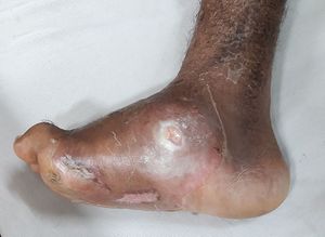
Charcot Foot
Original Editor - Chelsea McLene
Top Contributors - Chelsea Mclene, Kim Jackson and Nikhil Benhur Abburi
What is Charcot Foot?[edit | edit source]
Charcot foot is a rare but serious complication. It can affect persons with peripheral neuropathy, especially those with diabetes mellitus. This is a condition in which the nerves in the lower legs and feet have been damaged. The damage causes a loss of sensation in the feet. It affects the bones, joints, and soft tissues of the foot and ankle. The bones become weak and can break. It has to be treated as early as possible or else the joints in the foot collapse and the foot eventually becomes deformed causing pressure sores to develop in the foot or ankle. An open wound with foot deformity can lead to an infection and even amputation.[1]
Symptoms[edit | edit source]
- Swelling or redness of the foot or ankle.
- Skin feeling warmer at the point of injury.
- A deep aching feeling.
- Deformation of the foot.
Causes[edit | edit source]
- Alcohol or drug abuse.
- An infection.
- Spinal cord injury or disease.
- Parkinson's disease.
- HIV.
- Syphilis.
There’s no specific cause for Charcot foot. Some things can trigger it:
- A sprain or broken bone that doesn’t get treatment quickly
- A sore on your foot that doesn’t heal
- An infection
- Foot surgery that heals slowly
Stages[edit | edit source]
Stage One: Fragmentation and destruction[edit | edit source]
- This is acute stage characterized by redness, swelling, warmth of foot and ankle.
- Internally, soft tissue swelling and small bone fractures are starting to occur. The result is destruction of the joints and surrounding bone. This causes the joints to lose stability, resulting in dislocation. The bones may even jellify, softening completely.
- Rocker bottom foot deformity
- Bony protrusions
- If not treated, this stage can last for up to one year.
Stage Two: Coalescence[edit | edit source]
The body attempts to heal the damage done during the first stage. Destruction of the joints and bones slows down, resulting in less swelling, redness, and warmth.
Stage Three: Reconstruction[edit | edit source]
- This is the final stage in which the joints and bones of the foot heal. Unfortunately, they do not go back to their original condition or shape on their own. While no further damage is being done to the foot, it is often left in a deformed, unstable condition.
- The foot may also be more prone to the formation of sores and ulcers, which might lead to further deformity or in some cases the need for amputation.
Diagnosis[edit | edit source]
At early stages, it may be difficult to diagnose Charcot Foot since the X-ray and lab tests may be normal.
Later stages, X-rays produces images of structures inside the body, to examine the foot's bones and joints. An X-ray can reveal a bone fracture or joint dislocation related to Charcot foot, as well as any change in the shape, or alignment, of the foot.
Other tests:
- Semmes-Weinstein 5.07/10 gram monofilament test analyzes sensitivity to pressure and touch in large nerve fibers
- Pinprick test assesses ability to feel pain
- Neurometer test identifies peripheral nerve dysfunction such as diabetic neuropathy
- Testing tendon reflexes and analyzing the muscle tone and strength in leg and foot.
Treatment[edit | edit source]
Complications[edit | edit source]
- Weak bones
- Deformity: Rocker Bottom
- Toe curls
- Ankle may become twisted and unsteady.
- Bones may press against shoes.
Prognosis[edit | edit source]
All persons with diabetes who have been treated for Charcot foot should have regular foot care with a foot and ankle specialist or a specialist in diabetic foot problems. Close watch should be done on new changes related to Charcot and other diabetic foot complications. Patients who have Charcot foot from other causes also should have regular follow up as recommended by the doctor.
References[edit | edit source]
- ↑ Rogers LC, Frykberg RG, Armstrong DG, Boulton AJ, Edmonds M, Van GH, Hartemann A, Game F, Jeffcoate W, Jirkovska A, Jude E. The Charcot foot in diabetes. Journal of the American Podiatric Medical Association. 2011 Sep;101(5):437-46.


