Transverse Ligament of the Atlas: Difference between revisions
No edit summary |
No edit summary |
||
| Line 7: | Line 7: | ||
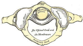
[[File:Transverse lig. of atlas.png|thumb|320x320px]] | [[File:Transverse lig. of atlas.png|thumb|320x320px]] | ||
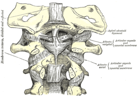
[[File:Tectorial membrane.png|thumb|280x280px]] | [[File:Tectorial membrane.png|thumb|280x280px]] | ||
The transverse | The transverse ligament of the atlas (TLA) is a thick, strong band, 20mm in length<ref name=":0">Vetter M, Oskouian RJ, Tubbs RS. [https://www.ncbi.nlm.nih.gov/pmc/articles/PMC5769988/ “False” ligaments: a review of anatomy, potential function, and pathology]. Cureus. 2017 Nov;9(11).</ref>. which arches across the ring of the atlas, and retains the odontoid process in contact with the anterior arch. It is concave in front, convex behind, broader and thicker in the middle than at the ends. The anterior surface of the ligament is covered with articular cartilage to allow articulation with the Odontoid process. | ||
== Attachments == | == Attachments == | ||
| Line 26: | Line 26: | ||
== Pathology == | == Pathology == | ||
[[ | Ossification of TLA isn't uncommon, however, it is presented in the elderly population<ref>Sasaji T, Kawahara C, Matsumoto F. [[Ossification of transverse ligament of atlas causing cervical myelopathy: a case report and review of the literature. Case reports in medicine]]. 2011;2011.</ref>, causes spinal cord myelopathy that may need surgical intervention. | ||
It may also be included in medical conditions such as rheumatoid arthritis. | |||
Laxity of the ligament present in 14%- 22% of Down syndrome. | |||
TLA used to classify if the Jefferson fracture (C1 fracture) is stable or non if it is involved in the fracture it is considered unstable and requires surgical intervention<ref name=":0" />. | |||
== Examination == | == Examination == | ||
Revision as of 00:52, 8 December 2020
Original Editor - Rachael Lowe
Top Contributors - Khloud Shreif, Rachael Lowe, Kim Jackson, Claire Knott, Evan Thomas, Tarina van der Stockt and WikiSysop
Description[edit | edit source]
The transverse ligament of the atlas (TLA) is a thick, strong band, 20mm in length[1]. which arches across the ring of the atlas, and retains the odontoid process in contact with the anterior arch. It is concave in front, convex behind, broader and thicker in the middle than at the ends. The anterior surface of the ligament is covered with articular cartilage to allow articulation with the Odontoid process.
Attachments[edit | edit source]
It is attached on either side to a small tubercle on the medial surface of the lateral mass of the Atlas.
As it crosses the odontoid process, a small fasciculus (crus superius) is prolonged upward, and another (crus inferius) downward, from the superficial or posterior fibers of the ligament. The former is attached to the basilar part of the occipital bone, in close relation with the Tectorial membrane; the latter is fixed to the posterior surface of the body of the Axis; hence, the whole ligament is named the cruciform ligament of the atlas.
Function[edit | edit source]
It is a very strong and primary stabilizer for upper cervical, we can say the odontoid process can be fractured before the ligament will tear.
It divides the ring of the Atlas into two unequal parts: of these, the posterior and larger serves for the transmission of the spinal cord, its membranes, and the accessory nerves; the anterior and smaller contains the Odontoid process.
It functions to prevent anterior displacement of the C1 (atlas) over C2 (axis).
It is the main stabilizer for the odontoid process preventing it from posterior migration.
Pathology[edit | edit source]
Ossification of TLA isn't uncommon, however, it is presented in the elderly population[2], causes spinal cord myelopathy that may need surgical intervention.
It may also be included in medical conditions such as rheumatoid arthritis.
Laxity of the ligament present in 14%- 22% of Down syndrome.
TLA used to classify if the Jefferson fracture (C1 fracture) is stable or non if it is involved in the fracture it is considered unstable and requires surgical intervention[1].
Examination[edit | edit source]
References[edit | edit source]
- ↑ 1.0 1.1 Vetter M, Oskouian RJ, Tubbs RS. “False” ligaments: a review of anatomy, potential function, and pathology. Cureus. 2017 Nov;9(11).
- ↑ Sasaji T, Kawahara C, Matsumoto F. Ossification of transverse ligament of atlas causing cervical myelopathy: a case report and review of the literature. Case reports in medicine. 2011;2011.


