Screw Home Mechanism of The Knee Joint: Difference between revisions
No edit summary |
No edit summary |
||
| (20 intermediate revisions by 5 users not shown) | |||
| Line 2: | Line 2: | ||
== Definition == | == Definition == | ||
Screw home mechanism (SHM) of knee joint is a critical mechanism that play an important role in terminal extension ( | Screw home mechanism (SHM) of [[Knee|knee joint]] is a critical mechanism that play an important role in terminal extension of the knee. | ||
* There is an observable rotation of the knee during flexion and extension. | |||
* This rotation is important for healthy movement of the knee. | |||
* During the last 30 degrees of knee extension, the tibia ([[Open Chain Exercise|open chain]]) or femur ([[Closed Chain Exercise|closed chain]]) must externally or internally rotate, respectively, about 10 degrees. | |||
* This slight rotation is due to inequality of the articular surface of femur condyles. | |||
* Rotation must occur to achieve full extension and then flexion from full extension.<ref>[https://www.ncbi.nlm.nih.gov/pubmed/581081 Goodfellow] J, O'Connor J. The mechanics of the knee and prosthesis design. J Bone Joint Surg Br. 1978;60(3):358–369.</ref><ref>[https://www.ncbi.nlm.nih.gov/pubmed/24619388 Bytyqi] D, Shabani B, Lustig S, Cheze L, Karahoda Gjurgjeala N, Neyret P. Gait knee kinematic alterations in medial osteoarthritis: three dimensional assessment. Int Orthop. 2014;38(6):1191–1198.</ref><ref>[https://www.ncbi.nlm.nih.gov/pubmed/9345219 Ishii Y, Terajima K, Terashima S, Koga Y. Three-dimensional kinematics of the human knee with intracortical pin fixation. Clin Orthop Relat Res. 1997;(343):144–150.] </ref><ref>https://www.ncbi.nlm.nih.gov/pubmed/12135550</ref><ref>[https://www.ncbi.nlm.nih.gov/pubmed/11451115 Asano T, Akagi M, Tanaka K, Tamura J, Nakamura T. In vivo three-dimensional knee kinematics using a biplanar imagematching technique. Clin Orthop Relat Res. 2001;(388):157–166]</ref><ref name=":0">[https://www.ncbi.nlm.nih.gov/pmc/articles/PMC4553277/# Kim, H. Y., Kim, K. J., Yang, D. S., Jeung, S. W., Choi, H. G., & Choy, W. S. (2015). Screw-Home Movement of the Tibiofemoral Joint during Normal Gait: Three-Dimensional Analysis. ''Clinics in orthopedic surgery'', ''7''(3), 303–309. doi:10.4055/cios.2015.7.3.303]</ref>. | |||
== Relevant Anatomy == | |||
</ref>. | [[File:Conyle inequality femur.jpg|Articular surface paradox of femur|right|frameless]]The knee joint is basically a [[Joint Classification|hinge joint]] with the main movement of flexion-extension. However, the radius and the length of the articular surface of the [[Femur|femu]]<nowiki/>r and [[tibia]] differ at the knee joint ie Articular surface of medial condyle of femur is greater than the articular surface of lateral condyle. | ||
* As a result, a complex movement (includes "sliding") occurs during the last 30 degrees of knee extension, in addition to rolling between the two bones this allows the knee joint to move smoothly. | |||
* At terminal extension, the knee joint is slightly hyperextended and stabilized with the tightening of the [[Anterior Cruciate Ligament (ACL)|cruciate]] and [[Medial Collateral Ligament Injury of the Knee|collateral]] ligaments. | |||
* As the length of the medial femoral condyle is longer than the length of the lateral condyle, the tibia rotates externally about 15° on the femur during the last 20° of extension. | |||
* This kinematic phenomenon is well known, and it is called the screw-home movement | |||
The shape of the condyles are not what brings about the movements. | |||
* Tibial-on-femoral rotation occurs in an open chain exercise like in the leg extension machine (tibia externally rotates). | |||
* Femoral-on-tibial rotation, as in a closed chain exercise like the squat (femur internally rotate). | |||
* During knee extension, tibia rolls anteriorly, elongating the PCL and the PCL’s pull on tibia, causes it to glide anteriorly. | |||
* During knee flexion tibia rolls posteriorly, elongating the ACL and it is the ACL’s pull on tibia, that causes it to glide posteriorly. | |||
* [[Popliteus Muscle|Popliteus]] - unlocks knee with open chain motions | |||
* [[Gluteus Medius|Hip external rotation]] - unlocks knee with closed chain motions<ref name=":1">Epomedicine [https://epomedicine.com/medical-students/screw-home-mechanism/ Screw home mechanism] Available from:https://epomedicine.com/medical-students/screw-home-mechanism/ (last accessed 12.9.2020)</ref>. | |||
[[File:Vmo terminal extention.jpg|thumb|VMO terminal extension exercise. From[https://www.rehabmypatient.com Rehab My Patient]]] | |||
== Clinical Significance == | |||
Restoration of terminal extension is an important goal of rehabilitation program. Terminal extension exercises and terminal extension mobilisations have important contributions on restoration of screw home movement /terminal extension. As does strengthening and stretching relevant muscles. | |||
# Loss of full knee extension range of motion (ROM) is a frequent finding in the population with [[Knee Osteoarthritis|knee OA]]. | |||
#* Such loss of normal terminal knee extension may have important effects on knee mechanics during [[Gait|walking]] and standing. | |||
#* A primary goal of physical therapy is to have a positive effect on the patient's ability to achieve terminal (end-range) knee extension during [[Activities of Daily Living|activities of daily living]]<ref>Taylor AL, Wilken JM, Deyle GD, Gill NW. [https://www.jospt.org/doi/full/10.2519/jospt.2014.4710 Knee extension and stiffness in osteoarthritic and normal knees: a videofluoroscopic analysis of the effect of a single session of manual therapy]. journal of orthopaedic & sports physical therapy. 2014 Apr;44(4):273-82. Available from: https://www.jospt.org/doi/full/10.2519/jospt.2014.4710 (last accessed 12.9.2020)</ref>. | |||
#The "screw-home" mechanism is considered to be a key element to knee stability for standing upright. The tibia rotates internally during the swing phase and externally during the stance phase. | |||
#* External rotation occurs during the terminal degrees of knee extension and results in tightening of both cruciate ligaments, which locks the knee. | |||
#* The tibia is then in the position of maximal stability with respect to the femur. | |||
#* Last 30 degrees of Extension causes a Medial rotation of Femur on Tibia will keep joint in closed packed position. The Knee is Unlocked by Lateral rotation of Femur. | |||
#* In open Kinematic chain Tibia laterally rotates on Femur during last 5 degrees of Extension to produce LOCKING. Unlocking by Medial rotation.<ref name=":1" /> | |||
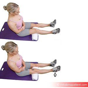
#In most of the knee problems generally there is an insufficiency on terminal extension and [[Vastus Medialis Oblique|vastus medialis obliquus]] function. | |||
In most of the knee problems generally there is an insufficiency on terminal extension and vastus medialis obliquus function | |||
== References == | == References == | ||
<references /> | <references /> | ||
[[Category:Knee - Anatomy]] | |||
[[Category:Knee - Interventions]] | |||
Latest revision as of 17:36, 17 January 2023
Definition[edit | edit source]
Screw home mechanism (SHM) of knee joint is a critical mechanism that play an important role in terminal extension of the knee.
- There is an observable rotation of the knee during flexion and extension.
- This rotation is important for healthy movement of the knee.
- During the last 30 degrees of knee extension, the tibia (open chain) or femur (closed chain) must externally or internally rotate, respectively, about 10 degrees.
- This slight rotation is due to inequality of the articular surface of femur condyles.
- Rotation must occur to achieve full extension and then flexion from full extension.[1][2][3][4][5][6].
Relevant Anatomy[edit | edit source]
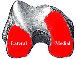
The knee joint is basically a hinge joint with the main movement of flexion-extension. However, the radius and the length of the articular surface of the femur and tibia differ at the knee joint ie Articular surface of medial condyle of femur is greater than the articular surface of lateral condyle.
- As a result, a complex movement (includes "sliding") occurs during the last 30 degrees of knee extension, in addition to rolling between the two bones this allows the knee joint to move smoothly.
- At terminal extension, the knee joint is slightly hyperextended and stabilized with the tightening of the cruciate and collateral ligaments.
- As the length of the medial femoral condyle is longer than the length of the lateral condyle, the tibia rotates externally about 15° on the femur during the last 20° of extension.
- This kinematic phenomenon is well known, and it is called the screw-home movement
The shape of the condyles are not what brings about the movements.
- Tibial-on-femoral rotation occurs in an open chain exercise like in the leg extension machine (tibia externally rotates).
- Femoral-on-tibial rotation, as in a closed chain exercise like the squat (femur internally rotate).
- During knee extension, tibia rolls anteriorly, elongating the PCL and the PCL’s pull on tibia, causes it to glide anteriorly.
- During knee flexion tibia rolls posteriorly, elongating the ACL and it is the ACL’s pull on tibia, that causes it to glide posteriorly.
- Popliteus - unlocks knee with open chain motions
- Hip external rotation - unlocks knee with closed chain motions[7].

Clinical Significance[edit | edit source]
Restoration of terminal extension is an important goal of rehabilitation program. Terminal extension exercises and terminal extension mobilisations have important contributions on restoration of screw home movement /terminal extension. As does strengthening and stretching relevant muscles.
- Loss of full knee extension range of motion (ROM) is a frequent finding in the population with knee OA.
- Such loss of normal terminal knee extension may have important effects on knee mechanics during walking and standing.
- A primary goal of physical therapy is to have a positive effect on the patient's ability to achieve terminal (end-range) knee extension during activities of daily living[8].
- The "screw-home" mechanism is considered to be a key element to knee stability for standing upright. The tibia rotates internally during the swing phase and externally during the stance phase.
- External rotation occurs during the terminal degrees of knee extension and results in tightening of both cruciate ligaments, which locks the knee.
- The tibia is then in the position of maximal stability with respect to the femur.
- Last 30 degrees of Extension causes a Medial rotation of Femur on Tibia will keep joint in closed packed position. The Knee is Unlocked by Lateral rotation of Femur.
- In open Kinematic chain Tibia laterally rotates on Femur during last 5 degrees of Extension to produce LOCKING. Unlocking by Medial rotation.[7]
- In most of the knee problems generally there is an insufficiency on terminal extension and vastus medialis obliquus function.
References[edit | edit source]
- ↑ Goodfellow J, O'Connor J. The mechanics of the knee and prosthesis design. J Bone Joint Surg Br. 1978;60(3):358–369.
- ↑ Bytyqi D, Shabani B, Lustig S, Cheze L, Karahoda Gjurgjeala N, Neyret P. Gait knee kinematic alterations in medial osteoarthritis: three dimensional assessment. Int Orthop. 2014;38(6):1191–1198.
- ↑ Ishii Y, Terajima K, Terashima S, Koga Y. Three-dimensional kinematics of the human knee with intracortical pin fixation. Clin Orthop Relat Res. 1997;(343):144–150.
- ↑ https://www.ncbi.nlm.nih.gov/pubmed/12135550
- ↑ Asano T, Akagi M, Tanaka K, Tamura J, Nakamura T. In vivo three-dimensional knee kinematics using a biplanar imagematching technique. Clin Orthop Relat Res. 2001;(388):157–166
- ↑ Kim, H. Y., Kim, K. J., Yang, D. S., Jeung, S. W., Choi, H. G., & Choy, W. S. (2015). Screw-Home Movement of the Tibiofemoral Joint during Normal Gait: Three-Dimensional Analysis. Clinics in orthopedic surgery, 7(3), 303–309. doi:10.4055/cios.2015.7.3.303
- ↑ 7.0 7.1 Epomedicine Screw home mechanism Available from:https://epomedicine.com/medical-students/screw-home-mechanism/ (last accessed 12.9.2020)
- ↑ Taylor AL, Wilken JM, Deyle GD, Gill NW. Knee extension and stiffness in osteoarthritic and normal knees: a videofluoroscopic analysis of the effect of a single session of manual therapy. journal of orthopaedic & sports physical therapy. 2014 Apr;44(4):273-82. Available from: https://www.jospt.org/doi/full/10.2519/jospt.2014.4710 (last accessed 12.9.2020)


