Rectus Sheath: Difference between revisions
(Created page with "{{subst:New Page}}") |
Kim Jackson (talk | contribs) m (Text replacement - "[[Pyramidalis muscle" to "[[Pyramidalis Muscle") |
||
| (8 intermediate revisions by one other user not shown) | |||
| Line 1: | Line 1: | ||
<div class="editorbox"> | <div class="editorbox"> | ||
'''Original Editor '''- [[User: | '''Original Editor '''- [[User:Lucinda hampton|lucinda hampton]] | ||
'''Top Contributors''' - {{Special:Contributors/{{FULLPAGENAME}}}} | '''Top Contributors''' - {{Special:Contributors/{{FULLPAGENAME}}}} | ||
</div> | </div> | ||
== Introduction == | == Introduction == | ||
[[File:Rectus abdominis sheath.jpeg|right|frameless]] | |||
The Rectus Sheath is a multilayered [[aponeurosis]], being a durable, resilient, fibrous compartment that contains both the [[Rectus Abdominis|rectus abdominis muscle]] and the [[Pyramidalis Muscle|pyramidalis muscle.]] <ref>Sevensma KE, Leavitt L, Pihl KD. [https://www.ncbi.nlm.nih.gov/books/NBK537153/ Anatomy, Abdomen and Pelvis, Rectus Sheath].Available:https://www.ncbi.nlm.nih.gov/books/NBK537153/ (accessed 19.12.2021)</ref> | |||
It covers the anterior and posterior surfaces of the upper three-quarters of the rectus abdominis muscle, and the lower quarter of its anterior surface. The lower quarter of the posterior surface of the rectus abdominis muscle isn’t covered with rectus sheath at all, but rather lays directly on the [[Transversus Abdominis|transversalis]] fascia.<ref>Ken hub Rectus Sheath Available:https://www.kenhub.com/en/library/anatomy/rectus-sheath (accessed 19.12.2021)</ref> | |||
Image 1: Dissection of a human Rectus abdominis sheath | |||
== | == Key Facts == | ||
{| class="wikitable" | |||
|[[File:Torso-rectus-abdominis-transversus-abdominis.png|right|frameless]] | |||
| | |||
|- | |||
|Definition | |||
|Multilayered aponeurosis that encloses the rectus abdominis and pyramidalis muscles | |||
|- | |||
|Walls of upper three-quarters | |||
|Anterior wall: Aponeurosis of [[External Abdominal Oblique|external abdominal oblique muscle]] and aponeurosis of [[Internal Abdominal Oblique|internal abdominal oblique muscle]] | |||
Posterior wall: Aponeurosis of internal abdominal oblique muscle and aponeurosis of transversus abdominis muscle | |||
|- | |||
|Walls of lower quarter | |||
|Anterior wall: As above | |||
Posterior wall: Absent | |||
|- | |||
|Function | |||
|Protection of anterior abdominal muscles and vessels. | |||
Provides maximal compression and support of [[Abdominal Muscle Anatomy|abdominal]] organs. | |||
|} | |||
# | == Contents == | ||
# | |||
# [[File:Rectus-sheath-grays-illustrations.jpeg|right|frameless|alt=|220x220px]]Rectus abdominis and Pyramidalis muscles | |||
# Lower 6 [[Thoracic Spinal Nerves|thoracic nerves]] and accompanying branches of the posterior intercostal vessels | |||
# Superior and inferior epigastric vessels<ref>Radiopedia Available: https://radiopaedia.org/articles/rectus-sheath<nowiki/>(accessed 19.12.2021)</ref> | |||
Image 3: Figure 2: Rectus sheath (Gray's illustrations) | |||
== References == | == References == | ||
<references /> | <references /> | ||
[[Category:Anatomy]] | |||
[[Category:Anatomical Landmarks]] | |||
[[Category:Muscles]] | |||
Latest revision as of 11:51, 23 December 2021
Original Editor - lucinda hampton
Top Contributors - Lucinda hampton and Kim Jackson
Introduction[edit | edit source]
The Rectus Sheath is a multilayered aponeurosis, being a durable, resilient, fibrous compartment that contains both the rectus abdominis muscle and the pyramidalis muscle. [1]
It covers the anterior and posterior surfaces of the upper three-quarters of the rectus abdominis muscle, and the lower quarter of its anterior surface. The lower quarter of the posterior surface of the rectus abdominis muscle isn’t covered with rectus sheath at all, but rather lays directly on the transversalis fascia.[2]
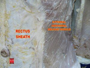
Image 1: Dissection of a human Rectus abdominis sheath
Key Facts[edit | edit source]
| Definition | Multilayered aponeurosis that encloses the rectus abdominis and pyramidalis muscles |
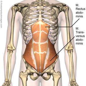
| Walls of upper three-quarters | Anterior wall: Aponeurosis of external abdominal oblique muscle and aponeurosis of internal abdominal oblique muscle
Posterior wall: Aponeurosis of internal abdominal oblique muscle and aponeurosis of transversus abdominis muscle |
| Walls of lower quarter | Anterior wall: As above
Posterior wall: Absent |
| Function | Protection of anterior abdominal muscles and vessels.
Provides maximal compression and support of abdominal organs. |
Contents[edit | edit source]
- Rectus abdominis and Pyramidalis muscles
- Lower 6 thoracic nerves and accompanying branches of the posterior intercostal vessels
- Superior and inferior epigastric vessels[3]
Image 3: Figure 2: Rectus sheath (Gray's illustrations)
References[edit | edit source]
- ↑ Sevensma KE, Leavitt L, Pihl KD. Anatomy, Abdomen and Pelvis, Rectus Sheath.Available:https://www.ncbi.nlm.nih.gov/books/NBK537153/ (accessed 19.12.2021)
- ↑ Ken hub Rectus Sheath Available:https://www.kenhub.com/en/library/anatomy/rectus-sheath (accessed 19.12.2021)
- ↑ Radiopedia Available: https://radiopaedia.org/articles/rectus-sheath(accessed 19.12.2021)


