Metatarsals: Difference between revisions
No edit summary |
(references and citations added, info updated) |
||
| Line 7: | Line 7: | ||
The metatarsals refer to the five long bones found in each foot. They are numbered I to V, from medial to lateral.<ref name=":1" /> | The metatarsals refer to the five long bones found in each foot. They are numbered I to V, from medial to lateral.<ref name=":1" /> | ||
Together, the metatarsal and tarsal bones help to form the main arches of the foot, which are essential for weight-bearing and walking. | Together, the metatarsal and tarsal bones help to form the main arches of the foot, which are essential for weight-bearing and [[Walking - Muscles Used|walking]]. | ||
== Gross Anatomy & Structure == | == Gross Anatomy & Structure == | ||
| Line 88: | Line 88: | ||
|} | |} | ||
== | == Ligaments == | ||
There are four deep transverse metatarsal ligaments (DTML) that bridge the head of the metatarsal bones. Specifically, they run "between the distal ends of adjacent metatarsal bones and intersect with the plantar ligaments of the metatarsophalangeal joint".<ref>Abdalbary SA, Elshaarawy EA, Khalid BE. T[https://www.ncbi.nlm.nih.gov/pmc/articles/PMC4779011/#:~:text=The%20deep%20transverse%20metatarsal%20ligament%20(DTML)%2C%20which%20is%20a,heads%20of%20the%20metatarsal%20bones. ensile properties of the deep transverse metatarsal ligament in hallux valgus]: a CONSORT-compliant article. Medicine. 2016 Feb;95(8).</ref> | There are four deep transverse metatarsal ligaments (DTML) that bridge the head of the metatarsal bones. Specifically, they run "between the distal ends of adjacent metatarsal bones and intersect with the plantar ligaments of the metatarsophalangeal joint".<ref>Abdalbary SA, Elshaarawy EA, Khalid BE. T[https://www.ncbi.nlm.nih.gov/pmc/articles/PMC4779011/#:~:text=The%20deep%20transverse%20metatarsal%20ligament%20(DTML)%2C%20which%20is%20a,heads%20of%20the%20metatarsal%20bones. ensile properties of the deep transverse metatarsal ligament in hallux valgus]: a CONSORT-compliant article. Medicine. 2016 Feb;95(8).</ref> | ||
| Line 94: | Line 94: | ||
The metatarsals contribute significantly to the three arches of the foot. The bones that contribute to each arch are as follows: | The metatarsals contribute significantly to the three arches of the foot. The bones that contribute to each arch are as follows: | ||
* Medial longitudinal arch: talus, calcaneus, navicular, all three cuneiforms and metatarsals 1-3 | * Medial longitudinal arch: talus, calcaneus, navicular, all three cuneiforms and metatarsals 1-3<ref>Babu D, Bordoni B. [https://www.ncbi.nlm.nih.gov/books/NBK562289/ Anatomy, Bony Pelvis and Lower Limb, Medial Longitudinal Arch of the Foot]. StatPearls [Internet]. 2020 Sep 1.</ref>. | ||
* Lateral longitudinal arch: calcaneus, cuboid and metatarsals 4 & 5<ref name=":3">Gwani AS, Asari MA, Ismail ZM. [https://journals.viamedica.pl/folia_morphologica/article/view/50999 How the three arches of the foot intercorrelate]. Folia morphologica. 2017;76(4):682-8.</ref>. | |||
* Transverse arch: bases of all five metatarsals, cuboid and cuneiforms 1-3.<ref name=":3" /> | |||
== Clinical | == Clinical significance == | ||
=== Metatarsalgia === | |||
Metatarsalgia is defined as forefoot pain under one or more of the metatarsal heads<ref name=":4">Besse JL. [https://pdf.sciencedirectassets.com/277782/1-s2.0-S1877056817X00029/1-s2.0-S187705681630189X/main.pdf?X-Amz-Security-Token=IQoJb3JpZ2luX2VjEOD%2F%2F%2F%2F%2F%2F%2F%2F%2F%2FwEaCXVzLWVhc3QtMSJIMEYCIQC3a6zwZxpXXXKg4SFBy3ICqMi1J6iU4jM5F%2FY6A2joygIhAI8NxB6AsX6ruP%2F2We797vWNDRAnNv0hmqbCYR3C7ir8KrsFCKn%2F%2F%2F%2F%2F%2F%2F%2F%2F%2FwEQBRoMMDU5MDAzNTQ2ODY1IgycQgQrRS7JKcvU5NEqjwV2FC%2BNRVLaXAQr9LtztOeLngV%2BiZRqYKFzsl5rU3cSZ64zGEB8%2FVyjBxHIEmdQE9kpRiATll%2F9p5LQxz1cUuoGerD272Dt25U9q9oWAcVHffjO8GLNr2PU9ElYv0I2ey30FtUtPGZnIL%2B73Hv923GLv7sneyFsljm2GC64Q6V7yL2ZBO1BNlQG1U%2FFfxI0KGHx3L%2Bc6BaMJ7lM%2F4PJLsxg%2BsnM8bmbkgejKcfsK1R%2Bw1Xi4XqmfYvJ0AnozBYZUxFHPQtXbn6AvrclK6IQqNt5ifBF1B6kX6Dr9fDsxIuSfTYhGdj%2F%2BMEgw84CiJ%2F6zdbpegAUtFSrhbgbwxUb6NGPSvsOtM1HzKwNGH0geu2JMSvOrafRPa8nFUJ%2BIwz1LrI3NJiBfinY2CZlnydCnCP1C%2Bch786vxfZw4rpW6XFcMRmIopFaxEeJqp8p7wrZ7BdFCp6EweXloTXo7KSpzUWJ3vBmPKnCiGkQGAkzXFDnkv1RVgoUbKN%2FuQPlsjxkynhXOmo8Jid1bNOlzhMx4HTwbthxWKpoE5iJU7HhcSYmulhyBDEIeHqKTZSEGFBjKf2SJaQ872RsSg1ragYudif%2FmuKuyJTqH0M0XTxNf6G34mScfEtzZofjmBVxSXyZv7It6Lr6x%2FVm52YypsmrQvCepls8s80%2BSv11gOutTztXlGVom0CwdEZvVRO4Bb1FWThtpUuNwWMJbpZoOk0iNJz63XnulVN%2F19M8f117sh79wa1Z%2BDc4RFzQt9U8O8mAvH5QdCv6wxtYLy5YZ2mYJ7w1WX3SKxDlOrDjUavdjNpMozR6Fw2wWa5rjkY2tFhpphl%2BCy8fR%2B0Y755ZV3TiPvflqIvIV%2FDG7mH2Je%2BFFYPyMO6guKcGOrABEom2H%2F3phqJ3zAeW7CgW7gTZ3AiA01yUYaZmMrLdeLT4Uzr%2Fasoaj0Eq2qAhnJ1I7a3s9qIc44JyD4B5bjplvI7iTk5mZL8hNlJSkeRuClT4ZOEHrtPO15mnQlXj7%2BJQCINZS165wPVsBScu%2B5aUDjKt7jL6s6acbcTQ%2B2C9W1NLs%2Fs4WUMDevIYJ8ZQ4YpRFCLIOf%2BBatQjaW46kqmt38wWR8zNFIWmxW8Cs4rm10U%3D&X-Amz-Algorithm=AWS4-HMAC-SHA256&X-Amz-Date=20230829T161952Z&X-Amz-SignedHeaders=host&X-Amz-Expires=300&X-Amz-Credential=ASIAQ3PHCVTY3EMQBKEZ%2F20230829%2Fus-east-1%2Fs3%2Faws4_request&X-Amz-Signature=19cdf3a6b45948f42a4926c469f53128698caa3bbbadaa3c44829014f55700ab&hash=6f5681b4d4ed43e0ef4f7bc575fcbf28d1954081e82b1d9e12988cc4803f713d&host=68042c943591013ac2b2430a89b270f6af2c76d8dfd086a07176afe7c76c2c61&pii=S187705681630189X&tid=spdf-437ef6d6-b636-4af8-b735-cb177af20f1f&sid=c08ce8986796224f9a7972479e24c5e1e7dbgxrqb&type=client&tsoh=d3d3LnNjaWVuY2VkaXJlY3QuY29t&ua=190759080452550750&rr=7fe61e3e2e5a0714&cc=za Metatarsalgia.] Orthopaedics & Traumatology: Surgery & Research. 2017 Feb 1;103(1):S29-39.</ref>. Primary metatarsalgia is "due to anatomical characteristics of the metatarsals that affect their relations to one another and to the rest of the foot" (Besse, 2017)<ref name=":4" />. | |||
=== Fractures === | === Fractures === | ||
| Line 113: | Line 115: | ||
=== Gout === | === Gout === | ||
[[Gout]] is a form of crystal-induced arthoropathy, formed by monosodium urate monohydrate crystals. The most common joint affected is the metatarsal-phalangeal joint of the big toe.<ref>Rothschild BM. Gout and Pseudogout [Internet]. [https://emedicine.medscape.com/article/329958-overview Practice Essentials, Background, Pathophysiology]. Medscape; 2021 [cited 2021May27]. Available from: <nowiki>https://emedicine.medscape.com/article/329958-overview</nowiki></ref>[[File:Gout big toe.jpeg|thumb|270x270px|Gout affecting the metatarsal-phalangeal joint of the big toe.|alt=|center]] | [[Gout]] is a form of crystal-induced arthoropathy, formed by monosodium urate monohydrate crystals. The most common joint affected is the metatarsal-phalangeal joint of the big toe.<ref>Rothschild BM. Gout and Pseudogout [Internet]. [https://emedicine.medscape.com/article/329958-overview Practice Essentials, Background, Pathophysiology]. Medscape; 2021 [cited 2021May27]. Available from: <nowiki>https://emedicine.medscape.com/article/329958-overview</nowiki></ref>[[File:Gout big toe.jpeg|thumb|270x270px|Gout affecting the metatarsal-phalangeal joint of the big toe.|alt=|center]] | ||
* | === Hallux valgus === | ||
[[File:Bunion 2.png|frameless|185x185px|alt=|right]][[Hallux Valgus|Hallux vagus]], sometimes referred to as a bunion, refers to malformation of the metatarsalphalangeal (MTP) joint of the big toe.<ref>Alzurahi A. Bunion Symptom Disorder: Surgical and Non-Surgical Treatment.</ref> The following is a more specific description of what is thought to be involved with hallux valgus: | |||
* Medial deviation of the first metatarsal | |||
* Lateral deviation, with or without rotation of the hallux | * Lateral deviation, with or without rotation of the hallux | ||
* Prominence | * Prominence with or without medial soft-tissue enlargement of the first metatarsal head.<ref name=":0">Frank CJ. Hallux Valgus [Internet]. Practice Essentials. Medscape; 2021 [cited 2021 May 27]. Available from: https://emedicine.medscape.com/article/1232902-overview</ref> | ||
[[File:Hallux valgus 2.jpeg|frameless|166x166px|alt=|right]] | [[File:Hallux valgus 2.jpeg|frameless|166x166px|alt=|right]] | ||
Etiologies for hallux valgus include biomechanical, traumatic, and metabolic causes, with biomechanical instability being the most common aetiology. Hallux valgus may also occur secondary to metabolic or inflammatory arthropathies such as [[Rheumatoid Arthritis|rheumatoid arthritis]], [[Psoriatic Arthritis|psoriatic arthritis]] and gouty arthritis. Connective tissue diseases including [[Ehlers-Danlos Syndrome|Ehlers-Danlos syndrome]], [[Marfan Syndrome|Marfan syndrome]], [[Down Syndrome (Trisomy 21)|Down syndrome]] | Etiologies for hallux valgus include biomechanical, traumatic, and metabolic causes, with biomechanical instability being the most common aetiology. Hallux valgus may also occur secondary to metabolic or inflammatory arthropathies such as [[Rheumatoid Arthritis|rheumatoid arthritis]], [[Psoriatic Arthritis|psoriatic arthritis]] and gouty arthritis. Connective tissue diseases including [[Ehlers-Danlos Syndrome|Ehlers-Danlos syndrome]], [[Marfan Syndrome|Marfan syndrome]], [[Down Syndrome (Trisomy 21)|Down syndrome]] and generalized ligamentous laxity may also cause hallux valgus.<ref name=":0" /> | ||
== References == | == References == | ||
Latest revision as of 18:34, 29 August 2023
Top Contributors - Shejza Mino, Lucinda hampton, Kim Jackson, Vidya Acharya and Wendy Snyders
Introduction[edit | edit source]
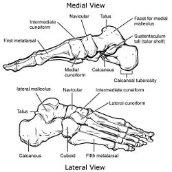
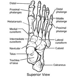
The metatarsals refer to the five long bones found in each foot. They are numbered I to V, from medial to lateral.[1]
Together, the metatarsal and tarsal bones help to form the main arches of the foot, which are essential for weight-bearing and walking.
Gross Anatomy & Structure[edit | edit source]
The metatarsal bones run from the tarsus to the phalanges, forming two joints: the tarsometatarsal joint & metatarsolphalangeal joint.[1]
Each metatarsal bone consists of the following:
Articulations[edit | edit source]
Each base of the metatarsal bone articulates with at least one of the tarsal bones, forming the tarsometatarsal joints. The head of the metatarsals articulate with the phalanges, making up the metatarsalphalangeal joints. Additionally, the base of the metatarsals also articulates with the base of the adjacent metatarsal, forming the intermetatarsal joints.[1][2]
The proximal and distal articulations, specific to each metatarsal bone, are listed in the chart below.
| Bone (metatarsal) | Proximal articulation | Distal articulation |
|---|---|---|
| Metatarsal I | medial cuneiform | 1st proximal phalanx |
| Metatarsal II | medial, intermediate & lateral cuneiforms | 2nd proximal phalanx |
| Metatarsal III | lateral cuneiform | 3rd proximal phalanx |
| Metatarsal IV | cuboid | 4th proximal phalanx |
| Metatarsal V | cuboid | 5th proximal phalanx |
Muscular Attachments[edit | edit source]
| Metatarsal | Muscle attachment |
|---|---|
| Metatarsal I | - Base: tibialis anterior, fibularis longus
- Tuberosity: fibularis longus - Lateral shaft: 1st dorsal interosseous |
| Metatarsal II | - Medial shaft: 1st dorsal interosseous
- Lateral shaft: 2nd doral interosseous |
| Metatarsal III | - Medial shaft: 2nd dorsal interosseus & 1st plantar interosseus
- Lateral shaft: 3rd dorsal interosseous |
| Metatarsal IV | - Medial shaft: 3rd dorsal interosseus & 2nd plantar interosseus
- Lateral shaft: 4th dorsal interosseus |
| Metatarsal V | - Medial shaft: 4th dorsal interosseus & 3rd plantar interosseus
- Dorsal base: fibularis tertius - Tuberosity: fibularis brevis - Plantar base: flexor digiti minimi brevis |
Ligaments[edit | edit source]
There are four deep transverse metatarsal ligaments (DTML) that bridge the head of the metatarsal bones. Specifically, they run "between the distal ends of adjacent metatarsal bones and intersect with the plantar ligaments of the metatarsophalangeal joint".[3]
Arches of the Foot[edit | edit source]
The metatarsals contribute significantly to the three arches of the foot. The bones that contribute to each arch are as follows:
- Medial longitudinal arch: talus, calcaneus, navicular, all three cuneiforms and metatarsals 1-3[4].
- Lateral longitudinal arch: calcaneus, cuboid and metatarsals 4 & 5[5].
- Transverse arch: bases of all five metatarsals, cuboid and cuneiforms 1-3.[5]
Clinical significance[edit | edit source]
Metatarsalgia[edit | edit source]
Metatarsalgia is defined as forefoot pain under one or more of the metatarsal heads[6]. Primary metatarsalgia is "due to anatomical characteristics of the metatarsals that affect their relations to one another and to the rest of the foot" (Besse, 2017)[6].
Fractures[edit | edit source]
Metatarsal fractures are common foot injuries that are caused by torsion forces or a direct strike. Many of these fractures can be treated successfully with good outcomes. Metatarsal fractures that progress to malunion or nonunion, on the other hand, can cause metatarsalgia or midfoot arthritis. Stress fractures, usually an overuse injury that leads to a thin crack in bone, can also occur in the metatarsals. Stress fractures can also be seen in patients with metabolic bone disease, rheumatoid arthritis.[7]
The following is a list of fractures specific to the metatarsals of the foot:
- Dancer’s Fractures (Avulsion fracture of the base of the 5th metatarsal)
- Jones Fractures (5th metadiaphyseal stress fractures)
- Metatarsal base fractures and Lisfranc injuries
- Metatarsal stress fractures
- Neuropathic metatarsal fractures
Gout[edit | edit source]
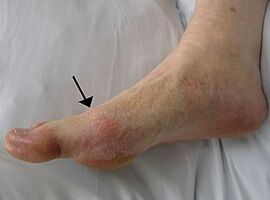
Gout is a form of crystal-induced arthoropathy, formed by monosodium urate monohydrate crystals. The most common joint affected is the metatarsal-phalangeal joint of the big toe.[8]
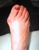
Hallux valgus[edit | edit source]
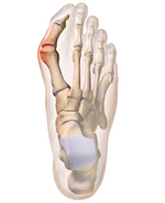
Hallux vagus, sometimes referred to as a bunion, refers to malformation of the metatarsalphalangeal (MTP) joint of the big toe.[9] The following is a more specific description of what is thought to be involved with hallux valgus:
- Medial deviation of the first metatarsal
- Lateral deviation, with or without rotation of the hallux
- Prominence with or without medial soft-tissue enlargement of the first metatarsal head.[10]
Etiologies for hallux valgus include biomechanical, traumatic, and metabolic causes, with biomechanical instability being the most common aetiology. Hallux valgus may also occur secondary to metabolic or inflammatory arthropathies such as rheumatoid arthritis, psoriatic arthritis and gouty arthritis. Connective tissue diseases including Ehlers-Danlos syndrome, Marfan syndrome, Down syndrome and generalized ligamentous laxity may also cause hallux valgus.[10]
References[edit | edit source]
- ↑ 1.0 1.1 1.2 1.3 Moore KL, Dalley AF. Clinically oriented anatomy. Wolters kluwer india Pvt Ltd; 2018 Jul 12.
- ↑ 2.0 2.1 Chummy SS. Last’s Anatomy: Regional and Applied. Churchill Livingstone.
- ↑ Abdalbary SA, Elshaarawy EA, Khalid BE. Tensile properties of the deep transverse metatarsal ligament in hallux valgus: a CONSORT-compliant article. Medicine. 2016 Feb;95(8).
- ↑ Babu D, Bordoni B. Anatomy, Bony Pelvis and Lower Limb, Medial Longitudinal Arch of the Foot. StatPearls [Internet]. 2020 Sep 1.
- ↑ 5.0 5.1 Gwani AS, Asari MA, Ismail ZM. How the three arches of the foot intercorrelate. Folia morphologica. 2017;76(4):682-8.
- ↑ 6.0 6.1 Besse JL. Metatarsalgia. Orthopaedics & Traumatology: Surgery & Research. 2017 Feb 1;103(1):S29-39.
- ↑ Cothran VE. Metatarsal Stress Fracture. Medscape 2019. https://emedicine.medscape.com/article/85746-overview (accessed May 29, 2021).
- ↑ Rothschild BM. Gout and Pseudogout [Internet]. Practice Essentials, Background, Pathophysiology. Medscape; 2021 [cited 2021May27]. Available from: https://emedicine.medscape.com/article/329958-overview
- ↑ Alzurahi A. Bunion Symptom Disorder: Surgical and Non-Surgical Treatment.
- ↑ 10.0 10.1 Frank CJ. Hallux Valgus [Internet]. Practice Essentials. Medscape; 2021 [cited 2021 May 27]. Available from: https://emedicine.medscape.com/article/1232902-overview
