Glenohumeral Joint: Difference between revisions
No edit summary |
(grammar corrections, PP links added, reference link added) |
||
| (21 intermediate revisions by 7 users not shown) | |||
| Line 8: | Line 8: | ||
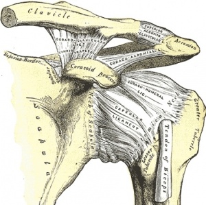
[[Image:Grays326.JPEG|thumb|right|Anterior view of the left shoulder and acromioclavicular joints, and proper scapular ligaments.]] | [[Image:Grays326.JPEG|thumb|right|Anterior view of the left shoulder and acromioclavicular joints, and proper scapular ligaments.]] | ||
The glenohumeral (GH) joint is a true synovial ball-and-socket style | The glenohumeral (GH) joint is a true synovial ball-and-socket style diarthrodial joint that is responsible for connecting the upper extremity to the trunk. It is one of four joints that comprise the [[Shoulder|shoulder]] complex. This joint is formed from the combination of the [[Humerus|humeral]] head and the glenoid fossa of the [[scapula]]. This joint is considered to be the most mobile and least stable joint in the body and is the most commonly dislocated diarthrodial joint <ref>.Christopher C. Dodson, Frank A. Cordasco, Anterior Glenohumeral Joint Dislocations, Orthopedic Clinics of North America,2008:39(4), 507-518. Available from:https://www.sciencedirect.com/science/article/abs/pii/S0030589808000461?via%3Dihub </ref>. | ||
== Motions Available == | == Motions Available == | ||
=== Abduction === | |||
Elevation of the humerus on the glenoid in the frontal (coronal) plane. | |||
=== Flexion === | |||
Forward and upward movement of the humerus on the glenoid in the sagittal plane. | |||
== | === Extension === | ||
Upward movement of the humerus on the glenoid in the sagittal plane towards the rear of the body. | |||
=== Internal rotation === | |||
rotation of the humerus on the glenoid in a medial direction. | |||
=== External rotation === | |||
Rotation of the humerus on the glenoid in a lateral direction. | |||
=== Scapular Plane Abduction === | |||
Elevation of the humerus on the glenoid in the scapular plane, which is midway between the coronal and sagittal planes. | |||
=== Horizontal Adduction === | |||
Movement of the humerus on the glenoid in a medial direction, usually accompanied with some degree of shoulder flexion. | |||
== Joint Capsule and Ligaments == | |||
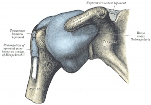
Together, the joint capsule and the [[Ligament|ligaments]] of the GH joint work to provide a passive restraint to keep the humeral head in contact with the glenoid fossa.<br>[[Image:Gray327.jpeg|thumb|right|Anterior view of the capsule of the right glenohumeral joint (distended).]] | |||
== | === Joint Capsule: === | ||
*The lateral attachment of the GH joint capsule attaches to the anatomical neck of the humerus. | |||
*The medial attachment of the joint capsule is the glenoid and the labrum. | |||
*According to some sources, the the overall strength of the capsule bears an inverse relationship to the patient's age; the older the patient, the weaker the joint capsule <ref name=":0">Dutton M. Dutton's Orthopaedic Examination Evaluation and Intervention. McGraw Hill Professional; 2012 Apr 13.</ref>. | |||
*With the arm in a resting position the inferior and anterior portions of the capsule are lax, while the superior portion is taut. | |||
*The anterior portion of the capsule is reinforced by the superior, middle, and inferior glenohumeral ligaments which form a Z-shaped pattern on the capsule. | |||
*The [[Muscle|muscles]] of the [[Rotator Cuff|rotator cuff]] act to reinforce the joint capsule superiorly, posteriorly, and anteriorly. | |||
*The joint capsule provides little support to the GH joint without the reinforcement of ligaments and the surrounding musculature <ref name=":1">Levangie PK, Norkin CC. Joint Structure and Function: A Comprehensive Analysis. FA Davis; 2011 Mar 9.</ref>.<br> | |||
=== Ligaments=== | |||
Superior glenohumeral ligament: <ref name=":0" /><ref name=":1" /> | |||
*Limits external rotation and inferior translation of the humeral head. | |||
*Arises from the glenoid and inserts on the anatomical neck of the humerus.<br> | |||
Middle glenohumeral ligament: <ref name=":0" /><ref name=":1" /> | |||
*Limits external rotation and anterior translation of the humeral head. | |||
*Arises from the glenoid and inserts on the anatomical neck of the humerus.<br> | |||
Inferior glenohumeral ligament: <ref name=":0" /><ref name=":1" /> | |||
*Limits external rotation and superior and anterior translation of the humeral head (anterior portion); | |||
*Limits internal rotation and anterior translation (posterior portion). | |||
*Arises from the glenoid and inserts on the humerus just beyond the lesser tuberosity.<br> | |||
Coracohumeral ligament: <ref name=":0" /><ref name=":1" /> | |||
*Split into anterior and posterior divisions by the biceps tendon. | |||
*Anterior portion limits extension while the posterior portion limits flexion. | |||
*Both divisions limit inferior and posterior translation of the humeral head. | |||
*Helps to support the weight of the resting arm against gravity. | |||
*Runs laterally from the coracoid process to the humerus, covering the superior glenohumeral ligament and blending with the superior joint capsule and supraspinatus tendon superiorly.<br> | |||
Transverse humeral ligament: <ref name=":0" /><ref name=":1" /> | |||
*This ligament serves to keep the tendon of the long head of the biceps in the bicipital groove. | |||
== Muscles == | |||
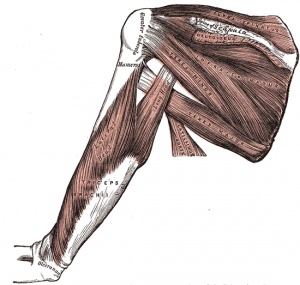
[[Image:Gray412.JPEG|thumb|right|Muscles on the dorsum of the scapula, and the Triceps brachii.]] | |||
*[http://www.rad.washington.edu/academics/academic-sections/msk/muscle-atlas/upper-body/deltoid Deltoid] ( | === Flexors === | ||
*[http://www.rad.washington.edu/academics/academic-sections/msk/muscle-atlas/upper-body/deltoid Deltoid] (Anterior Portion) | |||
=== Extensors === | |||
*[http://www.rad.washington.edu/academics/academic-sections/msk/muscle-atlas/upper-body/triceps-brachii Triceps Brachii] | |||
*[http://www.rad.washington.edu/academics/academic-sections/msk/muscle-atlas/upper-body/teres-major Teres Major] | |||
*[http://www.rad.washington.edu/academics/academic-sections/msk/muscle-atlas/upper-body/deltoid Deltoid] (Posterior Portion) | |||
*[http://www.rad.washington.edu/academics/academic-sections/msk/muscle-atlas/upper-body/latissimus-dorsi Latissimus Dorsi] | |||
=== Rotator Cuff === | |||
*[http://www.rad.washington.edu/academics/academic-sections/msk/muscle-atlas/upper-body/supraspinatus Supraspinatus] | *[http://www.rad.washington.edu/academics/academic-sections/msk/muscle-atlas/upper-body/supraspinatus Supraspinatus] | ||
*[http://www.rad.washington.edu/academics/academic-sections/msk/muscle-atlas/upper-body/infraspinatus Infraspinatus] | *[http://www.rad.washington.edu/academics/academic-sections/msk/muscle-atlas/upper-body/infraspinatus Infraspinatus] | ||
| Line 54: | Line 83: | ||
*[http://www.rad.washington.edu/academics/academic-sections/msk/muscle-atlas/upper-body/subscapularis Subscapularis] | *[http://www.rad.washington.edu/academics/academic-sections/msk/muscle-atlas/upper-body/subscapularis Subscapularis] | ||
=== Internal Rotators === | |||
*[http://www.rad.washington.edu/academics/academic-sections/msk/muscle-atlas/upper-body/subscapularis Subscapularis] | *[http://www.rad.washington.edu/academics/academic-sections/msk/muscle-atlas/upper-body/subscapularis Subscapularis] | ||
*[http://www.rad.washington.edu/academics/academic-sections/msk/muscle-atlas/upper-body/teres-major Teres Major] | *[http://www.rad.washington.edu/academics/academic-sections/msk/muscle-atlas/upper-body/teres-major Teres Major] | ||
*[http://www.rad.washington.edu/academics/academic-sections/msk/muscle-atlas/upper-body/latissimus-dorsi Latissimus Dorsi] | *[http://www.rad.washington.edu/academics/academic-sections/msk/muscle-atlas/upper-body/latissimus-dorsi Latissimus Dorsi] | ||
*[http://www.rad.washington.edu/academics/academic-sections/msk/muscle-atlas/upper-body/pectoralis-major Pectoralis Major] | *[http://www.rad.washington.edu/academics/academic-sections/msk/muscle-atlas/upper-body/pectoralis-major Pectoralis Major] | ||
=== External Rotators === | |||
*[http://www.rad.washington.edu/academics/academic-sections/msk/muscle-atlas/upper-body/teres-minor Teres Minor] | *[http://www.rad.washington.edu/academics/academic-sections/msk/muscle-atlas/upper-body/teres-minor Teres Minor] | ||
*[http://www.rad.washington.edu/academics/academic-sections/msk/muscle-atlas/upper-body/infraspinatus Infraspinatus] | *[http://www.rad.washington.edu/academics/academic-sections/msk/muscle-atlas/upper-body/infraspinatus Infraspinatus] | ||
=== Abductors === | |||
*[http://www.rad.washington.edu/academics/academic-sections/msk/muscle-atlas/upper-body/deltoid Deltoid] | |||
*[http://www.rad.washington.edu/academics/academic-sections/msk/muscle-atlas/upper-body/supraspinatus Supraspinatus] | |||
=== Adductors === | |||
*[http://www.rad.washington.edu/academics/academic-sections/msk/muscle-atlas/upper-body/ | *[http://www.rad.washington.edu/academics/academic-sections/msk/muscle-atlas/upper-body/pectoralis-major Pectoralis Major] | ||
== Video == | |||
{{#ev:youtube|https://youtu.be/eXlPBm38Wyg|width}}<ref>The Noted Anatomist. Glenohumeral joint: Structure and actions. Available from: https://youtu.be/eXlPBm38Wyg. [last accessed: 2020/05/31]</ref> | |||
== Closed Packed Position == | == Closed Packed Position == | ||
| Line 83: | Line 109: | ||
== Open Packed Position == | == Open Packed Position == | ||
The open packed position of the GH joint is around 50 degrees of abduction with slight horizontal adduction and external rotation. However, the point of maximal capsular laxity has been found to be 39 degrees of abduction in the scapular plane, which suggests that the open packed position may be close to neutral position of the shoulder.<ref> | The open packed position of the GH joint is around 50 degrees of abduction with slight horizontal adduction and external rotation. However, the point of maximal capsular laxity has been found to be 39 degrees of abduction in the scapular plane, which suggests that the open packed position may be close to neutral position of the shoulder.<ref>Hsu AT, Chang JH, Chang CH. [https://www.jospt.org/doi/abs/10.2519/jospt.2002.32.12.605 Determining the Resting Position of the Glenohumeral Joint: A Cadaver Study.] Journal of Orthopaedic & Sports Physical Therapy. 2002 Dec;32(12):605-12.</ref> | ||
== | ==Capsular Pattern== | ||
Capsular pattern of the GH joint is characterised by external rotation being the most limited, followed by abduction, internal rotation, and flexion. | |||
==Labrum== | |||
The labrum serves to deepen the glenoid fossa by around 50%, allowing for more contact area between the surface of glenoid and the humeral head. The increase in contact area also enhances joint stability.<ref name=":0" /> Common pathologies of the labrum include [[SLAP Lesion|SLAP]] lesions and [[Bankart lesion|Bankart lesions]].<br> | |||
== | ==Bursae== | ||
Multiple bursae are distributed throughout the [[shoulder]] complex, however, the subacromial bursa is one of the largest bursae in the body. The subacromial bursa is composed of the subdeltoid and subacromial bursa because they are often continuous. This bursa serves to allow the [[Rotator Cuff|rotator cuff]] to slide easily beneath the [[deltoid]] muscle. Common problems may include [[Shoulder Bursitis|shoulder bursitis]].<ref name=":0" /> | |||
== Arthrokinematics == | |||
* | The arthrokinematics below are described for the open kinematic chain since most functional tasks of the glenohumeral joint occur as a movement of the humerus on the glenoid. | ||
* Flexion | |||
** Pure spin of the humerus on glenoid (posterior spin when following greater tuberosity) | |||
* Extension | |||
** Pure spin of the humerus on glenoid (anterior spin when following greater tuberosity) | |||
* Abduction | |||
** Superior roll of the Humerus | |||
** Inferior glide of the humerus | |||
* Adduction | |||
** Inferior roll of the humerus | |||
** Superior glide of the humerus | |||
* Internal rotation | |||
** Anterior roll of the humerus | |||
** Posterior glide of the humerus | |||
* External rotation | |||
** Posterior roll of the humerus | |||
** Anterior glide of the humerus | |||
{{#ev:youtube|X4ozqdbAp-E}} | |||
== References == | == References == | ||
<references /><br> | <references /><br> | ||
[[Category:Anatomy]] [[Category:Shoulder]] [[Category:Joints]] [[Category: | [[Category:Anatomy]] | ||
[[Category:Shoulder]] | |||
[[Category:Joints]] | |||
[[Category:Shoulder - Anatomy]] | |||
[[Category:Shoulder - Anatomy]] | |||
[[Category:Shoulder - Joints]] | |||
[[Category:Musculoskeletal/Orthopaedics]] | |||
Latest revision as of 14:13, 26 July 2023
Original Editor - Tyler Shultz
Top Contributors - Tyler Shultz, Admin, Rachael Lowe, Kim Jackson, Redisha Jakibanjar, Naomi O'Reilly, Alexandra Kopelovich, 127.0.0.1, Evan Thomas, WikiSysop, Wendy Snyders and Shreya Pavaskar
Description[edit | edit source]
The glenohumeral (GH) joint is a true synovial ball-and-socket style diarthrodial joint that is responsible for connecting the upper extremity to the trunk. It is one of four joints that comprise the shoulder complex. This joint is formed from the combination of the humeral head and the glenoid fossa of the scapula. This joint is considered to be the most mobile and least stable joint in the body and is the most commonly dislocated diarthrodial joint [1].
Motions Available[edit | edit source]
Abduction[edit | edit source]
Elevation of the humerus on the glenoid in the frontal (coronal) plane.
Flexion[edit | edit source]
Forward and upward movement of the humerus on the glenoid in the sagittal plane.
Extension[edit | edit source]
Upward movement of the humerus on the glenoid in the sagittal plane towards the rear of the body.
Internal rotation[edit | edit source]
rotation of the humerus on the glenoid in a medial direction.
External rotation[edit | edit source]
Rotation of the humerus on the glenoid in a lateral direction.
Scapular Plane Abduction[edit | edit source]
Elevation of the humerus on the glenoid in the scapular plane, which is midway between the coronal and sagittal planes.
Horizontal Adduction[edit | edit source]
Movement of the humerus on the glenoid in a medial direction, usually accompanied with some degree of shoulder flexion.
Joint Capsule and Ligaments[edit | edit source]
Together, the joint capsule and the ligaments of the GH joint work to provide a passive restraint to keep the humeral head in contact with the glenoid fossa.
Joint Capsule:[edit | edit source]
- The lateral attachment of the GH joint capsule attaches to the anatomical neck of the humerus.
- The medial attachment of the joint capsule is the glenoid and the labrum.
- According to some sources, the the overall strength of the capsule bears an inverse relationship to the patient's age; the older the patient, the weaker the joint capsule [2].
- With the arm in a resting position the inferior and anterior portions of the capsule are lax, while the superior portion is taut.
- The anterior portion of the capsule is reinforced by the superior, middle, and inferior glenohumeral ligaments which form a Z-shaped pattern on the capsule.
- The muscles of the rotator cuff act to reinforce the joint capsule superiorly, posteriorly, and anteriorly.
- The joint capsule provides little support to the GH joint without the reinforcement of ligaments and the surrounding musculature [3].
Ligaments[edit | edit source]
Superior glenohumeral ligament: [2][3]
- Limits external rotation and inferior translation of the humeral head.
- Arises from the glenoid and inserts on the anatomical neck of the humerus.
Middle glenohumeral ligament: [2][3]
- Limits external rotation and anterior translation of the humeral head.
- Arises from the glenoid and inserts on the anatomical neck of the humerus.
Inferior glenohumeral ligament: [2][3]
- Limits external rotation and superior and anterior translation of the humeral head (anterior portion);
- Limits internal rotation and anterior translation (posterior portion).
- Arises from the glenoid and inserts on the humerus just beyond the lesser tuberosity.
Coracohumeral ligament: [2][3]
- Split into anterior and posterior divisions by the biceps tendon.
- Anterior portion limits extension while the posterior portion limits flexion.
- Both divisions limit inferior and posterior translation of the humeral head.
- Helps to support the weight of the resting arm against gravity.
- Runs laterally from the coracoid process to the humerus, covering the superior glenohumeral ligament and blending with the superior joint capsule and supraspinatus tendon superiorly.
Transverse humeral ligament: [2][3]
- This ligament serves to keep the tendon of the long head of the biceps in the bicipital groove.
Muscles[edit | edit source]
Flexors[edit | edit source]
- Deltoid (Anterior Portion)
Extensors[edit | edit source]
- Triceps Brachii
- Teres Major
- Deltoid (Posterior Portion)
- Latissimus Dorsi
Rotator Cuff[edit | edit source]
Internal Rotators[edit | edit source]
External Rotators[edit | edit source]
Abductors[edit | edit source]
Adductors[edit | edit source]
Video[edit | edit source]
Closed Packed Position[edit | edit source]
The closed packed position of the GH joint is abduction and external rotation.
Open Packed Position[edit | edit source]
The open packed position of the GH joint is around 50 degrees of abduction with slight horizontal adduction and external rotation. However, the point of maximal capsular laxity has been found to be 39 degrees of abduction in the scapular plane, which suggests that the open packed position may be close to neutral position of the shoulder.[5]
Capsular Pattern[edit | edit source]
Capsular pattern of the GH joint is characterised by external rotation being the most limited, followed by abduction, internal rotation, and flexion.
Labrum[edit | edit source]
The labrum serves to deepen the glenoid fossa by around 50%, allowing for more contact area between the surface of glenoid and the humeral head. The increase in contact area also enhances joint stability.[2] Common pathologies of the labrum include SLAP lesions and Bankart lesions.
Bursae[edit | edit source]
Multiple bursae are distributed throughout the shoulder complex, however, the subacromial bursa is one of the largest bursae in the body. The subacromial bursa is composed of the subdeltoid and subacromial bursa because they are often continuous. This bursa serves to allow the rotator cuff to slide easily beneath the deltoid muscle. Common problems may include shoulder bursitis.[2]
Arthrokinematics[edit | edit source]
The arthrokinematics below are described for the open kinematic chain since most functional tasks of the glenohumeral joint occur as a movement of the humerus on the glenoid.
- Flexion
- Pure spin of the humerus on glenoid (posterior spin when following greater tuberosity)
- Extension
- Pure spin of the humerus on glenoid (anterior spin when following greater tuberosity)
- Abduction
- Superior roll of the Humerus
- Inferior glide of the humerus
- Adduction
- Inferior roll of the humerus
- Superior glide of the humerus
- Internal rotation
- Anterior roll of the humerus
- Posterior glide of the humerus
- External rotation
- Posterior roll of the humerus
- Anterior glide of the humerus
References[edit | edit source]
- ↑ .Christopher C. Dodson, Frank A. Cordasco, Anterior Glenohumeral Joint Dislocations, Orthopedic Clinics of North America,2008:39(4), 507-518. Available from:https://www.sciencedirect.com/science/article/abs/pii/S0030589808000461?via%3Dihub
- ↑ 2.0 2.1 2.2 2.3 2.4 2.5 2.6 2.7 Dutton M. Dutton's Orthopaedic Examination Evaluation and Intervention. McGraw Hill Professional; 2012 Apr 13.
- ↑ 3.0 3.1 3.2 3.3 3.4 3.5 Levangie PK, Norkin CC. Joint Structure and Function: A Comprehensive Analysis. FA Davis; 2011 Mar 9.
- ↑ The Noted Anatomist. Glenohumeral joint: Structure and actions. Available from: https://youtu.be/eXlPBm38Wyg. [last accessed: 2020/05/31]
- ↑ Hsu AT, Chang JH, Chang CH. Determining the Resting Position of the Glenohumeral Joint: A Cadaver Study. Journal of Orthopaedic & Sports Physical Therapy. 2002 Dec;32(12):605-12.


