Foot and Ankle Structure and Function: Difference between revisions
No edit summary |
No edit summary |
||
| Line 23: | Line 23: | ||
#Talonavicular (TN) JointIt is formed between anterior talar head and concavity on navicular bone. It does not have its own capsule and is the same one as of two anterior talocalcaneal articulations. | #Talonavicular (TN) JointIt is formed between anterior talar head and concavity on navicular bone. It does not have its own capsule and is the same one as of two anterior talocalcaneal articulations. | ||
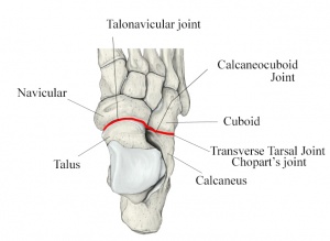
#[[Image:Transverse-tarsal-joint.jpg|right| | #[[Image:Transverse-tarsal-joint.jpg|right|300x350px]]Calcaneocuboid (CC) Joint – It is formed between anterior facet of calcaneus and posterior cuboid. Both articulating surface present convex and concave surface, with joint being convex vertically and concave transversely. Very little movement occur between joint segments. | ||
====== Tarsometatarsal (TMT) Joint complex ====== | ====== Tarsometatarsal (TMT) Joint complex ====== | ||
Revision as of 04:35, 1 May 2016
Original Editor - Vinit Kothekar.
Top Contributors - Vinit Kothekar, Wanda van Niekerk, Kim Jackson, Admin, Evan Thomas, Lucinda hampton, Chelsea Mclene, Rachael Lowe, Candace Goh, Simisola Ajeyalemi, Jess Bell, Khloud Shreif, 127.0.0.1, Ewa Jaraczewska, Cath Young, Priyanka Chugh, WikiSysop and Rucha Gadgil
Anatomy[edit | edit source]

Foot & ankle form a complex system which consists of 26 bones, 33 joints and more than 100 muscles, tendons and ligaments. It functions as a rigid structure for weight bearing and it can also function as a flexible structure to conform the uneven terrain. Foot and ankle provide various important functions which may include: support of body’s weight, providing balance, acts as a shock absorption, is the first contact to ground and transfer point for ground reaction force, compensates for proximal malalignment and may also substitute hand function in individuals with upper extremity amputations and paralysisCite error: Invalid <ref> tag; name cannot be a simple integer. Use a descriptive title. Foot is subdivided into rearfoot, midfoot and forefoot.
Talocrural (TC) joint[edit | edit source]
It is commonly known as ankle joint. Talocrural joint is formed between Tibia-fibula and Talus. Distal and inferior aspect of tibia – known as plafond – is connected to fibula via tibiofibular ligaments forming a strong mortise which articulates with the talar dome distally. Type of joint – hinge joint. Movements allowed – in sagittal plane, dorsiflexion and plantarflexion.
Subtalar (ST) joint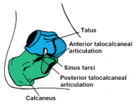 [edit | edit source]

It is also known as talocalcaneal joint and is formed between talus and calcaneus. Three facets of talus - anterior, middle and posterior facets, articulates inferiorly with calcaneus.
Midtarsal (MT) joint[edit | edit source]
Also known as transverse tarsal joints or Chopart’s joint. It is S-shaped joint when viewed from above, and it consists of two joints – Talonavicular joint and Calcaneocuboid joint.
- Talonavicular (TN) JointIt is formed between anterior talar head and concavity on navicular bone. It does not have its own capsule and is the same one as of two anterior talocalcaneal articulations.
- Calcaneocuboid (CC) Joint – It is formed between anterior facet of calcaneus and posterior cuboid. Both articulating surface present convex and concave surface, with joint being convex vertically and concave transversely. Very little movement occur between joint segments.

Tarsometatarsal (TMT) Joint complex[edit | edit source]
Also known as Lisfranc’s joint. The distal tarsal rows including three cuneiform bones and cuboid articulate with base of metatarsals to form TMT complex. It is S-shaped joint and divided into 3 distinct columns11.
Medial – composed of 1st Metatarsal and medial Cuneiform.
Middle – composed of 2nd and 3rd Metatarsal and intermediate and lateral Cuneiform respectively.
Lateral – composed of 4th and 5th Metatarsal and Cuboid. It also divides the midfoot from forefoot.
Metatarsophalangeal (MTP) joints and Interphalangeal (IP) joints[edit | edit source]
MTP Joints are formed between metatarsal heads and corresponding base of proximal phalanx. Interphalangeal joints of the toes are formed between the phalanges of the toes. Each toe has proximal and distal IP joints except for the great toe which only has one IP joint.

