Dysphagia: Difference between revisions
mNo edit summary |
mNo edit summary |
||
| Line 130: | Line 130: | ||
* Instruction: ‘‘lie in the supine position; complete 3 head lifts sustained for 1 min each; 1 min rest period between each head lift; then complete 30 consecutive head lifts holding for 2 s each’’ The suggested frequency is three times each day for 6 consecutive weeks. | * Instruction: ‘‘lie in the supine position; complete 3 head lifts sustained for 1 min each; 1 min rest period between each head lift; then complete 30 consecutive head lifts holding for 2 s each’’ The suggested frequency is three times each day for 6 consecutive weeks. | ||
* Physiological benefits:It increases anterior hyolaryngeal excursion, UES opening, strengthens suprahyoid muscles, and enhances thyrohyoid shortening.<ref name=":4" /> | * Physiological benefits:It increases anterior hyolaryngeal excursion, UES opening, strengthens suprahyoid muscles, and enhances thyrohyoid shortening.<ref name=":4" /> | ||
=== Masako === | |||
This manoeuvre involves swallowing while protruding the tongue beyond the lips and holding it between one’s teeth. It is intended to target the base of tongue and pharyngeal walls at that level.<ref name=":3" /> | |||
=== Neuromuscular Electrical Stimulation (NMES) === | === Neuromuscular Electrical Stimulation (NMES) === | ||
Revision as of 07:21, 20 April 2020
Introduction[edit | edit source]
Dysphagia is a difficulty in swallowing liquid or solid food due to disruption in swallowing mechanism from mouth to pharynx.[1] Dysphagia leads to severe complications [1][2]:
- Aspiration pneumonia
- Dehydration
- Malnutrition
- Can lead to death because of choking
Physiology of swallowing[edit | edit source]
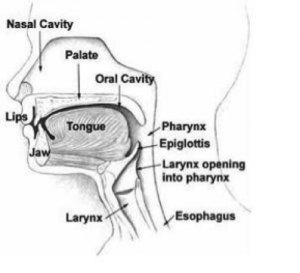
Having a thorough knowledge of anatomy and physiology of swallowing and eating is essential while evaluating and treating disorders of eating and swallowing,[3]There are four stages while describing the physiology of swallowing :
- Oral preparatory stage
- Oral propulsive stage
- Pharyngeal stage : Main feature of this stage are food passage, propelling the food bolus through the pharynx and UES to the esophagus; and airway protection, insulating the larynx and trachea from the pharynx during food passage to prevent the food from entering the airway.
- Esophageal stage
Eating, swallowing and breathing are tightly coordinated during the normal process. Swallowing is dominant to respiration in normal individuals.Breathing ceases briefly during swallowing due to physical closure of the airway by elevation of the soft palate and tilting of the epiglottis and also of neural suppression of respiration in the brainstem. [3]
When drinking a liquid bolus, swallowing usually starts during the expiratory phase of breathing. The respiratory pause continues for 0.5 to 1.5 s during swallowing, and respiration usually resumes with expiration. This resumption is regarded as one of the mechanisms that prevents inhalation of food remaining in the pharynx after swallowing. When performing sequential swallows while drinking from a cup, respiration can resume with inspiration. [3]
Eating solid food also alters the respiratory rhythm. The rhythm is perturbed with onset of mastication. Respiratory cycle duration decreases during mastication, but with swallowing. The “exhale – swallow – exhale” temporal relationship persists during eating. However, respiratory pauses are longer, often beginning substantially before swallow onset.[3]
Causes that affect the normal swallowing physiology[edit | edit source]
There are various causes for alteration in normal swallowing physiology. Broadly it can be categories into two heading :
- Structural abnormalities
- Functional abnormalities
Structural abnormalities[edit | edit source]
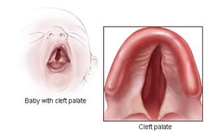
It can be acquired or congenital. Cleft palate, cervical osteophytes, webs or strictures in the passage are some of the examples of the structural abnormalities. The abnormalities might affect in any stage of the swallowing and alter the normal physiology. [3]
Functional abnormalities[edit | edit source]
Impairments affecting the jaw, lips, tongue, or cheek can hamper the oral phase or food processing. Reduced closing pressure of the lips may lead to drooling. In weakness of the buccal or labial muscles, food can be trapped in the buccal or labial sulci (between the lower teeth and the cheeks or gums, respectively). Tongue dysfunction produces impaired mastication and bolus formation, and bolus transport. These usually result from tongue weakness or in-coordination, but sensory impairments can produce similar effects including excessive retention of food in the oral cavity after eating and swallowing.
Loss of teeth reduces masticatory performance. Chewing can be prolonged by missing teeth, and particle size of the triturated bolus becomes larger due to lower efficiency of mastication.
Dysfunction of the pharynx can produce impaired swallow initiation, ineffective bolus propulsion, and retention of a portion of the bolus in the pharynx after swallowing. Insufficient velopharyngeal closure may result in nasal regurgitation and reduce pharyngeal pressure in swallow, hampering transport through the Upper Esophageal Sphincter (UES).
Impaired opening of the UES can cause partial or even total obstruction of the food-way with retention in the piriform sinuses and hypo-pharynx, increasing risk of aspiration after the swallow. Insufficient UES opening can be caused by increased stiffness of the UES, as in fibrosis or inflammation, or failure to relax the sphincter musculature.
Esophageal dysfunction is common and is often asymptomatic. Esophageal motor disorders include conditions of either hyperactivity (e.g., esophageal spasm), hypo-activity (e.g.weakness), or in-coordination of the esophageal musculature.Either of these can lead to ineffective peristalsis with retention of material in the esophagus after swallowing. Retention can result in regurgitation of material from the esophagus back into the pharynx, with risk of aspirating the regurgitated material. Esophageal motor disorders are sometimes provoked by gastroesophageal reflux disease, and in some cases, can respond to treatment with proton pump inhibitors[3].
Diagnosing dysphagia[edit | edit source]
There are many bedside and instrumental tools available for the diagnosis and treatment of dysphagia. Dysphagia evaluation tools can be grouped broadly as
- Imaging (Ultrasound, Videofluroscopy, Fiberoptic endoscopic evaluation of swallowing, and Fiberoptic endoscopic evaluation of swallowing with sensory testing)
- Non imaging(beside assessment tools, and pharyngeal manometry).[2]
Rehabilitation of dysphagia[edit | edit source]
Rehabilitative exercises changes and improves the swallowing physiology in force, speed or timing, with the goal being to produce a long-term effect, as compared to compensatory interventions used for a short-term effect. Rehabilitative exercises also involve retraining the neuromuscular systems to bring about neuroplasticity, since pushing any muscular system in an intense and persistent way will bring about changes in neural innervation and patterns of movement.[4] Rehabilitation exercise can be broadly divided into :
- Swallowing exercises
- Non-swallowing exercises
Swallowing Exercises[edit | edit source]
Swallowing exercises often are used to treat dysphagia with the goal of altering swallowing physiology and promoting long-term changes. Exercises are expected to impact swallowing mechanics and impact bolus flow.[5] Effortful swallow, Mendelsohn, super-supraglottic, Masako are some of the swallowing exercises.Swallowing exercises follow many of the neuroplasticity principles listed below[4]:
- Use it or loose it
- Use it and improve it
- Specificity
- Transference
- Intensity
Non-Swallowing Exercises[edit | edit source]
Non-swallowing exercises are those that do not involve the act of swallowing, for example tongue strengthening exercises.Non-swallowing exercises can be
done by patients who cannot eat orally (are tube fed) or those post-surgery who are temporarily restricted from eating orally. Shaker head lift, tongue strengthening, Lee Silverman voice treatment, expiratory muscle strength training are some of the non-swallowing exercises. Non-swallowing exercises follow few neuroplasticity principles and they are[4]:
- Transference
- Intensity
Therapeutic techniques for dysphagia management[edit | edit source]
Therapeutic techniques can be divided into those used as :
- Compensatory strategies (Head Rotation (Head Turn), Chin Tuck (Head Flexion). Head Tilt and Bolus Viscosity, Texture, and Volume Modifications)
- Exercises (Tongue Hold, Shaker Exercise)
- Those used as both compensatory strategies and/or exercises (Supraglottic Swallow, Super-Supraglottic Swallow, Effortful Swallow, Mendelsohn Maneuver)
- Alternate methods (Neuromuscular Electrical Stimulation (NMES), Oral Stimulation and Other Interventions )
Head Rotation (head turn)[edit | edit source]
Head rotation is a compensatory strategy used for patients with unilateral pharyngeal and/or laryngeal weakness as well as reduced UES opening.Head rotation toward the side of impairment effectively redirects the bolus to the side of the pharynx opposite the rotation (the stronger side).[5]
- Instruction given:‘‘turn your head to the side as if you are looking over your shoulder.’’
- Physiological benefits: It redirects the bolus to the side of the pharynx opposite the rotation (the stronger side). It drops UES pressure on the side opposite to the head turn thus allowing for increased extension and duration of UES opening.
Chin Tuck (Head Flexion)[edit | edit source]
The chin tuck (head flexion) has been a technique used for patients who have decreased airway protection associated with delayed swallow initiation and/or reduced tongue base retraction.[5]
- Instruction: ‘‘bring your chin to their chest and maintain this posture throughout the duration of the swallow".
- Physiological benefits: It leads to expansion of vallecular recesses, approximation of tongue base toward pharyngeal wall, narrowing entrance to the laryngeal vestibule, reduction in distance between hyoid and larynx, and increased duration of swallowing apnea during the swallow.
Head tilt[edit | edit source]
The head tilt is used for patients with unilateral oral weakness. [5]
- Instruction: ‘‘tilt your head like you’re trying to touch your ear to your shoulder.’’
- Physiological benefits: it directs the bolus to the stronger side of the oral cavity
Bolus Viscosity, Texture, and Volume Modifications[edit | edit source]
- Increasing the volume and/or viscosity for liquids is another technique used to reduce dysphagia symptoms for some patients. Thickening liquids may be used for patients who have poor oral control of thin liquids and/or demonstrate reduced airway protection.
- Increasing bolus volume increases bolus transit time as exemplified by sustained laryngeal elevation and hyoid excursion.
- Some patients may benefit from texture-modified foods.[5]
Supraglottic swallow[edit | edit source]
The supraglottic swallow is used for patients who demonstrate reduced airway protection during the swallow.
- Instruction: ‘‘First, inhale deep then hold your breath, continue to hold your breath and swallow immediately after you swallow (before you inhale), cough then immediately swallow again’’.
- The physiologic benefits of this strategy: increased airway closure by increasing arytenoid approximation and true vocal fold closure as well is increasing UES opening during the swallow. The airway is protected earlier in the swallow and hyolaryngeal excursion is prolonged which may be beneficial for patients with delayed swallow initiation.
Super-supraglottic swallow[edit | edit source]
Super-supraglottic swallow is also used for patients with reduced airway closure as for supraglottic swallow ; however, the difference with the super-supraglottic is patients are instructed to implement an effortful breath hold,
- Instruction: ‘‘take a breath and hold it tightly while bearing down; continue to hold your breath and bear down as you swallow; immediately after your swallow (before you inhale) cough then immediately swallow hard again (before you inhale).’’
- Physiological benefit: With this technique, the patient has earlier tongue base movement, higher hyoid position at swallow onset, increased hyoid movement as well as longer bolus transit time, tongue base and pharyngeal wall contact, and airway closure.
- Note: Both the supraglottic swallow and the supra-supraglottic swallow maneuvers may result in Valsalva and can result to arrhythmia in stroke patients during treatment sessions.Hence, clinicians should be mindful of using these maneuvers in stroke patients especially in those with coexisting heart disease.[5]
Effortful Swallow[edit | edit source]
The effortful swallow is used for patients who present with clinically significant residue in the valleculae and/or pyriform sinuses as well as for patients who may have decreased airway closure.
- Instructions:‘‘squeeze your throat muscles as hard as you can while swallowing’’.
- Physiological benefits: It increases hyolaryngeal excursion, duration of hyoid elevation and UES opening, laryngeal closure, lingual pressures, peristaltic amplitudes in the distal esophagus, and pressure and duration of tongue base retraction[5]
Mendelsohn maneuver[edit | edit source]
This technique is used for patients with decreased hyolaryngeal excursion and/or decreased duration of UES opening.
Prior to instructions, it is suggested that patients should first feel laryngeal elevation by palpation of thyroid cartilage during swallows.
- Instruction: ‘‘Swallow and when you feel your thyroid cartilage elevate hold it there for several seconds before finishing the swallow’’.
- Physiologic benefits: This technique increases extent and duration of hyolaryngeal excursion, UES opening, pharyngeal peak contractions, bolus transit time and duration, and pressure of tongue base contact.[5]
Tongue Hold[edit | edit source]
The tongue hold is used for reduced tongue base, and pharyngeal wall contact.
- Instruction: "hold the anterior tongue (slightly posterior to the tongue tip) between the teeth while swallowing.’’
- Physiological benefits:It increases anterior bulging of the posterior pharyngeal wall.[5]
Shaker Exercise[edit | edit source]
The Shaker Exercise is used for patients who have decreased UES opening and weakness of the suprahyoid muscles.
- Instruction: ‘‘lie in the supine position; complete 3 head lifts sustained for 1 min each; 1 min rest period between each head lift; then complete 30 consecutive head lifts holding for 2 s each’’ The suggested frequency is three times each day for 6 consecutive weeks.
- Physiological benefits:It increases anterior hyolaryngeal excursion, UES opening, strengthens suprahyoid muscles, and enhances thyrohyoid shortening.[5]
Masako[edit | edit source]
This manoeuvre involves swallowing while protruding the tongue beyond the lips and holding it between one’s teeth. It is intended to target the base of tongue and pharyngeal walls at that level.[4]
Neuromuscular Electrical Stimulation (NMES)[edit | edit source]
Neuromuscular electrical stimulation (NMES) is a treatment where electrodes are placed on the anterior neck and an electrical current evokes a muscle contraction. NMES treatment is typically used as an adjunct modality concurrently while the patient swallows and/or performs a traditional exercise. [5]
Oral Stimulation and Other Interventions[edit | edit source]
- Oral exercises to include tactile-thermal stimulation, lingual, and labial strengthening are used as a treatment modality for stroke patients with dysphagia.
- Sensory stimulation is assumed to increase corticobular excitability which has been associated with swallowing recovery after stroke,
- Massage with an ice stick is applied to the throat, base of the anterior faucial arches, base of tongue, and the posterior pharyngeal wall for 10 s with rubbing and light compression.Ice massage shortened the latency for triggering the swallow after the command and the massage in itself had an immediate effect on triggering a swallow response even in patients who could not swallow voluntarily. The effectiveness of ice massage was found to be more significant in subjects with supra-nuclear lesions than in those with nuclear lesions. Ice massage could activate a damaged supranuclear tract of swallowing and/or a normal nuclear and sub-nuclear tract.
- Lip muscle training has also shown good effect in stroke patients with dysphagia.
- lingual exercise following I-PRO (isometric progressive resistance oropharyngeal) has also shown good result in stroke patients with swallowing difficulty.
- Transcranial magnetic stimulation (TMS) as well as transcranial direct current stimulation (tDCS) are also used for dysphasia management.[5]
References[edit | edit source]
- ↑ 1.0 1.1 Balamurali K, Sekar D, Thangaraj M, Kumar MA. Dysphagia in Patients with Stroke: A Prospective Study. available from:https://www.ijcmsr.com/uploads/1/0/2/7/102704056/ijcmsr_96.pdf
- ↑ 2.0 2.1 González-Fernández M, Ottenstein L, Atanelov L, Christian AB. Dysphagia after stroke: an overview. Current physical medicine and rehabilitation reports. 2013 Sep 1;1(3):187-96.
- ↑ 3.0 3.1 3.2 3.3 3.4 3.5 Matsuo K, Palmer JB. Anatomy and physiology of feeding and swallowing: normal and abnormal. Physical medicine and rehabilitation clinics of North America. 2008 Nov 1;19(4):691-707.
- ↑ 4.0 4.1 4.2 4.3 Langmore SE, Pisegna JM. Efficacy of exercises to rehabilitate dysphagia: a critique of the literature. International Journal of Speech-Language Pathology. 2015 May 4;17(3):222-9.
- ↑ 5.00 5.01 5.02 5.03 5.04 5.05 5.06 5.07 5.08 5.09 5.10 5.11 Vose A, Nonnenmacher J, Singer ML, González-Fernández M. Dysphagia management in acute and sub-acute stroke. Current physical medicine and rehabilitation reports. 2014 Dec 1;2(4):197-206.


