Differentiating Buttock Pain - Gluteal Tendinopathy
Original Editors - Mariam Hashem
Top Contributors - Mariam Hashem, Kim Jackson, Tarina van der Stockt, Lucinda hampton and Ewa Jaraczewska
This article or area is currently under construction and may only be partially complete. Please come back soon to see the finished work! (14/10/2020)
Introduction[edit | edit source]
Deep Gluteal pain is a complex condition that could be the manifestation of different soft tissue pathologies. Due to the complexity of the anatomy the symptoms are overlapping. Scans such as MRI and ultrasound don't rule out the irritating source of pain and the special tests used in physical examination often don't discriminate the underlying structures that contribute to the symptoms[1].
Gluteal Tendinopathy is a potential source of deep gluteal pain. The condition has been introduced as a concept very recently in the research compared to other structures that can cause gluteal pain such as Sacroiliac Joint and the Lumbar region.
Gluteal Tendinopathy[edit | edit source]
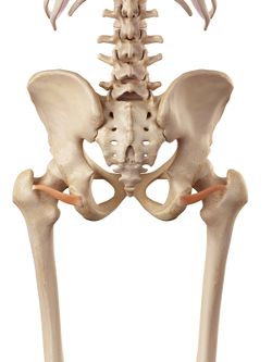
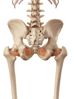
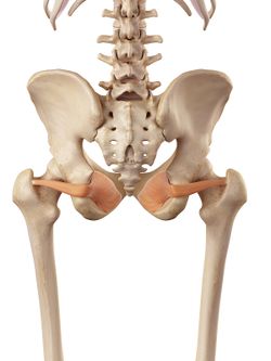
Previously known as Greater Trochanteric Pain Syndrome is a pain that starts in the greater trochanter region and may radiate to the lateral thigh and/or leg. Trochanteric Pain primarily is caused by the gluteal tendons and a secondary cause of this pain is the bursal inflammation that used to be thought as the main source of pain. Other structures that could be involved int he pathology are the posterior hip capsule, Gemelli's and the Obturators[1].
| Inferior Gemellus | Obturator Internus | Obturator Externus |
|---|
This condition has significant impacts on sleep quality, physical activity, work participation and the quality of life similar to patients waiting for hip replacement for severe hip osteoarthritis.[2], The pain of Gluteal Tendon origin can refer to the sacroiliac region, the buttock, the groin and into the anterior thigh. This overlap of referral pattern doesn't help in differentiating other pathologies.
Gluteal Tendinopathy is more relevant in females over 40 years old and believed to be present in 23.5% of women who are at a risk of knee osteoarthritis[3] . A percentage of 73 of the patients are believed to be either menopausal or peri-menopausal indicating a link between hormonal changes and tendinopathy. Certain medications are also shown to influence the tendon structural changes such as quinolone antibiotics, oestrogen inhibitors such as Tamoxifen for patients who had breast cancer[1].
Other factors that were found to affect the presence and prognosis of Tendinopathy are:
- Smoking
- Diabetes
- Steriods
- Changes in load either underload or overload.
Factors such as greater psychological stress, poorer quality of life, greater waist girth and a higher BMI were relevant in sever cases. However, these factors might also develop as a result of pain[4].
Risk factors are[5]:
- Female gender
- Older age
- Higher BMI
- Back pain
- Lower femoral neck angle
Diagnosis[edit | edit source]
There are important measures in diagnosing Gluteal Tendinopathy[1]:
- Patient's history such as gender, BMI, age and history of loading
- Pain location: lateral hip pain.
- Pain severity: usually 4/10 on most days
- Palpation: tenderness over the superior aspect of the greater trochanter (the insertional point of glute medius and glute minimus)
- FADER or FADER resisted tests ( flexion adduction external rotation position). Pain over the lateral hip region is a positive test and could be diagnostic of gluteal tendinopathy. If the pain was reported in the deep gluteal region it could indicate deep gluteal pain syndrome. When the leg is flexion> 60 degrees, adduction, external rotation, the Piriformis becomes an internal rotator. Also, glute med and glute max swap roles from being an external rotator to being an internal rotator.
- A common sign is a reported difficulty in walking after sitting for a period of time often described as'' hobbling''.
To rule out osteoarthritis of the hip, the subjective history of the patient and FADIR ( flexion adduction internal rotation test) are used. A positive FADIR test might not rule out OA completely but a negative test is recognised to rule out intra-articular hip pathologies such as OA, Femoral Acetabular Impingement or Labral tear. Also, in hip Osteoarthritis hip flexion ROM tends to be reduced.
Hip External Rotator; Gemelli's, Obturators and Quadratus femori have been included in Lateral hip pain pathology[6]. They are positioned at 90 degrees to the long axis of the hip and serve important functions[1]:
- Compress the hip joint and to provide stability at the joint.
- Reinforce the posterior capsule of the hip
Management of Gluteal Tendinopathy[edit | edit source]
Patient education is considered the most important part of management[1]. Education includes the patient in the management and puts them in control with their treatment plan.
Managing associated or provoking factors such as hormonal changes, co-morbidities, sleep, smoking and medications should be explained and discussed with the patient. Although some of these factors might not be modifiable however patients should be aware of them[7].
Promotion of healthy lifestyle changes such as quitting smoking and weight loss is integral in the management of Tendinopathy[8].
Load management is used in the management of tendinopathy by gently increasing the load on the tendon and managing the ''weekend-warrior exercise'' or load and no-load scenario.
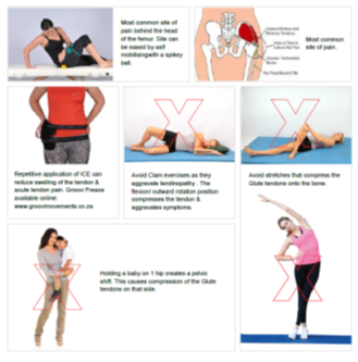
The Gluteal tendon is believed to be under a significant compression load of the hip flexors and internal rotators and this needs to be addressed by avoiding further compression. Stretching, especially in the early phases, of the Tensor Fascia Lata or Piriformis should be avoided to prevent the compression onto the insertional area of the Gluteal tendons.
Advise the patient to sleep with a firm pillow between their knees.
They should not sit with their legs crossed and they should avoid hanging on that hip if they standing.
So, for example, if you have a baby on one hip, you want to avoid that type of compressive and tensile loading onto those tendons.
Posture and functional loading mechanics. Dynamic valgus (hip adduction and internal rotation) during loading activities as it increases compression and tensile loading on the damaged tendon.
The Visa G questionnaire was validated by Fearon et al [5] as an outcome measure for disability resulting from lateral hip pain. The questionnaire looks at activities such as lying on the affected side, stairs negotiation and the overall severity of the hip pain.
Selection of Exercises[edit | edit source]
Compression forces can aggravate the lateral hip pain. An exercise such as Clam can provoke the tendon pain due to the high compressive force. Similarly, sitting with crossed legs and sleeping on the affected side place the gluteal tendon under a high compression force of Tensor Facia lata.
An important finding is that hip abductor strength wasn't associated with the severity of the tendinopathy[4].
Progressive exercises that increase the load starting with isometric exercises. Ebonie Rio has shown that isometrics can be very hypoalgesic and can be used in season. So that might be a way to start to try and help the patients self-manage their pain and presentation.
Progressing from isometric to bridging exercises.
The clam is not recommended, instead, a modified clam exercise which effectively allows for the top shin to be lifted off the bottom shin to get a pure abduction motion without bringing in the external rotation component, which will increase the compressive load on the insertional painful tendon.
Other exercises such as monster walks and Sumo walks can be used in this stage. Slow, controlled abduction, external rotation with a loop around both hips.
Some studies have been done on split squats with the weight in the contralateral hand has shown to be very effective in isolating and strengthening the glute med tendon and muscle.
Loading should be monitored. Pain can be used to monitor the load on the tendon. A four out of ten pain during the exercise and no worse over 24 hours.
Abductor slides using slidos, to get more of an abduction component.
Non-discogenic Sciatica Pain
the deep gluteal space, a highly nociceptive area, that is superiorly bounded by the greater sciatic notch, inferiorly by the ischial tuberosity, laterally by the linear aspersa and the greater trochanter and medially by the sacrotuberous ligament.
The sciatic nerve can get entrapped in this area.
The hamstring origin tendinopathy, ischiofemoral impingement.
People with GT are shown to have excessive hip adduction.
Adduction or movement of the leg across the midline should be avoided. This can be achieved by avoiding[1]:
- Stretches should be avoided especially Tensor Fascia Lata muscle to avoid the compression/pressure on the Gluteus medieus and minimus tendons.
- Crossing the legs especially with the affected leg on top of the non-affected
- Standing or hanging on one leg
- Lying on the affected side or non-affected without a firm pillow in between the legs.
Rio et al[9] recommended the use of external motor control cues such as visual (mirror, video and pressure feedback) auditory (exercise metronome) and mental (mental rehearsing of the task) to stimulate neuroplastic changes.
Advising patients to perform the exercises slowly to stimulate the deep stability system of the muscle. You can use cues such as 2 seconds out, 2 seconds hold and 2 seconds return for a rate of 6 seconds per repetition.
The intensity of isometric exercises is recommended to be at 4/10 intensity and symptoms shouldn't last more than 24 hours to monitor the load. No pain should be allowed during functional tasks such as lunges, step-ups as it reflects the poor control of optimal alignment and increased compression on the Gluteal Tendon.
Hip flexion should be avoided as it can negatively affect motor control and optimal muscle activation when TFL and superficial hip flexors dominate over hip abductors and external rotators.
Posture such as prolonged seated that provoke small hip flexors causes anterior hip tilting (lordodsis) increases hip internal rotation and encourage medial collapse during loading. Encourage exercises from a neutral lumbar spine.
Progressive increase the load. This should be individualised to the patient.Isometric exercises are recommended to help with pain and corticospinal excitability [9] and can be used as an entry into rehabilitation and as 'in-season' for athletes to reduce pain and control symptoms.
Build up resistance to 4/10. Hold for 30 seconds. repeat 5 times daily[1].
The load can be done by adjusting frequency, repetitions, resistance.
The use of logbook can help in monitoring the load.
Example of exercises:
Loop exercises (Abduction, ER, and extension) 1 set of 8 reps x4/weekly. monitor the symptoms for 24 hours post exercises. Progression in 2 weeks to 3 sets of 12 reps then change to a stronger exercise band and reduce either sets or reps and monitor the symptoms for the following 24 hours.
If an exercise flares the patient, it might be the correct exercise but you need to adjust the load. Either return to this exercise later in the rehab stages or reduce the load and monitor the symptoms.
Correction of excessive adduction/Internal rotation often known as valgus or medial collapse is recommended when performing functional activities such as lunges, sit to stand, step downs. Cues: keep your nose in your line with your knee and knee in line with your middle of your foot.
Recommended Exercise Programme by Tanya Bell-Jenje:
| Week/Stage | Exercise Type | Examples | Load |
|---|---|---|---|
| Week 1 Early | Isometric Abduction
Isomteric Extension |
In standing or Lying, against wall, bridging with resisted abduction
In standing or Lying, against wall, supine into ball |
Low effort. Build up resistance slowly to a 4/10 pain max. 30-45 sec hold. 5-8 reps. 1-2 sets throughout the day. |
| Week 2 | Isotonic:side-lying against gravity
Standing Abduction/External Rotation Motor Control |
Modified clam using elastic band to increase resistance
Bilateral. Starting with light resistance Glute Max over bed |
Moderate effort. 8-12 reps. 2 sets once daily
Light effort to activate Glutes Max before hamstrings or back extensor with 10 seconds holds |
| Week 3 | Neuroplastic Training
Bridge Loading |
Fire Hydrant-progress to doing the exercise with resistance
Off-set Bridge, Single Leg Bridge, Hip Drop Bridge |
High load. 6-8 reps. 1 set.Once Daily
10 reps. 1-3 sets |
| Week 4-6 | Increase loading to incorporate functional high loading with heavy slow resistance | Abduction slides
Proprioception functional loading The Bird Dog Lateral Step Down |
Progressive. Increase resistance.8-12 Reps, 3 sets. 3-4 times weekly. Stop modified clam and optimal Glute Max |
| Week 6-12 Late Rehab | Increase loading. Sports specific exercises | Single-Leg squat
Backward Lunges on ball Contralateral split squat Step-up |
Increase weight-slow and heavy resistance. reduce reps as you increase the weight. 8-12 reps. 3 sets. 3-4 times weekly |
References[edit | edit source]
- ↑ 1.0 1.1 1.2 1.3 1.4 1.5 1.6 1.7 Bell-Jenje T. Greater Trochanteric Pain Syndrome – Recommended Exercises & Progressions. Part 2. Groovimovements website. (Feb)2020. Available from: https://www.groovimovements.co.za/greater-trochanteric-pain-syndrome-recommended-exercises-progressions/
- ↑ Fearon AM, et al. Greater Trochanteric Pain Syndrome Negatively Affects Work, Physical Activity and Quality of Life: A Case Control Study. J Arthroplast. 2014;29(2):383–86.
- ↑ Segal NA, Felson DT, Torner JC, Zhu Y, Curtis JR, Niu J, et al. Greater trochanteric pain syndrome: epidemiology and associated factors. Arch Phys Med Rehab 2007;88(8):988e92. http://dx.doi.org/10.1016/j.apmr.2007.04.014
- ↑ 4.0 4.1 Mellor R, Grimaldi A, Wajswelner H, Hodges P, Abbott JH, Bennell K, Vicenzino B. Exercise and load modification versus corticosteroid injection versus ‘wait and see’for persistent gluteus medius/minimus tendinopathy (the LEAP trial): a protocol for a randomised clinical trial. BMC musculoskeletal disorders. 2016 Dec 1;17(1):196.
- ↑ 5.0 5.1 Fearon AM, Ganderton C, Scarvell JM, Smith PN, Neeman T, Nash C, Cook JL. Development and validation of a VISA tendinopathy questionnaire for greater trochanteric pain syndrome, the VISA-G. Manual therapy. 2015 Dec 1;20(6):805-13.
- ↑ Cox JM, Bakkum BW. Possible generators of retrotrochanteric gluteal and thigh pain: the gemelli–obturator internus complex. Journal of manipulative and physiological therapeutics. 2005 Sep 1;28(7):534-8.
- ↑ Bell-Jenje T. Differentiating Buttock Pain (Part 2). Physioplus Course 2020.
- ↑ Cuff A. Tennis Elbow Management. Physioplus Course 2019.
- ↑ 9.0 9.1 Rio E, Kidgell D, Moseley GL, Gaida J, Docking S, Purdam C, Cook J. Tendon neuroplastic training: changing the way we think about tendon rehabilitation: a narrative review. British journal of sports medicine. 2016 Feb 1;50(4):209-15.


