Anterior Sacroiliac Ligament: Difference between revisions
Kim Jackson (talk | contribs) m (Removed resource section) |
(references and citations added, info updated, grammar corrected) |
||
| Line 5: | Line 5: | ||
== Description == | == Description == | ||
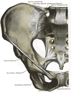
[[File:Pelvis ligaments anterior view.png|thumb|294x294px|Anterior sacroiliac ligament]] | [[File:Pelvis ligaments anterior view.png|thumb|294x294px|Anterior sacroiliac ligament]] | ||
Anterior sacroiliac | Anterior sacroiliac [[ligament]] (ASL) is comprised of many thin strands and forms from a thickened part of the anterior joint capsule<ref name=":0" /><ref name=":1">WONG M, SINKLER M, KIEL J. [https://www.ncbi.nlm.nih.gov/books/NBK507801/ Anatomy, Abdomen and Pelvis, Sacroiliac Joint]. StatPearls. Treasure Island (FL).</ref> . It is a smooth sheet of dense connective tissue stretching between the ventral surfaces of the sacral alar and ilium<ref name=":0" />. It is the thinnest sacroiliac joint ligament<ref name=":1" /> and is larger in males<ref name=":0" />. | ||
=== Attachments === | === Attachments === | ||
It | It runs from the iliac ala, anterior to the auricular surface, to the sacrum's pelvic surface<ref>1. MD GS. Sacroiliac joint [Internet]. Kenhub; 2022 [cited 2023 Aug 7]. Available from: https://www.kenhub.com/en/library/anatomy/sacroiliac-joint</ref>. | ||
== Function == | == Function == | ||
| Line 18: | Line 18: | ||
It is located close to the trunk of the lumbosacral plexus(especially fibers from L4–L5) and to the [[Obturator Nerve|obturator nerve]]. | It is located close to the trunk of the lumbosacral plexus(especially fibers from L4–L5) and to the [[Obturator Nerve|obturator nerve]]. | ||
Leakage of fluid to the surrounding structures at the site of the anterior capsule may happen because it is a relatively thin capsule<ref>Vleeming A, Schuenke MD, Masi AT, Carreiro JE, Danneels L, Willard FH. [https://www.ncbi.nlm.nih.gov/pmc/articles/PMC3512279/ The sacroiliac joint: an overview of its anatomy, function and potential clinical implications]. Journal of anatomy. 2012 Dec;221(6):537-67.</ref>. | Leakage of fluid to the surrounding structures at the site of the anterior capsule may happen because it is a relatively thin capsule<ref name=":0">Vleeming A, Schuenke MD, Masi AT, Carreiro JE, Danneels L, Willard FH. [https://www.ncbi.nlm.nih.gov/pmc/articles/PMC3512279/ The sacroiliac joint: an overview of its anatomy, function and potential clinical implications]. Journal of anatomy. 2012 Dec;221(6):537-67.</ref>. | ||
== Assessment == | == Assessment == | ||
There are various provocation tests for the [[Sacroiliac Joint|sacroiliac joint]] but none of them isolate the anterior sacroiliac joint ligament. | |||
== References == | == References == | ||
| Line 34: | Line 30: | ||
[[Category:Ligaments]] | [[Category:Ligaments]] | ||
<div class="noeditbox"> | <div class="noeditbox"> | ||
</div> | </div> | ||
[[Category:Pelvis - Anatomy]] | [[Category:Pelvis - Anatomy]] | ||
[[Category:Pelvis]] | [[Category:Pelvis]] | ||
Revision as of 13:34, 7 August 2023
Original Editor - Khloud Shreif Top Contributors - Khloud Shreif, Memoona Awan, Wendy Snyders and Kim Jackson
Description[edit | edit source]
Anterior sacroiliac ligament (ASL) is comprised of many thin strands and forms from a thickened part of the anterior joint capsule[1][2] . It is a smooth sheet of dense connective tissue stretching between the ventral surfaces of the sacral alar and ilium[1]. It is the thinnest sacroiliac joint ligament[2] and is larger in males[1].
Attachments[edit | edit source]
It runs from the iliac ala, anterior to the auricular surface, to the sacrum's pelvic surface[3].
Function[edit | edit source]
Limiting sacral nutation.
Clinical relevance[edit | edit source]
It is associated with sacroiliitis
It is located close to the trunk of the lumbosacral plexus(especially fibers from L4–L5) and to the obturator nerve.
Leakage of fluid to the surrounding structures at the site of the anterior capsule may happen because it is a relatively thin capsule[1].
Assessment[edit | edit source]
There are various provocation tests for the sacroiliac joint but none of them isolate the anterior sacroiliac joint ligament.
References[edit | edit source]
- ↑ 1.0 1.1 1.2 1.3 Vleeming A, Schuenke MD, Masi AT, Carreiro JE, Danneels L, Willard FH. The sacroiliac joint: an overview of its anatomy, function and potential clinical implications. Journal of anatomy. 2012 Dec;221(6):537-67.
- ↑ 2.0 2.1 WONG M, SINKLER M, KIEL J. Anatomy, Abdomen and Pelvis, Sacroiliac Joint. StatPearls. Treasure Island (FL).
- ↑ 1. MD GS. Sacroiliac joint [Internet]. Kenhub; 2022 [cited 2023 Aug 7]. Available from: https://www.kenhub.com/en/library/anatomy/sacroiliac-joint


