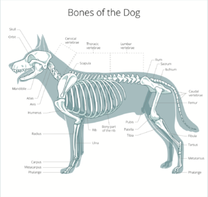
Canine Hindlimb Anatomy
Original Editor - User Name
Top Contributors - Vidya Acharya, Jess Bell, Chelsea Mclene, Lucinda hampton, Kim Jackson and Olajumoke Ogunleye
This article or area is currently under construction and may only be partially complete. Please come back soon to see the finished work! (7/05/2021)
Introduction[edit | edit source]
The hindlimb skeleton of the canine includes the pelvic girdle, consisting of the fused ilium, ischium, and pubis, and the bones of the hindlimb. The bones of the hindlimb are femur, patella, fabella, tibia, tarsus and meta-tarsus. The size of hindlimb bones varies due to the significant variation in size for breeds of dogs.
Osteology of Pelvic Limb[edit | edit source]
The Pelvic limb bears 40-45% of the weight and provides the majority of the propulsion for locomotion.[2]
Os coxae[edit | edit source]
- Tuber coxae end tubes sacrale both palpable[2]
- Tuber ischii underneath hamstrings
Femur[edit | edit source]
- Heaviest and largest canine bone[3]
- Has a relatively thick and short femoral neck with relatively short and wide shaft with a narrow isthmus in the middle
- Greater trochanter lateral to the head of the humerus[1]
- Lesser trochanter distal to head of humerus[1]
- Trochanteric fossa: Caudal depression between the trochanters[1]
- Third trochanter: Lateral aspect distal to greater trochanter[1]
- Extensor fossa: depression on lat condyle for insertion of long digital extensor.[1]
- Femoral trochlea: Groove on cran femur for articulating with patella bound by two ridges of which the medial is thicker.[1]
Patella[edit | edit source]
- The canine patella, or kneecap, is the largest sesamoid bone in the body.[3]
- It is an ossification in the quadriceps femoris muscle.[3]
- The patella alters the pull, increases the moment arm, and protects the quadriceps tendon, and provides a greater contact surface for the tendon on the trochlea of the femur than would exist without the patella. The canine patellar articular surface is mildly convex[3]
Fabellae[edit | edit source]
- Two small sesamoid bones embedded in the heads of the gastrocnemius muscle[3]
- Sesamoid in the lateral head is the largest, is palpable, and articulates with the lateral femoral condyle,
- Sesamoid in the medial head is smaller and may not have a distinct facet on the medial femoral condyle.
Tibia[edit | edit source]
- Major bone in the crus. [3]
- Proximal tibia is wider than the distal cylindrical tibia.
- Medial and lateral tibial condyles, an intercondylar eminence, and a tibial tuberosity are on the proximal tibia.
- Large tibial tuberosity - patellar ligament
- Tibial plateau slopes distally from cranial to caudal. The extensor groove, on the cranial tibia and lateral to the tibial tuberosity, provides a pathway for the long digital extensor muscle. There is a popliteal notch on the caudal tibia in the midline, where the popliteal vessels course.
- Tibia articulates with the fibula proximally, along the interosseous crest, and distally.
- The tibial cochlea articulate with the trochlea of the talus to form the talocrural joint.
- Cochlea = two grooves seperated by a ridge.[2]
Fibula[edit | edit source]
- Does not bear much weight.[2]
- Is a long, slender bone that articulates with the tibia and also serves as a site for muscle attachment.[3]
- A distinctive groove exists in the lateral malleolus, the sulcus malleolaris lateralis, through which course the tendons of the lateral digital extensor and peroneus brevis muscles.[3]
Tarsals and metatarsals[edit | edit source]
- Dogs and cats have 7 tarsal bones[2]
- Tarsus, or hock, consists of the talus, calcaneus, a central tarsal bone, and tarsal bones I to IV. [3]
- Talus articulates with the distal tibia and has prominent ridges. At the talocrural joint, two convex ridges of the trochlea of the talus articulate with two reciprocal concave grooves of the cochlea of the tibia.[3]
- Orientation of the grooves and ridges deviates laterally approximately 25 degrees from the sagittal plane. This deviation allows the hindpaws to pass lateral to the forepaws when dogs gallop.[3]
- Calcaneus is large and serves as the insertion of the common calcaneal tendon.[3]
- Central tarsal bone lies between the talus and the numbered tarsal bones I to III.[3]
- Tarsal IV is large and articulates with the calcaneus and metatarsal bones, spanning this entire region.[3]
- Hindpaw has five metatarsal bone[3]
- Reduced first MT and digit (dew claw) often absent[2]
Joints of the pelvic limb[edit | edit source]
Sacro-iliac[2][edit | edit source]
Type:
- Synchondrosis ( synovial joint - sacropelvic surface of ilium
- Joint capsule present
ROM:
- Minimal Designed for stability
- Accessory movements = rotations
Supporting structures:
- Dorsal and ventral Sacroiliac ligaments
- Sacrotuberous
Hip[2][edit | edit source]
Type:
- Ball and socket
- Femoral head and acetabulum of the ilium, ischium and pubis
- A band of fibrocartilage on the rim of the acetabulum deepens acetabulium
ROM:
- Flexion & Extension
- Minimal - adduction & abduction
Supporting structures:
- Acetabular lip (fibrocartilage) continues as transverse ligament
- Ligament of the femoral head
- Synovial structures and tendon sheaths:
- Large joint capsule
- Internal obturator
Stifle[edit | edit source]
Type:
- Hinge joint with two cartilages/menisci
- Femur & tibia - femorotibial (condylar)
- Femur & patella – femoropatellar (gliding joint)
ROM:
- Flexion and Extension
- At the end of flexion there is int rot
- At the end of extension there is external rotation
Supporting structures:
- Patellar ligament (patella is a sesamoid within the quadriceps tendon)
- Medial collateral ligament fused with joint capsule and med meniscus
- Lateral collateral ligament: Separated from lat meniscus by popliteus tendon
- Cranial cruciate ligament: Caudolateral femur to cranial tibial. Prevents anterior translation of tibial relative to femur
- Caudal cruciate ligament: Craniomedial femur to caudal tibia. Prevents candal translation of tibial relative to femur
- Lateral meniscus has extra ligament— meniscofemoral lig
- Connects caudal lat meniscus to femur
- Femoropatellar ligaments – lat and med
- Extend from epicondyles to patella
Hock (tarsal)[edit | edit source]
Type:
- Tarsocrucial joint (TCJ)- greatest movt
- Prox & distal intertarsal joint
- Tarsometatarsal joint
- Intertarsal
ROM:
- TCJ - Flexion & Extension and Lateral & rotatory accessory movts
- Others - small amount or translatory and rotatory ROM:
Ligaments of the tarsal joint:
- Medial & lat collateral (both have long and short parts)
- Intertarsal ligaments
- Prox extensor retinaculum
- Transverse ligament on distal tibia
- Holds down tendons of long digital extensor and cranial tibial
- Distal extensor retinaculum
- Hold down tendon of long digital extensor
- Long plantar ligament - connect calcaneus to metatarsus
- Flexor retinaculum - thickening of deep fascia over plantar aspect of tarsus
- Form the tarsal canal with the tarsal bones containing:
- Tendon and sheath of Deep digital flexor
- Plantar branch of saphenous artery & vein
- Medial & Lateral plantar nerves
Muscles of the Hind Limb[edit | edit source]
Muscles of the Hip[edit | edit source]
| Muscle | Origin | Insertion | Action | Nerve Supply |
|---|---|---|---|---|
| Pectineus | Pubis | Femur | Adduct the limb and flexes the hip | Obturator |
| Iliopsoas
Iliacus Psoas |
Ventral surface of the wing of Ilium |
Lesser of trochanter of Femur | Flexes Hip and externally rotates the thigh | Ventral branches of Lumbar Spinal Nerves |
| Gracilis | Pubis Symphysis | Medial Tibia | Adduct limb | Obturator |
| Adductors | Pubic surface limb symphysis | Adduct limb | Obturator | |
| Internal Obturator | Pelvic floor | Trochanteric fossa of the femur | Lateral rotation of femur | Ischiatic nerve |
| Gemelli | Ischium | Trochanteric fossa of the femur | Lateral rotation of femur | Ischiatic nerve |
| External Obturator | Ventral Surface of pubis Ischium | Trochanteric fossa of the femur | Adduct thigh | Ischiatic |
| Quadratus femoris | Ischium | Caudal surface of femur | Extenf hip and adduct thigh | Obturator |
| Deep Gluteal | Body of ischium | On or near greater trochanter | Extension
Abduction |
Gluteal |
Muscles of the Hip and Stifle 1[edit | edit source]
| Muscle | Origin | Insertion | Action | Nerve Supply |
|---|---|---|---|---|
| Superficial gluteal | Dorsal to hip joint | Third trochanter | Abduct hip | Gluteal |
| Tensore fascia lata | Tuber coxae | Lateral femoral fascia | Hip flexion and stifle extension via
tension on the lateral femoral fascia |
Gluteal |
| Biceps femoris | Ischiatic tuberosity | Greater trochanter | Extend and Abduct hip | Gluteal |
| Semitendinosus | Ischiatic tuberosity | Tibia and calcaneal tub | Extend hip, flex stifle, extend tarsus | Ischiatic |
| Semimembranosus | Ischiatic tuberosity | Femur and Tibia | Extends hip, Flexes or extends stifle | Ischiatic |
Muscles of the Hip and Stifle 2[edit | edit source]
| Muscles | Origin | Insertion | Action | Nerve supply |
|---|---|---|---|---|
| Middle gluteal | Wing of ilium | Greater trochanter | Extension and abduction | Gluteal |
| Rectus femoris | Ilium (cranioventral iliac spine) | Patella and patellar tuberosity | Flexes the hip and extends the stifle | Femoral |
| Vastus lateralis | Proximal femur | Patella and patellar tuberosity | Extends the stifle | Femoral |
| Vastus medialis | Proximal femur | Patella and patellar tuberosity | Extends | Femoral |
| Vastus intermedius | Proximal femur | Patella and patellar tuberosity | Extends | Femoral |
Muscles of the Stifle, Hock and Pes[edit | edit source]
| Gastrocnemius | Distocaudal surface of the femur | Calcaneal tuberosity | Extends tarsus and flexes stifle | Tibial |
| Long digital extensors | Extensor fossa of femur | extensor processes of distal phalanges | Flexes hip and extends stifle | Peroneal |
| Soleus | Head of fibula | Tendon and lateral head of gastrocnemius | Extends tarsus | Tibial |
| Lateral digital extensor | Fibula | Lateral aspect of digit | Extend digit and flexes tarsus | Peroneal |
| Deep digital flexor | Tibia and fibula | Distal phalanges | Extends tarsus and flexes digit | Tibial |
| Superficial digital flexor | Caudal distal femur, deep to gastrocnemius | Calcaneal tuberosity and middle phalanx | Flexes stifle, extends tarsus and flexes digit | Tibial |
Nerves of the Pelvic Limb[edit | edit source]
Femoral nerve (L4, L5 sometimes L3, L6)[edit | edit source]
- Musle Innervation - Stifle extensors (quadriceps), llio-psoas.
- Cutaneous Innervation - Medial aspect of limb
Obturator nerve (L5, L6)[edit | edit source]
- Muscle Innervation - Adductors - obturator, add, gracilis
- Cutaneous Innervation - nil
Gluteal nerve (L6, L7, S1)[edit | edit source]
- Muscle Innervation - gluteals, TFL, biceps femoris, semitenndinosus.
- Cutaneous Innervation - nil
Sciatic nerve (L6, L7, S1, S2)[edit | edit source]
- Muscle InnervationMI - biceps femoris, semitendinosus, semimembranosis
- Cutaneous Innervation - see tibial and fibula branches
Tibial nerve (S1, S2)[edit | edit source]
- Muscle InnervationMI - extensors of hock, flexors of digits
- Cutaneous Innervation - caudal aspects o1 the limb below stifle
Fibular nerve[edit | edit source]
- Muscle InnervationMI - flexors of the hock, extensors of the digits
- Cutaneous Innervation - cranial and lat aspects of limb
Resources[edit | edit source]
- bulleted list
- x
or
- numbered list
- x
References[edit | edit source]
- ↑ 1.0 1.1 1.2 1.3 1.4 1.5 1.6 Pinoy Vet AnatomistSkeletal System (Part 5)- Bones of the pelvic limbAvailable from https://www.youtube.com/watch?v=IWSzVpTTYpQ on 5/5/21
- ↑ 2.0 2.1 2.2 2.3 2.4 2.5 2.6 2.7 Van Der Walt, A. Managing Disorders of the Canine Hind Limb. Physioplus Course, 2021.
- ↑ 3.00 3.01 3.02 3.03 3.04 3.05 3.06 3.07 3.08 3.09 3.10 3.11 3.12 3.13 3.14 Canine Anatomy.Basic Science of Veterinary Rehabilitation. Cheryl Riegger-Krugh, Darryl L. Millis, and Joseph P. Weigel


