Basic Anatomy of the Dancer's Ankle and Foot: Difference between revisions
Carin Hunter (talk | contribs) No edit summary |
Kim Jackson (talk | contribs) m (Text replacement - "Physioplus" to "Plus ") |
||
| (22 intermediate revisions by 5 users not shown) | |||
| Line 1: | Line 1: | ||
<div class="editorbox"> '''Original Editor '''- [[User:Carin Hunter|Carin Hunter]] based on the course by [https://members.physio-pedia.com/course_tutor/michelle-green-smerdon/ Michelle Green-Smerdon]<br>'''Top Contributors''' - {{Special:Contributors/{{FULLPAGENAME}}}}</div> | <div class="editorbox"> '''Original Editor '''- [[User:Carin Hunter|Carin Hunter]] based on the course by [https://members.physio-pedia.com/course_tutor/michelle-green-smerdon/ Michelle Green-Smerdon]<br>'''Top Contributors''' - {{Special:Contributors/{{FULLPAGENAME}}}}</div> | ||
[[File:Talus bone - animation01.gif|left|thumb|Tibia, Fibula and Talus]] | |||
The ankle and foot are complex and detailed structures that bear the weight of the whole body, and are designed to showcase a beautiful work of art. The ankle and foot | == Introduction == | ||
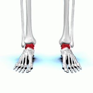
[[File:Talus bone - animation01.gif|left|thumb|Tibia, Fibula and Talus]]"The ankle and foot are complex and detailed structures that bear the weight of the whole body, and are designed to showcase a beautiful work of art."<ref name=":3">Green-Smerdon M. Basic Anatomy of the Dancer’s Ankle and Foot course. Plus , 2022.</ref> | |||
The ankle and foot are part of a very complex system.<ref>Brockett CL, Chapman GJ. [https://www.sciencedirect.com/science/article/pii/S1877132716300483 Biomechanics of the ankle.] Orthopaedics and trauma. 2016 Jun 1;30(3):232-8.</ref> This part of the body has to cope with high compressive and shearing forces and, at the same time, it has to offer a high degree of stability. The ankle is the kinetic link between the foot and structures that are higher up in the body. Moreover, the foot is the point where the body interacts with the ground and has to control multi-axial motions, which occur simultaneously.<ref>Houglum PA, Bertoti DB. [https://books.google.co.uk/books?hl=en&lr=&id=offuAwAAQBAJ&oi=fnd&pg=PR1&dq=Houglum+PA,+Bertoti+DB.+Brunnstrom%27s+clinical+kinesiology.+FA+Davis%3B+2012&ots=IxM_DDsDiu&sig=In1KgRYuAUxjjylNd2KuoISLCAw&redir_esc=y#v=onepage&q&f=false Brunnstrom's clinical kinesiology.] FA Davis; 2012</ref> | |||
[[File:Transverse-tarsal-joint.jpg|thumb|Transverse-tarsal joint]] | [[File:Transverse-tarsal-joint.jpg|thumb|Transverse-tarsal joint]] | ||
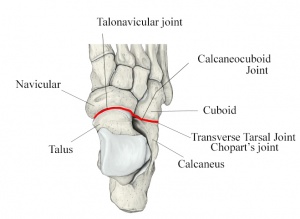
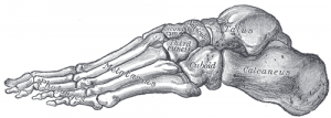
Very simply, the ankle is made up of three bones, the [[tibia]], [[fibula]] and [[talus]] and three joints:<ref name=":3" /> | |||
# The '''transverse-tarsal joint''' which works together with the talocalcaneal joint | |||
# The '''talocalcaneal joint''' which controls inversion and eversion | |||
# The '''tibiotalar joint''' which helps with stability, flexion and extension | |||
== Foot Summary == | == Foot Summary == | ||
# '''<u>Hind foot</u>''' | # '''<u>Hind foot</u>'''<ref name=":0">Ficke J, Byerly DW. Anatomy, [https://www.ncbi.nlm.nih.gov/books/NBK546698/ Bony Pelvis and Lower Limb, Foot.] StatPearls [Internet]. 2021 Aug 11.</ref> | ||
## '''Bones''' | ##'''Bones''' | ||
### Talus | ###'''Talus''' | ||
### Calcaneous | #### This is the highest foot bone | ||
### Lateral and medial malleolus | #### There are no tendons attached to it, only the deltoid ligament | ||
#### Approximately 60 percent of the surface of the talus is covered by articular cartilage<ref>Bell DJ. Talus [Internet]. Radiopedia. 2021 [cited 15/02/2022]. Available from: https://radiopaedia.org/articles/talus</ref> | |||
#### It has a poor blood supply and therefore relatively poor healing | |||
###'''Calcaneous''' | |||
###'''Lateral and medial malleolus''' | |||
## '''Joints''' | ## '''Joints''' | ||
### | ### Tibiotalar Joint: | ||
# | #### The talus is at its widest anteriorly, meaning the joint is more stable in dorsiflexion | ||
#### The conforming geometry of the tibiotalar joint is believed to contribute to the stability of the joint - in stance phase, the geometry of the joint alone is sufficient to provide resistance to eversion; otherwise stability is derived from the soft tissue structures. | |||
### The talus is at its widest anteriorly, meaning the joint is more stable in dorsiflexion | ### Subtalar joint: | ||
### The conforming geometry of the tibiotalar joint is | #### Absorbs rotational stress from structures higher up in the body | ||
### | ### Transverse tarsal joint: | ||
### | #### Transitional link between the hindfoot and forefoot | ||
### Transverse tarsal joint | # '''<u>Mid Foot</u>'''<ref name=":0" /> | ||
# '''<u>Mid Foot</u>''' | ##'''Bones''' | ||
## '''Bones''' | ###'''Navicular''' | ||
### Navicular | #### The navicular has a poor blood supply | ||
### Cuboid | #### The main attachment for the posterior tibial tendon is on medial side | ||
### | ### '''Cuboid''' | ||
### '''Three cuneiforms''' (medial, intermedius, lateral) | |||
#### These bones are important for stability along with the plantar and dorsal ligaments | |||
## '''Joints''' | ## '''Joints''' | ||
### | ### Five tarsal-metatarsal joints, also known as the Lisfranc joint | ||
## ''' | ## '''Connective tissue, ligaments, muscles and tendons''' | ||
### Plantar fascia | ### Plantar fascia | ||
### | #### The plantar fascia is responsible for forming the arches of the feet and is a shock absorber when dancing<ref name=":3" /> | ||
# '''<u>Forefoot</u>''' | # '''<u>Forefoot</u>'''<ref name=":0" /> | ||
## '''Bones''' | ##'''Bones''' | ||
### 14 | ### 14 phalanges | ||
### | ### Five metatarsals | ||
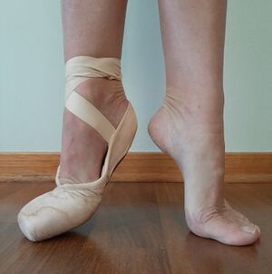
### | #### The metatarsal heads are the main weightbearing surface in the following ballet positions: Releve,<ref>Veirs KP, Rippetoe JR, Baldwin JD, Lutz K, Haleem AM, Dionne CP. [https://apta.confex.com/apta/csm2020/meetingapp.cgi/Paper/23082 Multi-Segment Assessment of Ankle and Foot Kinematics during Elevé Barefoot Demi-Pointe and En Pointe.] In2020 Combined Sections Meeting (CSM) 2020 Feb 13. APTA.</ref> quarter pointe, demi-pointe, and three quarter pointe. | ||
### Two sesamoid bones: | |||
#### These are located inside the [[Flexor Hallucis Brevis|flexor hallucis brevis]] tendon and allow the toe to move up and down. | |||
## '''Joints''' | ## '''Joints''' | ||
### | ### Metatarsophalangeal joints | ||
## '''Ligaments, muscles and tendons''' | ## '''Ligaments, muscles and tendons''' | ||
### | ### The first metatarsal bone is the location for the attachment of several tendons and is important for its role in propulsion and weight bearing. | ||
== Ligaments of the Foot and Ankle == | == Ligaments of the Foot and Ankle == | ||
# '''<u>Medial Ligaments</u>''' | # '''<u>Medial Ligaments</u>'''<ref name=":3" /><ref name=":1">Das A, Bhuyan D. [https://d1wqtxts1xzle7.cloudfront.net/56895719/418-Article_Text-998-1-10-20180416-with-cover-page-v2.pdf?Expires=1640129178&Signature=COmS1F5vXyQiBCXussXr3odQAKKWH4ZHaYam1gN5WxHRBYUVwxTuXStXOXfqhaA6JODIsdCj6xP5vUTEpe0S9NQNNgys4ZG~MD7oHiazGnroTjS4XLCT~C3tpDzJoJ2t64jJJpvMj5VXwZ6SKHMlLk51nVbx4QCZYBqgRabtDT-IhBqK5SkMc1WQAJSEllHz9QeLMKXMvzI61TrQiTZnWK7TvGfeIKSN5qfMaxGiwEM-yuPbuZYuabLkO~NDxvqvDlkp2LpwHLhXkBlmxJpiuwpd7QvNOQFB1otnLadXY9NiFpdO6YqFndAIEW1OJZGi1C4g6UYG-eE5w6kqtHBe9Q__&Key-Pair-Id=APKAJLOHF5GGSLRBV4ZA A Review on the Anatomy and Biomechanics of the Foot-Ankle Complex.] Asian Journal For Convergence In Technology (AJCT). 2018 Apr 15.</ref> | ||
## | ## The deltoid ligament is fan shaped, comprising of four ligaments and resists eversion: | ||
### | ### Anterior tibiotalar ligament (deep component) | ||
### | ### Tibiocalcaneal ligament | ||
### | ### Tibionavicular ligament | ||
### | ### Posterior tibiotalar ligament (deep component) | ||
## '''Expansion of joint capsule''' | ## '''Expansion of the joint capsule''' | ||
### Spring ligament | ###'''Spring ligament:''' Cradles and supports the talar head | ||
### Lisfranc ligaments | ### '''Lisfranc ligaments:''' Series of ligaments that stabilise the tarsometatarsal joints and provide stability to the arch of the foot. The plantar ligament is stronger than the dorsal ligament. | ||
### Inter-metatarsal ligament | ### '''Inter-metatarsal ligament:''' This is found between the tarsal bones and keeps the metatarsals moving in sync. If the nerve running between these joints gets irritated, this can result in a [[Morton's Neuroma|Morton's neuroma]].<ref name=":3" />[[File:On pointe good alignment.jpg|thumb|Pointe with Good Alignment]] | ||
# '''<u>Lateral Ligaments</u>''' | # '''<u>Lateral Ligaments</u>'''<ref name=":3" /><ref name=":1" /> | ||
## | ## The lateral ankle is commonly injured in ballet dancers <ref>SAH SA, MR ES, Srijit D, Norzana AG. [https://core.ac.uk/download/pdf/96113838.pdf Ankle Injuries in Sports: Anatomical Considerations and Clinical Implications.]</ref><ref name=":3" /> | ||
## | ## Anterior talofibular ligament | ||
## | ### Tightens in plantarflexion | ||
## | ### Many authors conclude that this is the weakest ligament<ref name=":3" /> | ||
### | ## Calcaneo-fibular ligament | ||
### | ### Tightens in dorsiflexion | ||
### | ## The following ligaments contribute to ankle stability | ||
### Posterior talofibular ligament | |||
### Anterior-inferior talofibular ligament | |||
### Posterior-inferior talofibular ligament<ref name=":4" /> | |||
==== '''Ballet Specific Ligament Anatomy''' ==== | |||
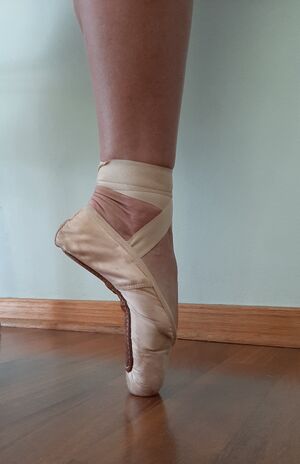
* In a demi-plié, when the ankle is in dorsiflexion, the anterior talofibular ligament relaxes and the calcaneo-fibular ligament is under tension | |||
* The opposite is expected en pointe (when the ankle is in plantarflexion), although no studies have been done to examine extreme position en pointe | |||
* Strain in the anterior talofibular ligament increases with increasing plantarflexion, which is further accentuated during compressive loading through the ankle | |||
* Maximum plantarflexion en pointe places the anterior talofibular ligament parallel to the fibula, which causes it to function as a primary stabiliser of the lateral ankle | |||
* This places the anterior talofibular ligament at particular risk (weakest and at its longest at maximal tension force)<ref name=":3" /><ref name=":4">Russell JA, McEwan IM, Koutedakis Y, Wyon MA. [https://www.ingentaconnect.com/content/jmrp/jdms/2008/00000012/00000003/art00002 Clinical anatomy and biomechanics of the ankle in dance.] Journal of dance medicine & science. 2008 Sep 1;12(3):75-82.</ref> | |||
== Syndesmosis == | == Syndesmosis == | ||
This ligament is made up of the: | |||
# | # Anterior-inferior talofibular ligament | ||
# Interosseus membrane | # Interosseus membrane | ||
# | # Posterior-inferior talofibular ligament | ||
# Transverse ligament | # Transverse ligament | ||
# Interosseus ligament | # Interosseus ligament | ||
The function of this ligament is to hold the tibia and fibula together at the appropriate distance and form a mortise where the talus sits.<ref>Norkus SA, Floyd RT. [https://www.ncbi.nlm.nih.gov/pmc/articles/PMC155405/ The anatomy and mechanisms of syndesmotic ankle sprains.] Journal of athletic training. 2001 Jan;36(1):68.</ref> | |||
== Muscles of the Foot and Ankle == | == Muscles of the Foot and Ankle == | ||
==== 1. Extrinsic Foot Muscles ==== | ==== 1. Extrinsic Foot Muscles ==== | ||
These muscles have contractile portions that lie outside the ankle, in the leg, but their tendons insert onto the bones of the foot in such a way that ankle motion occurs when these muscles contract. There are four four muscle compartments, separated by fascia: the Superficial Posterior Compartment, Deep Posterior Compartment and the Lateral Compartment, which are all plantar flexors, and the Anterior Compartment, which are dorsiflexors.<ref name=":3" /> | |||
These muscles have contractile portions that lie outside the ankle, in the leg, | |||
# '''Superficial Posterior | # '''Superficial Posterior Compartment (plantar flexors)'''<ref name=":3" /> | ||
## | ## Gastrocnemius | ||
## Soleus ( | ### Action: Plantarflexion when knee is extended, flexion of the knee and also raises the heel during walking | ||
### Insertion: Posterior surface of the calcaneus via Calcaneal Tendon (Achilles Tendon) | |||
## Soleus | |||
### Action: Plantarflexion and steadies the leg on the foot | |||
### Insertion: Posterior surface of the calcaneus via Calcaneal Tendon (Achilles Tendon) | |||
## Plantaris | ## Plantaris | ||
# '''Deep Posterior Compartment ( | ### Action: Weakly assists gastrocnemius in plantarflexion | ||
## | ### Insertion: Posterior surface of the calcaneus via Calcaneal Tendon (Achilles Tendon) | ||
## | # '''Deep Posterior Compartment (plantar flexors)'''<ref name=":3" /> | ||
## Tibialis Posterior | ## Flexor hallucis longus<ref>Murdock CJ, Munjal A, Agyeman K. [https://www.ncbi.nlm.nih.gov/books/NBK539776/ Anatomy, Bony Pelvis and Lower Limb, Calf Flexor Hallucis Longus Muscle.] StatPearls [Internet]. 2021 Aug 6.</ref> | ||
# '''Lateral Compartment ( | ### Action: Inversion | ||
## | ### Insertion: On the plantar surface of the first toe | ||
## | ## Flexor digitorum longus | ||
# '''Anterior | ### Action: Inversion | ||
## Tibialis anterior | ### Insertion: On the plantar surface second to fifth toes | ||
## | ## Tibialis posterior<ref>Corcoran NM, Varacallo M. Anatomy, [https://www.ncbi.nlm.nih.gov/books/NBK539913/ Bony Pelvis and Lower Limb, Tibialis Posterior Muscle.] StatPearls [Internet]. 2020 Sep 17.</ref> | ||
## | ### Action: Inversion | ||
## | ### Insertion: Navicular, medial cuneiform, second to fourth toes, other cuneiforms, cuboid | ||
# '''Lateral Compartment (plantar flexors)'''<ref name=":3" /> | |||
## Peroneus longus | |||
### Action: Eversion and plantar flexion | |||
### Insertion: First metatarsal, medial cuneiform and first toe | |||
## Peroneus brevis | |||
### Action: Eversion | |||
### Insertion: Proximal end of the fifth metatarsal<ref>Basit H, Eovaldi BJ, Siccardi MA. [https://www.ncbi.nlm.nih.gov/books/NBK535427/ Anatomy, Bony Pelvis and Lower Limb, Foot Peroneus Brevis Muscle]. In StatPearls [Internet] 2019 May 19. StatPearls Publishing.</ref> | |||
# '''Anterior Compartment (dorsiflexors)'''<ref name=":3" /> | |||
## Tibialis anterior | |||
### Action: Inversion | |||
### Insertion: First toe and medial cuneiform | |||
## Extensor hallucis longus | |||
### Action: Inversion | |||
### Insertion: Dorsal aspect of base of distal phalanx of big toe | |||
## Extensor digitorum longus | |||
### Action: Dorsiflexion and extends lateral four digits | |||
### Insertion: Middle & distal phalanges of lateral four digits | |||
## Peroneals | |||
### Action: Dorsiflexion, aids eversion | |||
### Insertion: Dorsum of the base of the 5th metatarsal | |||
==== 2. Intrinsic Foot Muscles ==== | |||
These muscles all originate and insert within the foot. They are known to move the toes and stabilise the foot. Dancers refer to these muscles as the “core” muscles of the foot.<ref>Farris DJ, Kelly LA, Cresswell AG, Lichtwark GA. [https://www.pnas.org/content/116/5/1645.short The functional importance of human foot muscles for bipedal locomotion.] Proceedings of the National Academy of Sciences. 2019 Jan 29;116(5):1645-50.</ref> | |||
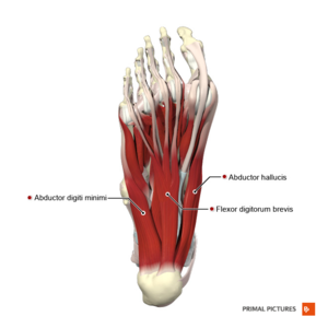
The three largest muscles are abductor hallucis, flexor digitorum brevis and quadratus plantae. They all provide support and stability to the arch. In dancers, these muscles require strength and control. A dancer needs to learn to work with “straight” toes, which includes providing counter-stability to the metatarsals when pointing.<ref name=":3" /> | |||
==== Four Muscle Layers of the Plantar Foot ==== | |||
{| class="wikitable" | {| class="wikitable" | ||
|+ | |+ | ||
! | ! | ||
! | !Muscles | ||
! | !Tendons | ||
! | ! | ||
|- | |- | ||
|Flexor | |'''Layer One''' | ||
| | | | ||
| | * Abductor hallucis | ||
| | * Flexor digitorum brevis (FDB) | ||
* Abductor digiti minimi | |||
| | | | ||
|[[File:Plantar muscles of the foot first layer Primal.png|thumb|First Layer]] | |||
|- | |- | ||
| | |'''Layer Two''' | ||
| | | | ||
* Quadratus plantae | |||
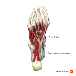
| | * Lumbrical muscles | ||
| | |||
| | * Flexor digitorum longus (FDL) | ||
* Flexor hallucis longus (FHL) | |||
|[[File:Plantar muscles of the foot second layer Primal.png|thumb|Second Layer]] | |||
|- | |- | ||
| | |'''Layer Three''' | ||
| | | | ||
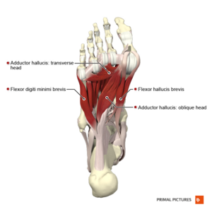
| | * Flexor hallucis brevis | ||
| | * Oblique and transverse heads of the adductor hallucis | ||
| | * Flexor digiti minimi brevis | ||
| | | | ||
|[[File:Plantar muscles of the foot third layer Primal.png|thumb|Third Layer]] | |||
|- | |- | ||
|Peroneus | |'''Layer Four''' | ||
| | |||
|Plantar | * Dorsal interosseous | ||
* Plantar interosseus | |||
| | | | ||
| | * Peroneus longus | ||
* Tibialis posterior | |||
|[[File:Plantar muscles of the foot fourth layer Primal.png|thumb|Fourth Layer]] | |||
|} | |} | ||
==== | == Plantar fascia == | ||
* This is made up of strong fibrous tissue | |||
* It originates deep within the plantar surface of the calcaneus and inserts on the base of each of the toes | |||
* When the toes are in dorsiflexion, the fascia tightens and supports the arch | |||
* Windlass mechanism:<ref>Metsavaht L, Leporace G. [https://jfootankle.com/JournalFootAnkle/article/view/1189/1440 Current trends in the biokinetic analysis of the foot and ankle.] Journal of the Foot & Ankle. 2020 Aug 30;14(2):191-6.</ref> The windlass mechanism is a mechanical model that describes the manner through which the plantar fascia supports the foot during weight-bearing activities and provides information regarding the biomechanical stresses placed on plantar fascia<ref>Bolgla LA, Malone TR. [https://www.ncbi.nlm.nih.gov/pmc/articles/PMC385265/ Plantar fasciitis and the windlass mechanism: a biomechanical link to clinical practice.] Journal of athletic training. 2004 Jan 1;39(1):77.</ref> | |||
** For more information, please see [[Windlass Test]] | |||
[[File:Arches of the Foot - shutterstock 478538809 smaller.jpg|thumb|Arches of the foot]] | |||
== Arches == | == Arches == | ||
The purpose of the three arches of the foot are:<ref>Batenhorst EZ. [https://digitalcommons.wou.edu/cgi/viewcontent.cgi?article=1225&context=honors_theses A Dancer’s View: Analysis and Prevention of Common Dance Injuries].</ref> | |||
# Spring | # Spring | ||
# Weight bearing | # Weight bearing | ||
# Shock absorption | # Shock absorption | ||
# | # To provides flexibility to the foot to facilitate function | ||
[[File:Medial arch of the foot.gif|thumb|Medial arch of the foot]] | |||
==== '''<u>1. Medial | ==== '''<u>1. Medial Longitudinal Arch (MLA)</u>'''<ref name=":2">Ahonen J. [https://www.ingentaconnect.com/content/jmrp/jdms/2008/00000012/00000003/art00005# Biomechanics of the foot in dance: a literature review.] Journal of Dance Medicine & Science. 2008 Sep 1;12(3):99-108.</ref><ref>Ozdinc SA, Turan FN. [https://www.researchgate.net/profile/Sevgi-Oezdinc/publication/305321572_Effects_of_ballet_training_of_children_in_Turkey_on_foot_anthropometric_measurements_and_medial_longitudinal_arc_development/links/5bed24f792851c6b27bfd3e1/Effects-of-ballet-training-of-children-in-Turkey-on-foot-anthropometric-measurements-and-medial-longitudinal-arc-development.pdf Effects of ballet training of children in Turkey on foot anthropometric measurements and medial longitudinal arc development.] J. Pak. Med. Assoc. 2016 Jul 1;66(7):869-74.</ref> ==== | ||
This is the highest arch due to the shape of bones. | |||
#'''Bones''' | |||
# ''' | ## The first three metatarsal | ||
## Three cuneiforms | |||
## | |||
## | |||
## Navicular | ## Navicular | ||
## Talus | ## Talus | ||
## | ## Calcaneus | ||
# ''' | #'''Ligaments''' | ||
## Spring ligament | ## Spring ligament | ||
## Deltoid ligament | ## Deltoid ligament | ||
| Line 227: | Line 225: | ||
## Plantar aponeurosis | ## Plantar aponeurosis | ||
## Long and short plantar ligaments | ## Long and short plantar ligaments | ||
# ''' | #'''Muscles''' | ||
## | ## Tibialis posterior | ||
## | ## Tibialis anterior | ||
## | ## Flexor hallucis longus | ||
## | ## Flexor digitorum longus | ||
## Short muscles of the big toe | ## Short muscles of the big toe | ||
==== '''<u>2. Lateral | ===='''<u>2. Lateral Longitudinal Arch (LLA)</u>'''<ref name=":2" />==== | ||
[[File:Lateral Arch of the foot.png|thumb|Lateral Arch of the foot]] | [[File:Lateral Arch of the foot.png|thumb|Lateral Arch of the foot]] | ||
The only arch to lie on the ground in a standing position. | |||
# ''' | # '''Bones''' | ||
## Calcaneus | ## Calcaneus | ||
## Cuboid | ## Cuboid | ||
## | ## Four and fifth metatarsals | ||
# ''' | # '''Ligaments''' | ||
## | ## Long and short plantar ligaments | ||
## Interosseus ligament | ## Interosseus ligament | ||
## Plantar aponeurosis | ## Plantar aponeurosis | ||
# ''' | # '''Muscles''' | ||
## Peroneus longus and brevis | ## Peroneus longus and brevis | ||
## Flexor digitorum longus | ## Flexor digitorum longus | ||
## Short muscles of the little toe | ## Short muscles of the little toe | ||
[[File:Demi pointe - correct, showing with shoe on&off.jpg|thumb|Demi Pointe with weight bearing surface on the Transverse Arch]] | [[File:Demi pointe - correct, showing with shoe on&off.jpg|thumb|Demi Pointe with weight bearing surface on the Transverse Arch]] | ||
# ''' | ==== 3. Transverse Arch<ref name=":2" /> ==== | ||
## Wedge shape of the lateral and intermediate | #'''Bones''' | ||
## Wedge shape of the lateral and intermediate cuneiform | |||
## Metatarsal bases | ## Metatarsal bases | ||
## Cuboid | ## Cuboid | ||
## | ## Three cuneiforms | ||
# ''' | #'''Ligaments''' | ||
## Deep transverse ligament | ## Deep transverse ligament | ||
## Dorsal and plantar ligament | ## Dorsal and plantar ligament | ||
# ''' | #'''Muscles''' | ||
## Peroneus longus and brevis | ## Peroneus longus and brevis | ||
## Transverse head of adductor hallucis | ## Transverse head of adductor hallucis | ||
## Slips of tibial posterior | ## Slips of tibial posterior | ||
== References == | |||
[[Category:Course Pages]] | [[Category:Course Pages]] | ||
[[Category: | [[Category:Plus Content]] | ||
[[Category:Foot - Anatomy]] | [[Category:Foot - Anatomy]] | ||
[[Category:Foot - Bones]] | [[Category:Foot - Bones]] | ||
Latest revision as of 11:32, 18 August 2022
Top Contributors - Carin Hunter, Jess Bell, Kim Jackson, Wanda van Niekerk and Olajumoke Ogunleye
Introduction[edit | edit source]
"The ankle and foot are complex and detailed structures that bear the weight of the whole body, and are designed to showcase a beautiful work of art."[1]
The ankle and foot are part of a very complex system.[2] This part of the body has to cope with high compressive and shearing forces and, at the same time, it has to offer a high degree of stability. The ankle is the kinetic link between the foot and structures that are higher up in the body. Moreover, the foot is the point where the body interacts with the ground and has to control multi-axial motions, which occur simultaneously.[3]
Very simply, the ankle is made up of three bones, the tibia, fibula and talus and three joints:[1]
- The transverse-tarsal joint which works together with the talocalcaneal joint
- The talocalcaneal joint which controls inversion and eversion
- The tibiotalar joint which helps with stability, flexion and extension
Foot Summary[edit | edit source]
- Hind foot[4]
- Bones
- Talus
- This is the highest foot bone
- There are no tendons attached to it, only the deltoid ligament
- Approximately 60 percent of the surface of the talus is covered by articular cartilage[5]
- It has a poor blood supply and therefore relatively poor healing
- Calcaneous
- Lateral and medial malleolus
- Talus
- Joints
- Tibiotalar Joint:
- The talus is at its widest anteriorly, meaning the joint is more stable in dorsiflexion
- The conforming geometry of the tibiotalar joint is believed to contribute to the stability of the joint - in stance phase, the geometry of the joint alone is sufficient to provide resistance to eversion; otherwise stability is derived from the soft tissue structures.
- Subtalar joint:
- Absorbs rotational stress from structures higher up in the body
- Transverse tarsal joint:
- Transitional link between the hindfoot and forefoot
- Tibiotalar Joint:
- Bones
- Mid Foot[4]
- Bones
- Navicular
- The navicular has a poor blood supply
- The main attachment for the posterior tibial tendon is on medial side
- Cuboid
- Three cuneiforms (medial, intermedius, lateral)
- These bones are important for stability along with the plantar and dorsal ligaments
- Navicular
- Joints
- Five tarsal-metatarsal joints, also known as the Lisfranc joint
- Connective tissue, ligaments, muscles and tendons
- Plantar fascia
- The plantar fascia is responsible for forming the arches of the feet and is a shock absorber when dancing[1]
- Plantar fascia
- Bones
- Forefoot[4]
- Bones
- 14 phalanges
- Five metatarsals
- The metatarsal heads are the main weightbearing surface in the following ballet positions: Releve,[6] quarter pointe, demi-pointe, and three quarter pointe.
- Two sesamoid bones:
- These are located inside the flexor hallucis brevis tendon and allow the toe to move up and down.
- Joints
- Metatarsophalangeal joints
- Ligaments, muscles and tendons
- The first metatarsal bone is the location for the attachment of several tendons and is important for its role in propulsion and weight bearing.
- Bones
Ligaments of the Foot and Ankle[edit | edit source]
- Medial Ligaments[1][7]
- The deltoid ligament is fan shaped, comprising of four ligaments and resists eversion:
- Anterior tibiotalar ligament (deep component)
- Tibiocalcaneal ligament
- Tibionavicular ligament
- Posterior tibiotalar ligament (deep component)
- Expansion of the joint capsule
- Spring ligament: Cradles and supports the talar head
- Lisfranc ligaments: Series of ligaments that stabilise the tarsometatarsal joints and provide stability to the arch of the foot. The plantar ligament is stronger than the dorsal ligament.
- Inter-metatarsal ligament: This is found between the tarsal bones and keeps the metatarsals moving in sync. If the nerve running between these joints gets irritated, this can result in a Morton's neuroma.[1]
- The deltoid ligament is fan shaped, comprising of four ligaments and resists eversion:
- Lateral Ligaments[1][7]
- The lateral ankle is commonly injured in ballet dancers [8][1]
- Anterior talofibular ligament
- Tightens in plantarflexion
- Many authors conclude that this is the weakest ligament[1]
- Calcaneo-fibular ligament
- Tightens in dorsiflexion
- The following ligaments contribute to ankle stability
- Posterior talofibular ligament
- Anterior-inferior talofibular ligament
- Posterior-inferior talofibular ligament[9]
Ballet Specific Ligament Anatomy[edit | edit source]
- In a demi-plié, when the ankle is in dorsiflexion, the anterior talofibular ligament relaxes and the calcaneo-fibular ligament is under tension
- The opposite is expected en pointe (when the ankle is in plantarflexion), although no studies have been done to examine extreme position en pointe
- Strain in the anterior talofibular ligament increases with increasing plantarflexion, which is further accentuated during compressive loading through the ankle
- Maximum plantarflexion en pointe places the anterior talofibular ligament parallel to the fibula, which causes it to function as a primary stabiliser of the lateral ankle
- This places the anterior talofibular ligament at particular risk (weakest and at its longest at maximal tension force)[1][9]
Syndesmosis[edit | edit source]
This ligament is made up of the:
- Anterior-inferior talofibular ligament
- Interosseus membrane
- Posterior-inferior talofibular ligament
- Transverse ligament
- Interosseus ligament
The function of this ligament is to hold the tibia and fibula together at the appropriate distance and form a mortise where the talus sits.[10]
Muscles of the Foot and Ankle[edit | edit source]
1. Extrinsic Foot Muscles[edit | edit source]
These muscles have contractile portions that lie outside the ankle, in the leg, but their tendons insert onto the bones of the foot in such a way that ankle motion occurs when these muscles contract. There are four four muscle compartments, separated by fascia: the Superficial Posterior Compartment, Deep Posterior Compartment and the Lateral Compartment, which are all plantar flexors, and the Anterior Compartment, which are dorsiflexors.[1]
- Superficial Posterior Compartment (plantar flexors)[1]
- Gastrocnemius
- Action: Plantarflexion when knee is extended, flexion of the knee and also raises the heel during walking
- Insertion: Posterior surface of the calcaneus via Calcaneal Tendon (Achilles Tendon)
- Soleus
- Action: Plantarflexion and steadies the leg on the foot
- Insertion: Posterior surface of the calcaneus via Calcaneal Tendon (Achilles Tendon)
- Plantaris
- Action: Weakly assists gastrocnemius in plantarflexion
- Insertion: Posterior surface of the calcaneus via Calcaneal Tendon (Achilles Tendon)
- Gastrocnemius
- Deep Posterior Compartment (plantar flexors)[1]
- Flexor hallucis longus[11]
- Action: Inversion
- Insertion: On the plantar surface of the first toe
- Flexor digitorum longus
- Action: Inversion
- Insertion: On the plantar surface second to fifth toes
- Tibialis posterior[12]
- Action: Inversion
- Insertion: Navicular, medial cuneiform, second to fourth toes, other cuneiforms, cuboid
- Flexor hallucis longus[11]
- Lateral Compartment (plantar flexors)[1]
- Peroneus longus
- Action: Eversion and plantar flexion
- Insertion: First metatarsal, medial cuneiform and first toe
- Peroneus brevis
- Action: Eversion
- Insertion: Proximal end of the fifth metatarsal[13]
- Peroneus longus
- Anterior Compartment (dorsiflexors)[1]
- Tibialis anterior
- Action: Inversion
- Insertion: First toe and medial cuneiform
- Extensor hallucis longus
- Action: Inversion
- Insertion: Dorsal aspect of base of distal phalanx of big toe
- Extensor digitorum longus
- Action: Dorsiflexion and extends lateral four digits
- Insertion: Middle & distal phalanges of lateral four digits
- Peroneals
- Action: Dorsiflexion, aids eversion
- Insertion: Dorsum of the base of the 5th metatarsal
- Tibialis anterior
2. Intrinsic Foot Muscles[edit | edit source]
These muscles all originate and insert within the foot. They are known to move the toes and stabilise the foot. Dancers refer to these muscles as the “core” muscles of the foot.[14]
The three largest muscles are abductor hallucis, flexor digitorum brevis and quadratus plantae. They all provide support and stability to the arch. In dancers, these muscles require strength and control. A dancer needs to learn to work with “straight” toes, which includes providing counter-stability to the metatarsals when pointing.[1]
Four Muscle Layers of the Plantar Foot[edit | edit source]
| Muscles | Tendons | ||
|---|---|---|---|
| Layer One |
|
||
| Layer Two |
|
|
|
| Layer Three |
|
||
| Layer Four |
|
|
Plantar fascia[edit | edit source]
- This is made up of strong fibrous tissue
- It originates deep within the plantar surface of the calcaneus and inserts on the base of each of the toes
- When the toes are in dorsiflexion, the fascia tightens and supports the arch
- Windlass mechanism:[15] The windlass mechanism is a mechanical model that describes the manner through which the plantar fascia supports the foot during weight-bearing activities and provides information regarding the biomechanical stresses placed on plantar fascia[16]
- For more information, please see Windlass Test
Arches[edit | edit source]
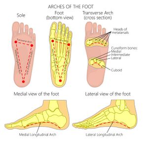
The purpose of the three arches of the foot are:[17]
- Spring
- Weight bearing
- Shock absorption
- To provides flexibility to the foot to facilitate function
1. Medial Longitudinal Arch (MLA)[18][19][edit | edit source]
This is the highest arch due to the shape of bones.
- Bones
- The first three metatarsal
- Three cuneiforms
- Navicular
- Talus
- Calcaneus
- Ligaments
- Spring ligament
- Deltoid ligament
- Interosseus ligament
- Plantar aponeurosis
- Long and short plantar ligaments
- Muscles
- Tibialis posterior
- Tibialis anterior
- Flexor hallucis longus
- Flexor digitorum longus
- Short muscles of the big toe
2. Lateral Longitudinal Arch (LLA)[18][edit | edit source]
The only arch to lie on the ground in a standing position.
- Bones
- Calcaneus
- Cuboid
- Four and fifth metatarsals
- Ligaments
- Long and short plantar ligaments
- Interosseus ligament
- Plantar aponeurosis
- Muscles
- Peroneus longus and brevis
- Flexor digitorum longus
- Short muscles of the little toe
3. Transverse Arch[18][edit | edit source]
- Bones
- Wedge shape of the lateral and intermediate cuneiform
- Metatarsal bases
- Cuboid
- Three cuneiforms
- Ligaments
- Deep transverse ligament
- Dorsal and plantar ligament
- Muscles
- Peroneus longus and brevis
- Transverse head of adductor hallucis
- Slips of tibial posterior
References[edit | edit source]
- ↑ 1.00 1.01 1.02 1.03 1.04 1.05 1.06 1.07 1.08 1.09 1.10 1.11 1.12 1.13 1.14 Green-Smerdon M. Basic Anatomy of the Dancer’s Ankle and Foot course. Plus , 2022.
- ↑ Brockett CL, Chapman GJ. Biomechanics of the ankle. Orthopaedics and trauma. 2016 Jun 1;30(3):232-8.
- ↑ Houglum PA, Bertoti DB. Brunnstrom's clinical kinesiology. FA Davis; 2012
- ↑ 4.0 4.1 4.2 Ficke J, Byerly DW. Anatomy, Bony Pelvis and Lower Limb, Foot. StatPearls [Internet]. 2021 Aug 11.
- ↑ Bell DJ. Talus [Internet]. Radiopedia. 2021 [cited 15/02/2022]. Available from: https://radiopaedia.org/articles/talus
- ↑ Veirs KP, Rippetoe JR, Baldwin JD, Lutz K, Haleem AM, Dionne CP. Multi-Segment Assessment of Ankle and Foot Kinematics during Elevé Barefoot Demi-Pointe and En Pointe. In2020 Combined Sections Meeting (CSM) 2020 Feb 13. APTA.
- ↑ 7.0 7.1 Das A, Bhuyan D. A Review on the Anatomy and Biomechanics of the Foot-Ankle Complex. Asian Journal For Convergence In Technology (AJCT). 2018 Apr 15.
- ↑ SAH SA, MR ES, Srijit D, Norzana AG. Ankle Injuries in Sports: Anatomical Considerations and Clinical Implications.
- ↑ 9.0 9.1 Russell JA, McEwan IM, Koutedakis Y, Wyon MA. Clinical anatomy and biomechanics of the ankle in dance. Journal of dance medicine & science. 2008 Sep 1;12(3):75-82.
- ↑ Norkus SA, Floyd RT. The anatomy and mechanisms of syndesmotic ankle sprains. Journal of athletic training. 2001 Jan;36(1):68.
- ↑ Murdock CJ, Munjal A, Agyeman K. Anatomy, Bony Pelvis and Lower Limb, Calf Flexor Hallucis Longus Muscle. StatPearls [Internet]. 2021 Aug 6.
- ↑ Corcoran NM, Varacallo M. Anatomy, Bony Pelvis and Lower Limb, Tibialis Posterior Muscle. StatPearls [Internet]. 2020 Sep 17.
- ↑ Basit H, Eovaldi BJ, Siccardi MA. Anatomy, Bony Pelvis and Lower Limb, Foot Peroneus Brevis Muscle. In StatPearls [Internet] 2019 May 19. StatPearls Publishing.
- ↑ Farris DJ, Kelly LA, Cresswell AG, Lichtwark GA. The functional importance of human foot muscles for bipedal locomotion. Proceedings of the National Academy of Sciences. 2019 Jan 29;116(5):1645-50.
- ↑ Metsavaht L, Leporace G. Current trends in the biokinetic analysis of the foot and ankle. Journal of the Foot & Ankle. 2020 Aug 30;14(2):191-6.
- ↑ Bolgla LA, Malone TR. Plantar fasciitis and the windlass mechanism: a biomechanical link to clinical practice. Journal of athletic training. 2004 Jan 1;39(1):77.
- ↑ Batenhorst EZ. A Dancer’s View: Analysis and Prevention of Common Dance Injuries.
- ↑ 18.0 18.1 18.2 Ahonen J. Biomechanics of the foot in dance: a literature review. Journal of Dance Medicine & Science. 2008 Sep 1;12(3):99-108.
- ↑ Ozdinc SA, Turan FN. Effects of ballet training of children in Turkey on foot anthropometric measurements and medial longitudinal arc development. J. Pak. Med. Assoc. 2016 Jul 1;66(7):869-74.
