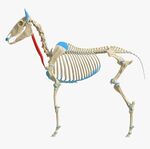
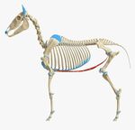
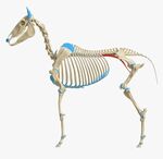
Equine Spine and Head Anatomy: Difference between revisions
No edit summary |
Kim Jackson (talk | contribs) m (Text replacement - "Physioplus" to "Plus ") |
||
| (38 intermediate revisions by 4 users not shown) | |||
| Line 1: | Line 1: | ||
<div class=" | <div class="editorbox"> '''Original Editor '''- [[User:Chelsea Mclene|Chelsea Mclene]] based on the course by | ||
[https://members.physio-pedia.com/course_tutor/petra-zikmann/ Petra Zikmann] | |||
'''Top Contributors''' - {{Special:Contributors/{{FULLPAGENAME}}}}</div> | |||
== Introduction == | == Introduction == | ||
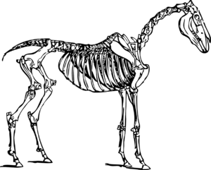
'''Equine | '''Equine anatomy''' refers to the gross and microscopic anatomy of horses and other equids (donkeys, and zebras). | ||
This page introduces the Anatomy of Equine Spine and Head. | This page introduces the Anatomy of Equine Spine and Head. | ||
[[File:Horseanatomy.png|thumb]] | |||
== Axial Skeleton == | == Axial Skeleton == | ||
The axial skeleton | The axial skeleton consists of the skull, vertebral column, sternum, and ribs. Multiple sternebrae fuse to form one bone, attached to the 8 "true" pairs of ribs, out of a total of 18.<ref name=":1">Van der Walt A. Equine Spine and Head Presentation. Plus , 2021.</ref> | ||
The vertebral column contains 54 bones: | The vertebral column contains 54 bones: | ||
* 7 | * 7 cervical vertebrae: includes the atlas (C1) and axis (C2) | ||
* 18-19 | * 18-19 thoracic vertebrae | ||
* 5-6 | * 5-6 lumbar vertebrae | ||
* 5 | * 5 sacral vertebrae | ||
* 15-25 | * 15-25 caudal vertebrae<ref>King, Christine, BVSc, MACVSc, and Mansmann, Richard, VMD, PhD. "[https://amp.en.google-info.org/12740439/1/equine-injury-and-lameness.html Equine Lameness]." ''Equine Research,'' Inc. 1997. | ||
</ref> | </ref> | ||
In certain breeds, there may be | In certain breeds, there may be variations in these numbers.<ref>Riegal, Ronald J. DVM, and Susan E. Hakola RN. [http://www.cep.unep.org/download/719640-file.pdf Illustrated Atlas of Clinical Equine Anatomy and Common Disorders of the Horse Vol. II. Equistar Publication, Limited. Marysville, OH. Copyright 2000.]</ref> | ||
=== Skull === | === Skull === | ||
The skull | The skull contains the brain and the most important organs of sense. | ||
==== Cranium ==== | |||
The Roof of the cranium is made up of frontal and parietal bones. | |||
The Floor is made up of sphenoid bone. | |||
The cranium consists of 5 orbital regions: | |||
* Frontal | |||
* Lacrimal | |||
* Palatine | |||
* Sphenoid | |||
* Zygomatic | |||
Interparietal bone: only found in horse and cat. | |||
The orbit is complete in horse and ruminants while it is incomplete in carnivores but completed by the orbital ligament. | |||
The lacrimal fossa collects tears and sends them through lacrimal canal into the nasal cavity. | |||
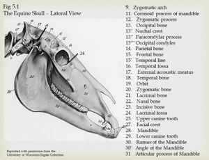
==== Bones in the | ==== Bones in the Equine Skull ==== | ||
There are 34 bones and most of them are flat. During the birth process, these bones overlap | [[File:Equine skull lateral view.jpeg|right|thumb]] | ||
There are 34 bones and most of them are flat. During the birth process, these bones overlap and allow the skull to compress as much as possible to allow for parturition. | |||
14 | The 14 major bones are:<ref name=":1" /> | ||
* Incisive bone (premaxillary): part of the upper jaw; where the incisors attach | * Incisive bone (premaxillary): part of the upper jaw; where the incisors attach | ||
| Line 43: | Line 60: | ||
* Occipital bone: forms the joint between the skull and the first vertebrae of the neck (the atlas) | * Occipital bone: forms the joint between the skull and the first vertebrae of the neck (the atlas) | ||
* Temporal bone: contains the eternal acoustic meatus, which transmits sound from the ear to the cochlea (eardrum) | * Temporal bone: contains the eternal acoustic meatus, which transmits sound from the ear to the cochlea (eardrum) | ||
* Zygomatic bone: attaches to the temporal bone to form the zygomatic arch ( | * Zygomatic bone: attaches to the temporal bone to form the zygomatic arch (cheekbone) | ||
* Palatine bone: forms the back of the hard palate | * Palatine bone: forms the back of the hard palate | ||
* Sphenoid: formed by fusion of the foetal basisphenoid and presphenoid bones, at the base of the skull. Can become fractured in horses that rear over backwards | * Sphenoid: formed by fusion of the foetal basisphenoid and presphenoid bones, at the base of the skull. Can become fractured in horses that rear over backwards | ||
* Vomer: forms the top of the inside of the nasal cavity | * Vomer: forms the top of the inside of the nasal cavity | ||
* Pterygoid: small bone attached to the sphenoid that extends downward | * Pterygoid: small bone attached to the sphenoid that extends downward | ||
==== Cavities ==== | ==== Cavities ==== | ||
The | The equine skull consists of 4 cavities: | ||
* '''The cranial cavity:''' Protects and encloses the brain, supports sense organs. The cranium consists of '''roof''' | * '''The cranial cavity:''' Protects and encloses the brain, supports sense organs. The cranium consists of a '''roof''' made up of the frontal and parietal bones and a '''floor''' made up of the sphenoid bone | ||
* '''The orbital cavity:''' | * '''The orbital cavity:''' Has 5 orbits: frontal, lacrimal, palatine, sphenoid and zygomatic. It protects and surrounds the eye.<ref>Budras, K. Sack, W.O., [https://www.academia.edu/43148945 Anatomy of the Horse, 6th Edition (2012)], Schlutersche Verlagsgesellschaft mbH & Co. KG</ref> Horses have both monocular and binocular vision: | ||
**'''Monocular vision:''' The horse can see objects with one eye. This means that the brain receives two images simultaneously | **'''Monocular vision:''' The horse can see objects with one eye. This means that the brain receives two images simultaneously | ||
** '''Binocular vision:''' The horse can focus with both eyes just like humans and the brain | ** '''Binocular vision:''' The horse can focus with both eyes just like humans and the brain receives only one signal | ||
* '''The oral cavity:''' | * '''The oral cavity:''' A passage into the respiratory and digestive system | ||
* '''The nasal cavity:''' | * '''The nasal cavity:''' Contains bone that protects the mucous membrane from inspired warm air | ||
==== Foramina of the | ==== Foramina of the Skull and the Structures Passing Through ==== | ||
{| class="wikitable" | {| class="wikitable" | ||
!Foramina | !Foramina | ||
| Line 88: | Line 104: | ||
CN V3 (horse and pig) | CN V3 (horse and pig) | ||
|- | |- | ||
|Internal | |Internal acoustic meatus | ||
|CNVIII | |CNVIII | ||
|- | |- | ||
| Line 105: | Line 121: | ||
== Joints and Ligaments == | == Joints and Ligaments == | ||
=== Joints and Ligaments of Skull === | === Joints and Ligaments of the Skull === | ||
''' | * '''The temporo-mandibular joint''' - a '''condylar joint''' between the mandibular condyles and the mandibular fossae of the temporal bones. It has a '''loose joint capsule''' with thickenings that form a lateral ligament, as well as an '''articular disc'''<ref name=":1" /> | ||
* '''Mandibular symphysis''' | |||
* '''Hyoid apparatus''' - consists of three joints: | |||
* Tympanohyoid cartilage- skull (syndesmosis) | ** Tympanohyoid cartilage- skull (syndesmosis) | ||
* Interhyoid joints (synovial) | ** Interhyoid joints (synovial) | ||
* Thyrohyoid bone- cranial cornu of thyroid cartilage (synovial) | ** Thyrohyoid bone- cranial cornu of thyroid cartilage (synovial)<ref name=":1" /> | ||
{{#ev:youtube|FhA-blWzC8Y|300}}<ref>Equine head. PeabodyDVM. Available from: https://www.youtube.com/watch?v=FhA-blWzC8Y[last accessed 01/05/2021]</ref> | |||
=== Cervical Spine === | === Cervical Spine === | ||
==== Joints ==== | ==== Joints ==== | ||
[[ File:Equine cervical spine.jpeg|right|thumb]] | |||
===== Atlanto-Occipital Joint ===== | |||
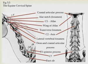
A '''condylar, modified synovial hinge joint.''' The articulating surfaces are the occipital condyles and the cranial articular surfaces of the atlas (C1). There are three thickenings that strengthen the spacious joint capsule: '''Dorsal, Ventral, Lateral'''. The '''transverse atlantal ligament''' holds the dens of the axis against the ventral arch of the atlas.<ref name=":1" /> | |||
===== Atlanto- | ===== Atlanto-Axial Joint ===== | ||
A '''pivot joint''' between the atlas and the saddle shaped surface of the axis (C2), which extends upon the dens. It has a '''loose joint capsule'''. The '''apical ligament of dens''' connects the apex of the dens to the occipital bone. Motion at this joint includes rotation of the atlas and head upon the axis and some accessory lateral flexion.<ref name=":1" /> Rotation at this joint makes up 73 percent of cervical rotation.<ref name=":2">Clayton HM, Townsend HG. Kinematics of the cervical spine of the adult horse. Equine Vet J. 1989;21(3):189-92.</ref> | |||
===== Cervical Spine C3-C4 ===== | |||
A planar, extensive, oval shaped joint that is obliquely oriented in transverse plane. The '''cranial articular processes''' face dorsomedially and the '''caudal articular processes''' face ventrolaterally. Spinous process height increases caudally from C6. Lateral flexion is the primary motion at these joints (25-45 degrees each joint - C1/C2 only has 3.9 degrees of lateral flexion).<ref name=":2" /> | |||
===== Cervical | |||
==== Ligaments ==== | ==== Ligaments ==== | ||
| Line 133: | Line 151: | ||
* Ligamentum flavum | * Ligamentum flavum | ||
* '''Nuchal ligament:''' | * '''Nuchal ligament:''' | ||
** This ligament connects the thoracic vertebra to the head and assists in supporting its weight. It consists of two paired parts:<ref name=":1" /> | |||
*** Funicular (cord) part - extends from the poll to +/- the second to the fourth thoracic spinous process | |||
*** Lamellar part - arises from the second and third thoracic spinous processes and the funicular part, and inserts on the C2-C6 spinous processes. The first digitation going to the axis is very strong, but it decreases in strength caudally | |||
It consists of | |||
* Funicular (cord) part- poll to +/- | |||
* Lamellar part- arises from | |||
=== Thoracic Spine (T1-T18) === | === Thoracic Spine (T1-T18) === | ||
==== Articular processes ==== | ==== Articular processes ==== | ||
'''Caudal articular processes''' face ventrally and at base of spinous process. ''' | '''Caudal articular processes''' face ventrally and are positioned at the base of the spinous process. The '''cranial articular processes''' are oval facets on the arch of the vertebra and face dorsally. Each thoracic vertebrae has a pair of costal facets on the dorsal body (except the last) forming the '''costal fovea'''.<ref name=":1" /> | ||
'''Anticlinal vertebrae''': | '''Anticlinal vertebrae''': This is the point in the caudal thoracic vertebral column at which the anatomic features of the vertebra start to change.<ref>Baines EA, Grandage J, Herrtage ME, Baines SJ. [https://pubmed.ncbi.nlm.nih.gov/19241757/ Radiographic definition of the anticlinal vertebra in the dog.] Veterinary Radiology & Ultrasound. 2009 Jan;50(1):69-73.</ref> This usually occurs at the 13th vertebra in horses. | ||
''' | '''Motion:''' | ||
==== Rib | * Flexion - most flexion occurs at T17/T18; least flexion occurs at T3-T9 | ||
* Extension - most extension occurs at T14-T18; least extension occurs at T2-9<ref name=":1" /> | |||
==== Rib Neck ==== | |||
Has 2 converse facets: '''Cranial and Caudal''' | |||
Rib 1 | Rib 1 attaches to C7, T1 and the associated IV disc | ||
Motion: rotation of the rib, which is greater caudally | |||
=== Costovertebral Joint === | === Costovertebral Joint === | ||
==== Joints ==== | ==== Joints ==== | ||
The costovertebral joints have two distinct articulations between most ribs and the vertebral column:<ref name=":1" /> | |||
# Head of the rib: Cranial and caudal costal facets of adjacent vertebrae; | # Head of the rib: Cranial and caudal costal facets of adjacent vertebrae; a ball and socket synovial joint | ||
# Tubercle of the rib: Transverse process of vertebrae; | # Tubercle of the rib: Transverse process of vertebrae; a plane synovial joint | ||
==== Ligaments ==== | ==== Ligaments ==== | ||
| Line 173: | Line 188: | ||
=== Lumbar Spine === | === Lumbar Spine === | ||
==== Joints ==== | ==== Joints ==== | ||
Horses usually have '''6 lumbar vertebrae (L1-L6)''', but some arabian horses only have 5 (L1-L5).<ref name=":1" /> | |||
===== Articular processes ===== | ===== Articular processes ===== | ||
'''Cranial articular processes''' | '''Cranial articular processes''' are fused with mammillary processes. They are concave dorsally and mostly in sagittal alignment. '''Caudal processes''' are convex ventrally and correspond with the convexity of the cranial articular processes. They are differentiated from the last thoracic vertebra by the lack of costal facets.<ref name=":1" /> | ||
'''Caudal processes''' are convex ventrally | |||
''' | '''Motion:''' The lumbar spine and caudal thoracic spine are the least mobile regions of a horse's back.<ref>Townsend HG, Leach DH, Fretz PB. [https://pubmed.ncbi.nlm.nih.gov/6873044/ Kinematics of the equine thoracolumbar spine]. Equine Vet J. 1983;15(2):117-22. </ref> Lateral flexion and rotation is very limited especially at L4-L6 due to intertransverse joints.<ref name=":1" /> | ||
==== Ligaments of Thoraco- | ==== Ligaments of Thoraco-Lumbar Spine ==== | ||
* Supraspinous ligament: | * Supraspinous ligament: A heavy band of connective tissue running over the top of spinous processes ( T2/T3 caudally). It prevents abnormal separation of spinous processes during flexion | ||
* Ventral | * Ventral longitudinal ligament: Marks the ventral surface of vertebrae from the axis to the sacrum. It is strongest and widest caudally. It plays a major role in preventing overextension of the spine | ||
* Dorsal longitudinal ligament: | * Dorsal longitudinal ligament: Extends from the floor of the vertebral canal from the axis to sacrum and helps to prevent spine hyper-flexion | ||
* Annulus fibrosis of IVD: | * Annulus fibrosis of IVD: Thick ventrally | ||
* Intertransverse ligament | * Intertransverse ligament | ||
* Interarcuate ligament/ yellow ligament/ ligamentum flavum: | * Interarcuate ligament/ yellow ligament/ ligamentum flavum: An elastic ligament that fills the dorsal space between the arch of the adjacent vertebra<ref name=":1" /> | ||
=== Lumbosacral Joint === | === Lumbosacral Joint === | ||
The cranial articular process of the first sacral vertebra are concave and face dorsomedially. | The cranial articular process of the first sacral vertebra are concave and face dorsomedially. | ||
''' | '''Motion:''' Flexion and Extension - 23.4 degrees<ref>Degueurce C, Chateau H, Denoix JM. [https://pubmed.ncbi.nlm.nih.gov/15656498/ In vitro assessment of movements of the sacroiliac joint in the horse.] Equine Vet J. 2004;36(8):694-8. </ref> | ||
=== Sacrum === | === Sacrum === | ||
The sacrum consists of '''fused sacral vertebrae''' and has '''dorsal''' and '''ventral''' sacral foramina.{{#ev:youtube|ektcPGjKJj0|300}}<ref>Equine spine. Dani Jay - Equestrian. Available from: https://www.youtube.com/watch?v=ektcPGjKJj0 [last accessed 01/05/2021]</ref> | |||
== Myology and Neurology == | == Myology and Neurology == | ||
| Line 207: | Line 218: | ||
==== Muscles of the Face ==== | ==== Muscles of the Face ==== | ||
The muscles of facial expressions are innervated by the motor fibers of CNVII (facial nerve).<ref name=":1" /> | |||
{| class="wikitable" | {| class="wikitable" | ||
! colspan="2" |Muscle | ! colspan="2" |Muscle | ||
| Line 223: | Line 234: | ||
|The lateral wing of nostril | |The lateral wing of nostril | ||
The maxillary lip | The maxillary lip | ||
| rowspan="2" | | | rowspan="2" |Elevates and retracts the angle of the mouth | ||
|- | |- | ||
| colspan="2" |M. Zygomaticus | | colspan="2" |M. Zygomaticus | ||
| Line 231: | Line 242: | ||
| colspan="2" |M. Buccinator | | colspan="2" |M. Buccinator | ||
|Maxilla and Mandible | |Maxilla and Mandible | ||
| | |Flattens the cheeks and thus presses food between the teeth | ||
|- | |- | ||
| colspan="2" |M. Depressor labii mandibulars | | colspan="2" |M. Depressor labii mandibulars | ||
|The alveolar border of the Mandible | |The alveolar border of the Mandible | ||
|The mandibular lip | |The mandibular lip | ||
| | |Depresses and retracts the mandibular lip | ||
|- | |- | ||
| colspan="2" |M. Orbicularis oris | | colspan="2" |M. Orbicularis oris | ||
| | |The sphincter muscle of the skin and the muscles of the lips | ||
Corner of the mouth | Corner of the mouth | ||
|Into the lips as it surrounds the mouth | |Into the lips as it surrounds the mouth | ||
| | |Closes the mouth | ||
|- | |- | ||
| colspan="2" |M. Risorius | | colspan="2" |M. Risorius | ||
|Part of M. cutaneous faciei | |Part of M. cutaneous faciei | ||
|The angle of the mouth | |The angle of the mouth | ||
| | |Retracts the angle of the mouth | ||
|- | |- | ||
| colspan="2" |M. Dilator naris | | colspan="2" |M. Dilator naris | ||
|Alar cartillage | |Alar cartillage | ||
|Alar cartillage | |Alar cartillage | ||
| | |Dilates the nostril | ||
|- | |- | ||
| rowspan="2" |M. Lateralis nasi | | rowspan="2" |M. Lateralis nasi | ||
| Line 263: | Line 274: | ||
Parietal cartilage | Parietal cartilage | ||
| rowspan="2" | | | rowspan="2" |Dilates the nostril and nasal vestibule | ||
|- | |- | ||
|Ventral part | |Ventral part | ||
| Line 272: | Line 283: | ||
|Maxilla close to the rostral extremity of the facial crest | |Maxilla close to the rostral extremity of the facial crest | ||
|Lateral wing of the nostril | |Lateral wing of the nostril | ||
| | |Dilates the nostril laterally | ||
|- | |- | ||
| colspan="2" |M. Levator nasolabialis | | colspan="2" |M. Levator nasolabialis | ||
|Frontal and Nasal bones | |Frontal and Nasal bones | ||
|Lateral wing of the nostril | |Lateral wing of the nostril | ||
| | |Elevates the maxillary lip and the commissure of the mouth | ||
Dilates the nostril | |||
|} | |} | ||
| Line 286: | Line 297: | ||
===== Outer Ear ===== | ===== Outer Ear ===== | ||
The '''outer ear''' includes: | The '''outer ear''' includes:<ref name=":1" /> | ||
* Pinna: | * Pinna: mobile and can move independently - can hear multiple sounds at the same time<ref name=":0">Aiello SE, Moses MA, Allen DG, editors. [https://vetu.pw/wy-dole.pdf The Merck veterinary manual]. Whitehouse Station, NJ, USA: Merck; 1998.</ref> | ||
* Ear canal | * Ear canal | ||
''' | '''Cartilage:''' Cartilages of the ear collect and transmit sound to the essential organ of hearing within the temporal bone. In order to achieve this, they (especially the concha) need to move.<ref name=":1" /> | ||
'''The muscles of outer ear:'''<ref name=":1" /> | |||
* Rostral | * Rostral | ||
* Dorsal | * Dorsal | ||
| Line 307: | Line 310: | ||
* Ventral | * Ventral | ||
There are 3 cartilages:<ref name=":1" /> | |||
* Conchal: Forms the framework of the portion of the ear which stands erect. It has a large vertical opening on one side to receive sound, and is attached below to the annular cartilage | |||
* Annular: A small ring of gristle connected to the auditory process of the petrous temporal bone | |||
* Scutiform: A small, flat and somewhat triangular cartilaginous plate situated in front of the base of concha, to which it is attached | |||
===== Middle Ear ===== | ===== Middle Ear ===== | ||
The '''middle ear''' includes: | The '''middle ear''' includes:<ref name=":1" /> | ||
* Eardrum | * Eardrum | ||
* Small, air-filled chamber containing 3 tiny bones: '''the hammer, anvil, and stirrup'''. It also includes '''2 muscles | * Small, air-filled chamber containing 3 tiny bones: '''the hammer, anvil, and stirrup'''. It also includes '''2 muscles: the oval window, and the eustachian tube.''' | ||
===== Inner Ear ===== | ===== Inner Ear ===== | ||
| Line 317: | Line 325: | ||
==== Muscles of Mastication ==== | ==== Muscles of Mastication ==== | ||
The muscles of mastication are innervated by the mandibular branch of trigeminal nerve CNV.<ref name=":1" /> | |||
{| class="wikitable" | {| class="wikitable" | ||
|+ | |+ | ||
| Line 327: | Line 335: | ||
|M. Masseter | |M. Masseter | ||
|The zygomatic arch and the facial crest | |The zygomatic arch and the facial crest | ||
|The lateral border of | |The lateral border of the ramus of the mandible | ||
| | |Closes the mouth | ||
|- | |- | ||
|M. Temporalis | |M. Temporalis | ||
|The temporal fossa and the temporal crest | |The temporal fossa and the temporal crest | ||
|The coronoid process of the | |The coronoid process of the mandible | ||
| rowspan="2" | | | rowspan="2" |Closes the mouth (to raise the mandible) | ||
|- | |- | ||
|M. Pterygoideus medialis | |M. Pterygoideus medialis | ||
|The crest formed by the pterygoid processes of the | |The crest formed by the pterygoid processes of the basisphenoid and the palatine bones | ||
|The medial surface of the | |The medial surface of the ramus of the mandible | ||
|- | |- | ||
|M. Pterygoideus lateralis | |M. Pterygoideus lateralis | ||
|The pterygoid process of the | |The pterygoid process of the sphenoid bone | ||
|Rostral border of the condyle of the | |Rostral border of the condyle of the mandible | ||
| | |Draws and moves the mandible rostrally | ||
|- | |- | ||
|M. Digastricus | |M. Digastricus | ||
|The jugular process of | |The jugular process of occipital bone | ||
|Medial surface of the ventral border of the molar part of the body of the | |Medial surface of the ventral border of the molar part of the body of the mandible | ||
| rowspan="2" | | | rowspan="2" |Opens the mouth | ||
|- | |- | ||
|M. Occipitomandibularis | |M. Occipitomandibularis | ||
|The jugular process | |The jugular process | ||
|The caudal border of the ramus of the | |The caudal border of the ramus of the mandible | ||
|} | |} | ||
==== Muscles of Eyes ==== | ==== Muscles of the Eyes ==== | ||
* '''M. Orbicularis oculi''' | * '''M. Orbicularis oculi''' - innervated by palpebral branch of CN VII | ||
* '''M. Levator palpebrae superioris''' originates from | * '''M. Levator palpebrae superioris''' - originates from the posterior orbit and inserts at orbicularis oculi fibers of the lower eyelid. It elevates the upper eyelid and is innervated by CN III (oculomotor nerve) | ||
* '''M. Malaris''' lowers the ventral eyelid. It is innervated by CN VII (facial nerve) | * '''M. Malaris''' - lowers the ventral eyelid. It is innervated by CN VII (facial nerve) | ||
* '''Muller's muscle''' | * '''Muller's muscle''' - innervated by sympathetic nerves | ||
* '''Ciliary muscles''' | * '''Ciliary muscles''' | ||
* '''M. Retractor anguli''' | * '''M. Retractor anguli''' - retracts and anchors the lateral canthus | ||
* '''M. Levator anguli oculi medialis''' and '''M. Frontalis''' slightly elevates of the upper eyelid | * '''M. Levator anguli oculi medialis''' and '''M. Frontalis''' - slightly elevates of the upper eyelid<ref>Gilger BC, Stoppini R[https://www.sciencedirect.com/science/article/pii/B0721605222500066 . Diseases of the eyelids, conjunctiva, and nasolacrimal system.] GILGER BC. 2005.</ref><ref>Brooks DE, Matthews AG. [https://scholar.google.com/scholar_url?url=http://citeseerx.ist.psu.edu/viewdoc/download%3Fdoi%3D10.1.1.470.6399%26rep%3Drep1%26type%3Dpdf&hl=en&sa=T&oi=gsb-ggp&ct=res&cd=1&d=10378886115965098544&ei=kwuNYJCzHYqWyQSZuLfQBA&scisig=AAGBfm3c5eqUPk8D0l4LPN6WiGVXeFv63Q Equine ophthalmology. Veterinary ophthalmology.] 1999;2:1108.</ref> | ||
==== Muscles of Tongue ==== | ==== Muscles of the Tongue ==== | ||
The equine tongue is made up of twelve different muscles<ref>Nicole Kitchener. Horse Sport. Available from<nowiki/>https://horsesport.com/magazine/health/horse-tongues-101/ | The equine tongue is made up of twelve different muscles<ref>Nicole Kitchener. Horse Sport. Available from<nowiki/>https://horsesport.com/magazine/health/horse-tongues-101/ | ||
[last accessed 01/05/2021]</ref> including '''styloglossus, genioglossus and hyoglossus'''. These muscles are covered by mucosa on the sides and underneath. | [last accessed 01/05/2021]</ref> including '''styloglossus, genioglossus and hyoglossus'''. These muscles are covered by mucosa on the sides and underneath.<ref name=":1" /> | ||
Action: prehension, mastication (i.e. chewing) | |||
Innervation: Hypoglossus (CNXII) | Innervation: Hypoglossus (CNXII) | ||
| Line 385: | Line 393: | ||
|- | |- | ||
| colspan="2" |M. Tensor veli palatini | | colspan="2" |M. Tensor veli palatini | ||
|Muscular process of the petrous part of the | |Muscular process of the petrous part of the temporal bone, pterygoid bone, and lateral lamina of the auditory tube | ||
|Palatine aponeurosis | |Palatine aponeurosis | ||
|Retracts the soft palate away from the | |Retracts the soft palate away from the dorsal pharyngeal wall, expanding the nasopharynx and slightly depressing it ventrad during inspiration | ||
|Mandibular branch of the | |Mandibular branch of the trigeminal nerve | ||
|- | |- | ||
| colspan="2" |M. Levator veli palatini | | colspan="2" |M. Levator veli palatini | ||
|Muscular process of the | |Muscular process of the petrous part of the temporal bone and the lateral lamina of the Auditory tube and passes along the lateral wall of the nasopharynx | ||
|Soft palate dorsal to the glandular layer | |Soft palate dorsal to the glandular layer | ||
|Elevates the soft palate during swallowing | |Elevates the soft palate during swallowing | ||
| Line 399: | Line 407: | ||
|Caudal aspect of the palatine aponeurosis | |Caudal aspect of the palatine aponeurosis | ||
|Caudal free margin of the soft palate | |Caudal free margin of the soft palate | ||
| rowspan="2" | | | rowspan="2" |Shortens the soft palate and depresses it towards the tongue | ||
|- | |- | ||
| colspan="2" |M. Palatopharyngeus | | colspan="2" |M. Palatopharyngeus | ||
|Palatine aponeurosis and from the palatine and | |Palatine aponeurosis and from the palatine and pterygoid bones | ||
|Upper edge of the | |Upper edge of the thyroid cartilage | ||
|- | |- | ||
| rowspan="2" |M. Stylopharyngeus | | rowspan="2" |M. Stylopharyngeus | ||
| Line 435: | Line 443: | ||
| colspan="2" |M. Thyrohyoideus | | colspan="2" |M. Thyrohyoideus | ||
|Lateral lamina of the Thyroid cartilage | |Lateral lamina of the Thyroid cartilage | ||
|Caudal aspect of the | |Caudal aspect of the thyrohyoid bone | ||
| | |Moves the larynx rostrad | ||
|- | |- | ||
| colspan="2" |M. Hyoglossus | | colspan="2" |M. Hyoglossus | ||
|Hyoid bones | |Hyoid bones | ||
|Median plane of the | |Median plane of the dorsum of the tongue | ||
| | |Retracts and depresses the base of the tongue | ||
|- | |- | ||
| colspan="5" |M. Hyoepiglotticus | | colspan="5" |M. Hyoepiglotticus | ||
| Line 452: | Line 460: | ||
| colspan="2" |M. Sternohyoideus | | colspan="2" |M. Sternohyoideus | ||
| rowspan="2" |Sternal manubrium | | rowspan="2" |Sternal manubrium | ||
|Basihyoid bone and lingual process of the | |Basihyoid bone and lingual process of the hyoid apparatus | ||
| rowspan="2" |Caudal traction | | rowspan="2" |Caudal traction | ||
| rowspan="2" |Branches of the first and second | | rowspan="2" |Branches of the first and second cervical nerves | ||
|- | |- | ||
| colspan="2" |M. Sternothyroideus | | colspan="2" |M. Sternothyroideus | ||
|Caudolateral aspect of the | |Caudolateral aspect of the thyroid cartilage | ||
|} | |} | ||
==== Muscles of Hyoid | ==== Muscles of the Hyoid Apparatus and Larynx ==== | ||
Muscles of | Muscles of the hyoid apparatus and larynx are innervated by CNX. | ||
===== Hyoid | ===== Hyoid Apparatus ===== | ||
The hyoid apparatus has muscular connections from the throat to the forelimbs, shoulder, and sternum. Sternohyoid and omohyoid provide a direct connection from the hyoid apparatus to the shoulder of the horse via the ventral neck. The tongue connects to the hyoid apparatus. Small muscles of the hyoid apparatus connect to the TMJ and the poll and the TMJ articulates with the hyoid apparatus.<ref>Cornelisse CJ, Rosenstein DS, Derksen FJ, Holcombe SJ. [https://scholar.google.com/scholar_url?url=https://avmajournals.avma.org/doi/abs/10.2460/ajvr.2001.62.1865&hl=en&sa=T&oi=gsb&ct=res&cd=1&d=8361378216004701372&ei=niuNYPPRE4vuygSir5CwBA&scisig=AAGBfm0JQnYNUi9DuBlmJBSoX5R0x9nNVg Computed tomographic study of the effect of a tongue-tie on hyoid apparatus position and nasopharyngeal dimensions in anaesthetised horses. American journal of veterinary research]. 2001 Dec 1;62(12):1865-9.</ref><ref>Chalmers HJ, Cheetham J, Yeager AE, Ducharme NG. [https://scholar.google.com/scholar_url?url=https://onlinelibrary.wiley.com/doi/abs/10.1111/j.1740-8261.2006.00170.x&hl=en&sa=T&oi=gsb&ct=res&cd=0&d=8287556430549573786&ei=xiuNYLuDCPqB6rQPvp6ByA0&scisig=AAGBfm05TOkMHItqKqLyMfz2EPOmuiUJKA Ultrasonography of the equine larynx]. Veterinary Radiology & Ultrasound. 2006 Sep;47(5):476-81.</ref> | |||
===== Larynx ===== | ===== Larynx ===== | ||
Intrinsic muscles: | Intrinsic muscles: | ||
* Cricoarytenoideus dorsalis | * Cricoarytenoideus dorsalis - abduction of arytenoids and tensing of vocal cords | ||
* Thyroarytenoideus | * Thyroarytenoideus - adduction of arytenoids | ||
* Arytenoideus transversus | * Arytenoideus transversus - adduction of arytenoids | ||
* Cricoarytenoideus lateralis | * Cricoarytenoideus lateralis - adduction of arytenoids<ref>Kelly PG. ''[http://theses.gla.ac.uk/7248/ Studies on serial resting and dynamic endoscopic examination] of thoroughbred yearlings'' (Doctoral dissertation, University of Glasgow).</ref> | ||
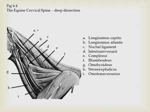
=== Muscles of Cervical spine === | === Muscles of Cervical spine === | ||
{| class="wikitable" | {| class="wikitable" | ||
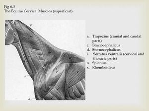
|+<ref>Payne RC, Veenman P, Wilson AM. [https://scholar.google.com/scholar_url?url=https://onlinelibrary.wiley.com/doi/abs/10.1111/j.1469-7580.2005.00353.x&hl=en&sa=T&oi=gsb&ct=res&cd=0&d=3057679320871654845&ei=IDSNYJfBCJz0yAS636q4Cg&scisig=AAGBfm21qD3VO0xVz_DwruKe27l1S2ozGw The role of the extrinsic thoracic limb muscles in equine locomotion. Journal of Anatomy]. 2005 Feb;206(2):193-204.</ref><ref>Wikivet. Online Veterinary Encyclopedia. Thoracic Limb Extrinsic Muscles - Horse Anatomy. Available from https://en.wikivet.net/Thoracic_Limb_Extrinsic_Muscles_-_Horse_Anatomy#:~:text=Innervation%3A%20Dorsal%20and%20ventral%20branches,cervical%20and%207th%20thoracic%20vertebrae [last accessed 01/05/2021]</ref> | |+<ref>Payne RC, Veenman P, Wilson AM. [https://scholar.google.com/scholar_url?url=https://onlinelibrary.wiley.com/doi/abs/10.1111/j.1469-7580.2005.00353.x&hl=en&sa=T&oi=gsb&ct=res&cd=0&d=3057679320871654845&ei=IDSNYJfBCJz0yAS636q4Cg&scisig=AAGBfm21qD3VO0xVz_DwruKe27l1S2ozGw The role of the extrinsic thoracic limb muscles in equine locomotion. Journal of Anatomy]. 2005 Feb;206(2):193-204.</ref><ref>Wikivet. Online Veterinary Encyclopedia. Thoracic Limb Extrinsic Muscles - Horse Anatomy. Available from https://en.wikivet.net/Thoracic_Limb_Extrinsic_Muscles_-_Horse_Anatomy#:~:text=Innervation%3A%20Dorsal%20and%20ventral%20branches,cervical%20and%207th%20thoracic%20vertebrae [last accessed 01/05/2021]</ref>[[File:Equine Cervical Muscles.jpeg|left|frameless]][[File:Equine Cervical Spine.jpeg|right|frameless]] | ||
! colspan="2" |Muscle | ! colspan="2" |Muscle | ||
!Origin | !Origin | ||
| Line 483: | Line 491: | ||
!Innervation | !Innervation | ||
|- | |- | ||
| colspan="2" |M. Omotransversarius | | colspan="2" |M. Omotransversarius [[File:Equine Omotransversarius.jpeg|150x150px|alt=|center]] | ||
|Fascia of shoulder | |Fascia of shoulder | ||
|Scapular cartilage and | |Scapular cartilage and transverse processes of C2-4 | ||
|Advances limb | |Advances limb | ||
Adducts limb | Adducts limb | ||
Moves neck laterally | Moves neck laterally | ||
|Ventral branch of | |Ventral branch of local cervical spinal nerve | ||
|- | |- | ||
| colspan="2" |M. Brachiocephalicus | | colspan="2" |M. Brachiocephalicus[[File:Equine Brachiocephalicus.jpeg|150x150px|alt=|center]] | ||
|Mastoid process of | |Mastoid process of temporal bone and first cervical vertebra | ||
|Deltoid tuberosity and | |Deltoid tuberosity and crest of the humerus | ||
|Shoulder extension | |Shoulder extension | ||
Protraction | Protraction | ||
Flexion of the neck towards the side of the protracting limb | |||
|Accessory nerve | |Accessory nerve | ||
|- | |- | ||
| colspan="2" |M. Cleidobrachialis | | colspan="2" |M. Cleidobrachialis [[File:Equine Cleidobrachialis.jpeg|150x150px|alt=|center]] | ||
|Inscription of clavicle | |Inscription of clavicle | ||
|Crest of the | |Crest of the humerus | ||
|Advances limb | |Advances limb | ||
| Line 511: | Line 519: | ||
| colspan="2" |M. Cleidomastoideus | | colspan="2" |M. Cleidomastoideus | ||
|Clavicular intersection | |Clavicular intersection | ||
|Mastoid process of | |Mastoid process of temporal bone | ||
|Advances limb | |Advances limb | ||
| Line 522: | Line 530: | ||
|- | |- | ||
| colspan="2" |M. Sternocephalicus | | colspan="2" |M. Sternocephalicus | ||
(Sternomandibularis) | (Sternomandibularis)[[File:Equine Sternocephalicus.jpeg|150x150px|alt=|center]] | ||
|Manubrium of the sternum | |Manubrium of the sternum | ||
|Caudal border of | |Caudal border of mandible | ||
|Turns head | |Turns head | ||
Opens mouth | Opens mouth | ||
|- | |- | ||
| colspan="2" |M. Omohyoideus | | colspan="2" |M. Omohyoideus [[File:Equinee omohyoid.jpeg|150x150px|alt=|center]] | ||
|Subscapular fascia | |Subscapular fascia | ||
|Lingual process of | |Lingual process of basihyoid bone | ||
|Retracts basihyoid bone and tongue | |Retracts basihyoid bone and tongue | ||
|Spinal nerve C1 | |Spinal nerve C1 | ||
|- | |- | ||
| colspan="2" rowspan="2" |M. Trapezius | | colspan="2" rowspan="2" |M. Trapezius [[File:Equine trapezius.jpeg|150x150px|alt=|center]] | ||
| rowspan="2" |Nuchal ligament and Supraspinous ligaments of C2-10 | | rowspan="2" |Nuchal ligament and Supraspinous ligaments of C2-10 | ||
|Cervical part: Entire scapular spine | |Cervical part: Entire scapular spine | ||
| Line 548: | Line 556: | ||
|Thoracic part: Dorsal third of Scapular spine | |Thoracic part: Dorsal third of Scapular spine | ||
|- | |- | ||
| colspan="2" |M. Rhomboideus | | colspan="2" |M. Rhomboideus [[File:Equine rhomboideus.jpeg|150x150px|alt=|center]] | ||
(cervicis and thoracis) | (cervicis and thoracis) | ||
|Nuchal ligament and | |Nuchal ligament and dorsoscapular ligaments of C2-T8 | ||
|Scapular cartilage | |Scapular cartilage | ||
|Elevates neck | |Elevates neck | ||
| Line 559: | Line 567: | ||
|Local thoracic nerve and Local cervical nerve | |Local thoracic nerve and Local cervical nerve | ||
|- | |- | ||
| colspan="2" |M. Serratus ventralis (cervicis) | | colspan="2" |M. Serratus ventralis (cervicis) [[File:EquineSerratus ventralis.jpeg|150x150px|alt=|center]] | ||
|Transverse processes of C4-7 | |Transverse processes of C4-7 | ||
|Scapular cartilage and | |Scapular cartilage and medial scapula | ||
|Supports trunk between forelimbs | |Supports trunk between forelimbs | ||
Raises neck when limb is fixed | Raises neck when the limb is fixed | ||
|Ventral branch of | |Ventral branch of local cervical nerve | ||
|- | |- | ||
| colspan="2" |M. Splenius | | colspan="2" |M. Splenius | ||
(capitus and cervicis) [[File:Equine Splenius.jpeg|150x150px|alt=|center]] | |||
(capitus and cervicis) | |Nuchal ligament and spinous processes of T3-T5 | ||
|Nuchal ligament and | |||
|Nuchal crest and mastoid process of temporal bone | |Nuchal crest and mastoid process of temporal bone | ||
|Extends neck | |Extends neck | ||
| Line 577: | Line 584: | ||
Bends neck laterally | Bends neck laterally | ||
|Dorsal branch of Accessory nerve and | |Dorsal branch of Accessory nerve and dorsal branch of local spinal nerve | ||
|- | |- | ||
| colspan="2" |M. Longissimus | | colspan="2" |M. Longissimus | ||
(cervicis, capitus, atlantis) [[File:Equine longissimus.jpeg|150x150px|alt=|center]] | |||
(cervicis, capitus, atlantis) | |||
|Transverse processes of cervical and thoracic vertebrae | |Transverse processes of cervical and thoracic vertebrae | ||
|Wing of | |Wing of atlas and mastoid process of temporal bone | ||
|Elevates head and neck | |Elevates head and neck | ||
| Line 589: | Line 595: | ||
Stabilizes and extends vertebral column | Stabilizes and extends vertebral column | ||
| rowspan="2" |Dorsal branch of | | rowspan="2" |Dorsal branch of local spinal nerve | ||
|- | |- | ||
| colspan="2" |M. Semispinalis capitis | | colspan="2" |M. Semispinalis capitis[[File:Equine semispinalis capitis.jpeg|150x150px|alt=|center]] | ||
|Articular processes of C2/3-7 and | |Articular processes of C2/3-7 and transverse processes of T1-6/7 | ||
|Occipital bone | |Occipital bone | ||
|Elevates head and neck | |Elevates head and neck | ||
| Line 598: | Line 604: | ||
Bends head and neck laterally | Bends head and neck laterally | ||
|- | |- | ||
| colspan="2" |M. Longus capitis | | colspan="2" |M. Longus capitis [[File:Equine longissimus capitis.jpeg|150x150px|alt=|center]] | ||
|Transverse processes of C3-5 | |Transverse processes of C3-5 | ||
|Base of skull | |Base of skull | ||
|Bends head and neck | |Bends head and neck | ||
| rowspan="3" |Ventral branch of | | rowspan="3" |Ventral branch of local spinal nerve | ||
|- | |- | ||
| rowspan="2" |M. Longus colli | | rowspan="2" |M. Longus colli [[File:Equine Longus Colli.jpeg|150x150px|alt=|center]] | ||
|Cervical part | |Cervical part | ||
|Transverse processes of C3-7 | |Transverse processes of C3-7 | ||
|Ventral tubercle of | |Ventral tubercle of atlas and bodies of cervical vertebrae | ||
|Flexes head | |Flexes head | ||
|- | |- | ||
| Line 630: | Line 636: | ||
|- | |- | ||
|Minor | |Minor | ||
|Dorsal arch of the | |Dorsal arch of the atlas | ||
|Occipital bone | |Occipital bone | ||
|- | |- | ||
| colspan="2" |M. Scalenes | | colspan="2" |M. Scalenes [[File:Equine scalenes.jpeg|150x150px|alt=|center]] | ||
|Transverse processes of the last 4 cervical vertebrae | |Transverse processes of the last 4 cervical vertebrae | ||
|Anterior border and Outer surface of first rib | |Anterior border and Outer surface of the first rib | ||
|Assists inspiration by drawing the first rib forward. | |Assists inspiration by drawing the first rib forward. | ||
| Line 644: | Line 650: | ||
=== Muscles of Trunk === | === Muscles of Trunk === | ||
{| class="wikitable" | {| class="wikitable" | ||
|+ | |+[[File:Equine Trunk muscles.jpeg|left|frameless]] | ||
! colspan="2" |Muscles | ! colspan="2" |Muscles | ||
!Origin | !Origin | ||
| Line 651: | Line 657: | ||
!Innervation | !Innervation | ||
|- | |- | ||
| colspan="2" |M. Latissimus dorsi | | colspan="2" |M. Latissimus dorsi [[File:Equine lattisimus dorsi.jpeg|150x150px|alt=|center]] | ||
|Supraspinous ligaments from T3 and | |Supraspinous ligaments from T3 and thoracolumbar fascia | ||
|Teres major tuberosity of | |Teres major tuberosity of humerus | ||
|Flexes shoulder and draws limb caudally. | |Flexes shoulder and draws limb caudally. | ||
Draws trunk cranially when limb is flexed. | Draws trunk cranially when the limb is flexed. | ||
|Thoracodorsal nerve | |Thoracodorsal nerve | ||
|- | |- | ||
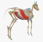
| colspan="2" |M. Serratus ventralis | | colspan="2" |M. Serratus ventralis | ||
(thoracis) | (thoracis) | ||
|Ribs 1-8/9 | |Ribs 1-8/9 | ||
|Scapular cartilage and Medial scapula | |Scapular cartilage and Medial scapula | ||
|Supports trunk between forelimbs. | |Supports trunk between forelimbs. | ||
Raises neck when limb is flexed. | Raises neck when the limb is flexed. | ||
|Long thoracic nerve | |Long thoracic nerve | ||
|- | |- | ||
| rowspan="2" |M. Serratus dorsalis | | rowspan="2" |M. Serratus dorsalis [[File:Equine serratus dorsalis.jpeg|150x150px|alt=|center]] | ||
|Cranialis | |Cranialis | ||
|Supraspinous ligament | |Supraspinous ligament | ||
|Cranial border of | |Cranial border of ribs 5-11 | ||
|Inspiration | |Inspiration | ||
| rowspan="2" |Intercostal nerve | | rowspan="2" |Intercostal nerve | ||
| Line 677: | Line 683: | ||
|Caudalis | |Caudalis | ||
|Thoracolumbar fascia | |Thoracolumbar fascia | ||
|Caudal borders of | |Caudal borders of ribs 11-18 | ||
|Expiration | |Expiration | ||
|- | |- | ||
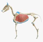
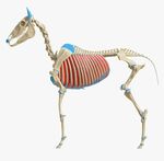
| colspan="2" |M. External intercostal | | colspan="2" |M. External intercostal [[File:Equine intercostal externi.jpeg|150x150px|alt=|center]] | ||
| colspan="2" |Muscles run caudodorsally in the intercostal spaces | | colspan="2" |Muscles run caudodorsally in the intercostal spaces | ||
|Inspiration | |Inspiration | ||
| rowspan="2" |Intercostal nerve | | rowspan="2" |Intercostal nerve | ||
|- | |- | ||
| colspan="2" |M. Internal intercostal | | colspan="2" |M. Internal intercostal [[File:Equine internal intercostals.jpeg|150x150px|alt=|center]] | ||
| colspan="2" |Muscles run cranioventrally in the intercostal spaces | | colspan="2" |Muscles run cranioventrally in the intercostal spaces | ||
|Expiration | |Expiration | ||
|- | |- | ||
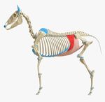
| colspan="2" |M. External abdominal oblique | | colspan="2" |M. External abdominal oblique [[File:Equine external abdominal oblique.jpeg|150x150px|alt=|center]] | ||
|Thoracolumbar fascia and | |Thoracolumbar fascia and lateral aspect of ribs 4-18 | ||
|Linea alba | |Linea alba | ||
| Line 700: | Line 706: | ||
Inguinal ligament | Inguinal ligament | ||
|Flexes trunk | |Flexes the trunk | ||
|Ventral branch of | |Ventral branch of lumbar nerve and local intercostal nerve | ||
|- | |- | ||
| colspan="2" |M. Internal abdominal oblique | | colspan="2" |M. Internal abdominal oblique | ||
|Coxal tuber and | |Coxal tuber and inguinal ligament | ||
|Linea alba | |Linea alba | ||
| Line 713: | Line 719: | ||
Cartilages of ribs 14-18 | Cartilages of ribs 14-18 | ||
| rowspan="2" |Flexes the trunk | | rowspan="2" |Flexes the trunk | ||
| rowspan="3" |Ventral branches of | | rowspan="3" |Ventral branches of lumbar nerve and local intercostal nerve | ||
|- | |- | ||
| colspan="2" |M. Transversus abdominis | | colspan="2" |M. Transversus abdominis [[File:Equine transverus abdominis.jpeg|150x150px|alt=|center]] | ||
|Medial surface of Costal cartilage 7-18 and | |Medial surface of Costal cartilage 7-18 and transverse processes of lumbar vertebrae | ||
|Linea alba | |Linea alba | ||
|- | |- | ||
| colspan="2" |M. Rectus abdominis | | colspan="2" |M. Rectus abdominis [[File:Equine rectus abdominis.jpeg|150x150px|alt=|center]] | ||
|Lateral surface of | |Lateral surface of costal cartilages 4-9 | ||
|Prepubic tendon and the | |Prepubic tendon and the head of the femur | ||
|Flexes the trunk | |Flexes the trunk | ||
Flexes lumbar spine and lumbosacral joint | Flexes lumbar spine and lumbosacral joint | ||
|- | |- | ||
| colspan="2" |M. Longissimus thoracis et lumborum | | colspan="2" |M. Longissimus thoracis et lumborum [[File:Equine longissimus.jpeg|150x150px|alt=|center]] | ||
|Spinous processes of | |Spinous processes of thoracic, lumbar and sacral vertebrae and wing of ilium | ||
|Transverse processes of | |Transverse processes of vertebrae and tubercles of ribs | ||
|Stabilizes and extends vertebral column | |Stabilizes and extends vertebral column | ||
| rowspan="3" |Dorsal branch of | | rowspan="3" |Dorsal branch of local spinal nerve | ||
|- | |- | ||
| colspan="2" |M. Semispinalis thoracis and lumborum | | colspan="2" |M. Semispinalis thoracis and lumborum | ||
| Line 746: | Line 752: | ||
| colspan="2" |M. Cutaneous trunci | | colspan="2" |M. Cutaneous trunci | ||
|Superficial trunk fascia | |Superficial trunk fascia | ||
|Superficial shoulder fascia and medial surface of | |Superficial shoulder fascia and medial surface of humerus | ||
|Moves the skin of the abdomen | |Moves the skin of the abdomen | ||
|Lateral thoracic nerve and | |Lateral thoracic nerve and intercostobrachial nerve | ||
|- | |- | ||
| colspan="2" |M. Multifidus lumborum | | colspan="2" |M. Multifidus lumborum | ||
|Articular processes of each vertebra | |Articular processes of each vertebra from C2 to sacrum | ||
|Spinous process of the preceding vertebrae | |Spinous process of the preceding vertebrae | ||
|Stabilizes and rotates vertebral column | |Stabilizes and rotates vertebral column | ||
|Dorsal branches of | |Dorsal branches of local spinal nerve | ||
|- | |- | ||
| colspan="2" |M. Psoas major | | colspan="2" |M. Psoas major [[File:Equnie psoas major.jpeg|150x150px|alt=|center]] | ||
|Lumbar transverse processes and | |Lumbar transverse processes and ventral surface of the last two ribs | ||
|Lesser trochanter of Femur | |Lesser trochanter of Femur | ||
|Rotates pelvic limb outward | |Rotates pelvic limb outward | ||
| Line 766: | Line 772: | ||
Stabilizes vertebral column when limb is fixed | Stabilizes vertebral column when limb is fixed | ||
|Ventral branches of | |Ventral branches of lumbar and local intercostal nerve and lumbar plexus | ||
|} | |} | ||
{{#ev:youtube|2UtMocTNrWQ|300}}<ref>Superficial muscles. Jessica Blackwell. Available from: https://www.youtube.com/watch?v=2UtMocTNrWQ [last accessed 01/05/2021]</ref> | |||
== References == | == References == | ||
<references /> | <references /> | ||
[[Category:Course Pages]] | [[Category:Course Pages]] | ||
[[Category:Plus Content]] | |||
[[Category:Animal Physiotherapy]] | [[Category:Animal Physiotherapy]] | ||
[[Category:Physiotherapy In Animals]] | [[Category:Physiotherapy In Animals]] | ||
[[Category:Anatomy Project]] | [[Category:Anatomy Project]] | ||
[[Category:Head - Anatomy]] | [[Category:Head - Anatomy]] | ||
Latest revision as of 11:30, 18 August 2022
Introduction[edit | edit source]
Equine anatomy refers to the gross and microscopic anatomy of horses and other equids (donkeys, and zebras).
This page introduces the Anatomy of Equine Spine and Head.
Axial Skeleton[edit | edit source]
The axial skeleton consists of the skull, vertebral column, sternum, and ribs. Multiple sternebrae fuse to form one bone, attached to the 8 "true" pairs of ribs, out of a total of 18.[1]
The vertebral column contains 54 bones:
- 7 cervical vertebrae: includes the atlas (C1) and axis (C2)
- 18-19 thoracic vertebrae
- 5-6 lumbar vertebrae
- 5 sacral vertebrae
- 15-25 caudal vertebrae[2]
In certain breeds, there may be variations in these numbers.[3]
Skull[edit | edit source]
The skull contains the brain and the most important organs of sense.
Cranium[edit | edit source]
The Roof of the cranium is made up of frontal and parietal bones.
The Floor is made up of sphenoid bone.
The cranium consists of 5 orbital regions:
- Frontal
- Lacrimal
- Palatine
- Sphenoid
- Zygomatic
Interparietal bone: only found in horse and cat.
The orbit is complete in horse and ruminants while it is incomplete in carnivores but completed by the orbital ligament.
The lacrimal fossa collects tears and sends them through lacrimal canal into the nasal cavity.
Bones in the Equine Skull[edit | edit source]
There are 34 bones and most of them are flat. During the birth process, these bones overlap and allow the skull to compress as much as possible to allow for parturition.
The 14 major bones are:[1]
- Incisive bone (premaxillary): part of the upper jaw; where the incisors attach
- Nasal bone: covers the nasal cavity
- Maxillary bone: a large bone that contains the roots of the molars
- Mandible: lower portion of the jaw; largest bone in the skull
- Lacrimal bone: contains the nasolacrimal duct, which carries fluid from the surface of the eye, to the nose
- Frontal bone: creates the forehead of the horse
- Parietal bone: extends from the forehead to the back of the skull
- Occipital bone: forms the joint between the skull and the first vertebrae of the neck (the atlas)
- Temporal bone: contains the eternal acoustic meatus, which transmits sound from the ear to the cochlea (eardrum)
- Zygomatic bone: attaches to the temporal bone to form the zygomatic arch (cheekbone)
- Palatine bone: forms the back of the hard palate
- Sphenoid: formed by fusion of the foetal basisphenoid and presphenoid bones, at the base of the skull. Can become fractured in horses that rear over backwards
- Vomer: forms the top of the inside of the nasal cavity
- Pterygoid: small bone attached to the sphenoid that extends downward
Cavities[edit | edit source]
The equine skull consists of 4 cavities:
- The cranial cavity: Protects and encloses the brain, supports sense organs. The cranium consists of a roof made up of the frontal and parietal bones and a floor made up of the sphenoid bone
- The orbital cavity: Has 5 orbits: frontal, lacrimal, palatine, sphenoid and zygomatic. It protects and surrounds the eye.[4] Horses have both monocular and binocular vision:
- Monocular vision: The horse can see objects with one eye. This means that the brain receives two images simultaneously
- Binocular vision: The horse can focus with both eyes just like humans and the brain receives only one signal
- The oral cavity: A passage into the respiratory and digestive system
- The nasal cavity: Contains bone that protects the mucous membrane from inspired warm air
Foramina of the Skull and the Structures Passing Through[edit | edit source]
| Foramina | Structures passing through |
|---|---|
| Infra-orbital foramen | Infra-orbital nerve. CNV |
| Maxillary foramen | |
| Cribriform foramen | Olfactory nerve. CNI |
| Optic canal | Optic nerve. CNII |
| Orbital fissure | CNVII, IV, V and VI (ophthalmic division) |
| Round foramen | CNV (maxillary division) |
| Oval foramen | CNV (mandibular division) |
| Foramen lacerum | Internal carotid artery
CN V3 (horse and pig) |
| Internal acoustic meatus | CNVIII |
| Jugular foramen | CNIX, X, XI |
| Stylomastoid foramen | CNVII |
| Mandibular foramen | CNV (mandibular alveolar nerve) |
| Mental foramen |
Joints and Ligaments[edit | edit source]
Joints and Ligaments of the Skull[edit | edit source]
- The temporo-mandibular joint - a condylar joint between the mandibular condyles and the mandibular fossae of the temporal bones. It has a loose joint capsule with thickenings that form a lateral ligament, as well as an articular disc[1]
- Mandibular symphysis
- Hyoid apparatus - consists of three joints:
- Tympanohyoid cartilage- skull (syndesmosis)
- Interhyoid joints (synovial)
- Thyrohyoid bone- cranial cornu of thyroid cartilage (synovial)[1]
Cervical Spine[edit | edit source]
Joints[edit | edit source]
Atlanto-Occipital Joint[edit | edit source]
A condylar, modified synovial hinge joint. The articulating surfaces are the occipital condyles and the cranial articular surfaces of the atlas (C1). There are three thickenings that strengthen the spacious joint capsule: Dorsal, Ventral, Lateral. The transverse atlantal ligament holds the dens of the axis against the ventral arch of the atlas.[1]
Atlanto-Axial Joint[edit | edit source]
A pivot joint between the atlas and the saddle shaped surface of the axis (C2), which extends upon the dens. It has a loose joint capsule. The apical ligament of dens connects the apex of the dens to the occipital bone. Motion at this joint includes rotation of the atlas and head upon the axis and some accessory lateral flexion.[1] Rotation at this joint makes up 73 percent of cervical rotation.[6]
Cervical Spine C3-C4[edit | edit source]
A planar, extensive, oval shaped joint that is obliquely oriented in transverse plane. The cranial articular processes face dorsomedially and the caudal articular processes face ventrolaterally. Spinous process height increases caudally from C6. Lateral flexion is the primary motion at these joints (25-45 degrees each joint - C1/C2 only has 3.9 degrees of lateral flexion).[6]
Ligaments[edit | edit source]
- Dorsal longitudinal ligament
- Ventral longitudinal ligament
- Ligamentum flavum
- Nuchal ligament:
- This ligament connects the thoracic vertebra to the head and assists in supporting its weight. It consists of two paired parts:[1]
- Funicular (cord) part - extends from the poll to +/- the second to the fourth thoracic spinous process
- Lamellar part - arises from the second and third thoracic spinous processes and the funicular part, and inserts on the C2-C6 spinous processes. The first digitation going to the axis is very strong, but it decreases in strength caudally
- This ligament connects the thoracic vertebra to the head and assists in supporting its weight. It consists of two paired parts:[1]
Thoracic Spine (T1-T18)[edit | edit source]
Articular processes[edit | edit source]
Caudal articular processes face ventrally and are positioned at the base of the spinous process. The cranial articular processes are oval facets on the arch of the vertebra and face dorsally. Each thoracic vertebrae has a pair of costal facets on the dorsal body (except the last) forming the costal fovea.[1]
Anticlinal vertebrae: This is the point in the caudal thoracic vertebral column at which the anatomic features of the vertebra start to change.[7] This usually occurs at the 13th vertebra in horses.
Motion:
- Flexion - most flexion occurs at T17/T18; least flexion occurs at T3-T9
- Extension - most extension occurs at T14-T18; least extension occurs at T2-9[1]
Rib Neck[edit | edit source]
Has 2 converse facets: Cranial and Caudal
Rib 1 attaches to C7, T1 and the associated IV disc
Motion: rotation of the rib, which is greater caudally
Costovertebral Joint[edit | edit source]
Joints[edit | edit source]
The costovertebral joints have two distinct articulations between most ribs and the vertebral column:[1]
- Head of the rib: Cranial and caudal costal facets of adjacent vertebrae; a ball and socket synovial joint
- Tubercle of the rib: Transverse process of vertebrae; a plane synovial joint
Ligaments[edit | edit source]
- Radiate longitudinal ligament
- Intercapital ligament
- Costotransverse ligament
- Ligament of the neck
Lumbar Spine[edit | edit source]
Joints[edit | edit source]
Horses usually have 6 lumbar vertebrae (L1-L6), but some arabian horses only have 5 (L1-L5).[1]
Articular processes[edit | edit source]
Cranial articular processes are fused with mammillary processes. They are concave dorsally and mostly in sagittal alignment. Caudal processes are convex ventrally and correspond with the convexity of the cranial articular processes. They are differentiated from the last thoracic vertebra by the lack of costal facets.[1]
Motion: The lumbar spine and caudal thoracic spine are the least mobile regions of a horse's back.[8] Lateral flexion and rotation is very limited especially at L4-L6 due to intertransverse joints.[1]
Ligaments of Thoraco-Lumbar Spine[edit | edit source]
- Supraspinous ligament: A heavy band of connective tissue running over the top of spinous processes ( T2/T3 caudally). It prevents abnormal separation of spinous processes during flexion
- Ventral longitudinal ligament: Marks the ventral surface of vertebrae from the axis to the sacrum. It is strongest and widest caudally. It plays a major role in preventing overextension of the spine
- Dorsal longitudinal ligament: Extends from the floor of the vertebral canal from the axis to sacrum and helps to prevent spine hyper-flexion
- Annulus fibrosis of IVD: Thick ventrally
- Intertransverse ligament
- Interarcuate ligament/ yellow ligament/ ligamentum flavum: An elastic ligament that fills the dorsal space between the arch of the adjacent vertebra[1]
Lumbosacral Joint[edit | edit source]
The cranial articular process of the first sacral vertebra are concave and face dorsomedially.
Motion: Flexion and Extension - 23.4 degrees[9]
Sacrum[edit | edit source]
The sacrum consists of fused sacral vertebrae and has dorsal and ventral sacral foramina.
Myology and Neurology[edit | edit source]
Muscles of the Head[edit | edit source]
Muscles of the Face[edit | edit source]
The muscles of facial expressions are innervated by the motor fibers of CNVII (facial nerve).[1]
| Muscle | Origin | Insertion | Action | |
|---|---|---|---|---|
| M. Levator labii maxillaris | Lacrimal, Zygomatic and Maxillary bones | The maxillary lip | Elevates the Maxillary lip | |
| M. Levator nasolabialis | Nasal and Frontal bones | The lateral wing of nostril
The maxillary lip |
Elevates and retracts the angle of the mouth | |
| M. Zygomaticus | The fascia covering the Masseter | The commissure of the lips | ||
| M. Buccinator | Maxilla and Mandible | Flattens the cheeks and thus presses food between the teeth | ||
| M. Depressor labii mandibulars | The alveolar border of the Mandible | The mandibular lip | Depresses and retracts the mandibular lip | |
| M. Orbicularis oris | The sphincter muscle of the skin and the muscles of the lips
Corner of the mouth |
Into the lips as it surrounds the mouth | Closes the mouth | |
| M. Risorius | Part of M. cutaneous faciei | The angle of the mouth | Retracts the angle of the mouth | |
| M. Dilator naris | Alar cartillage | Alar cartillage | Dilates the nostril | |
| M. Lateralis nasi | Dorsal part |
Nasal bone |
Parietal cartilage |
Dilates the nostril and nasal vestibule |
| Ventral part | Nasal process of Incisive bone | Lateral wall of the Nasal vestibule | ||
| M. Caninus | Maxilla close to the rostral extremity of the facial crest | Lateral wing of the nostril | Dilates the nostril laterally | |
| M. Levator nasolabialis | Frontal and Nasal bones | Lateral wing of the nostril | Elevates the maxillary lip and the commissure of the mouth
Dilates the nostril | |
Ear[edit | edit source]
The ear is an organ of hearing and balance. It consists of the outer, middle, and inner ear.
Outer Ear[edit | edit source]
The outer ear includes:[1]
- Pinna: mobile and can move independently - can hear multiple sounds at the same time[11]
- Ear canal
Cartilage: Cartilages of the ear collect and transmit sound to the essential organ of hearing within the temporal bone. In order to achieve this, they (especially the concha) need to move.[1]
The muscles of outer ear:[1]
- Rostral
- Dorsal
- Caudal
- Ventral
There are 3 cartilages:[1]
- Conchal: Forms the framework of the portion of the ear which stands erect. It has a large vertical opening on one side to receive sound, and is attached below to the annular cartilage
- Annular: A small ring of gristle connected to the auditory process of the petrous temporal bone
- Scutiform: A small, flat and somewhat triangular cartilaginous plate situated in front of the base of concha, to which it is attached
Middle Ear[edit | edit source]
The middle ear includes:[1]
- Eardrum
- Small, air-filled chamber containing 3 tiny bones: the hammer, anvil, and stirrup. It also includes 2 muscles: the oval window, and the eustachian tube.
Inner Ear[edit | edit source]
The inner ear is a complex structure that includes the cochlea and the vestibular system.[11]
Muscles of Mastication[edit | edit source]
The muscles of mastication are innervated by the mandibular branch of trigeminal nerve CNV.[1]
| Muscle | Origin | Insertion | Action |
|---|---|---|---|
| M. Masseter | The zygomatic arch and the facial crest | The lateral border of the ramus of the mandible | Closes the mouth |
| M. Temporalis | The temporal fossa and the temporal crest | The coronoid process of the mandible | Closes the mouth (to raise the mandible) |
| M. Pterygoideus medialis | The crest formed by the pterygoid processes of the basisphenoid and the palatine bones | The medial surface of the ramus of the mandible | |
| M. Pterygoideus lateralis | The pterygoid process of the sphenoid bone | Rostral border of the condyle of the mandible | Draws and moves the mandible rostrally |
| M. Digastricus | The jugular process of occipital bone | Medial surface of the ventral border of the molar part of the body of the mandible | Opens the mouth |
| M. Occipitomandibularis | The jugular process | The caudal border of the ramus of the mandible |
Muscles of the Eyes[edit | edit source]
- M. Orbicularis oculi - innervated by palpebral branch of CN VII
- M. Levator palpebrae superioris - originates from the posterior orbit and inserts at orbicularis oculi fibers of the lower eyelid. It elevates the upper eyelid and is innervated by CN III (oculomotor nerve)
- M. Malaris - lowers the ventral eyelid. It is innervated by CN VII (facial nerve)
- Muller's muscle - innervated by sympathetic nerves
- Ciliary muscles
- M. Retractor anguli - retracts and anchors the lateral canthus
- M. Levator anguli oculi medialis and M. Frontalis - slightly elevates of the upper eyelid[12][13]
Muscles of the Tongue[edit | edit source]
The equine tongue is made up of twelve different muscles[14] including styloglossus, genioglossus and hyoglossus. These muscles are covered by mucosa on the sides and underneath.[1]
Action: prehension, mastication (i.e. chewing)
Innervation: Hypoglossus (CNXII)
Muscles of Pharynx and Soft palate.[edit | edit source]
| Muscle | Origin | Insertion | Action | Innervation | |
|---|---|---|---|---|---|
| INTRINSIC MUSCLES | |||||
| M. Tensor veli palatini | Muscular process of the petrous part of the temporal bone, pterygoid bone, and lateral lamina of the auditory tube | Palatine aponeurosis | Retracts the soft palate away from the dorsal pharyngeal wall, expanding the nasopharynx and slightly depressing it ventrad during inspiration | Mandibular branch of the trigeminal nerve | |
| M. Levator veli palatini | Muscular process of the petrous part of the temporal bone and the lateral lamina of the Auditory tube and passes along the lateral wall of the nasopharynx | Soft palate dorsal to the glandular layer | Elevates the soft palate during swallowing | Pharyngeal branch of the Vagus nerve | |
| M. Palatinus | Caudal aspect of the palatine aponeurosis | Caudal free margin of the soft palate | Shortens the soft palate and depresses it towards the tongue | ||
| M. Palatopharyngeus | Palatine aponeurosis and from the palatine and pterygoid bones | Upper edge of the thyroid cartilage | |||
| M. Stylopharyngeus | Rostral | Medial surface of the rostral end of the Stylohyoid bone | Pharyngeal raphe | Pharyngeal constrictor | Glossopharyngeal nerve |
| Caudal | Medial aspect of the caudal third of the Stylohyoid bone | Dorsolateral wall of the pharynx | Pharyngeal dilator | ||
| EXTRINSIC MUSCLES | |||||
| M. Genioglossus | Median plane of the Tongue | Oral surface of the Mandible | Protracts the tongue | Hypoglossal nerve | |
| M. Geniohyoideus | Medial surface of the Mandible | Basihyoid bone | Protrudes the tongue | ||
| M. Thyrohyoideus | Lateral lamina of the Thyroid cartilage | Caudal aspect of the thyrohyoid bone | Moves the larynx rostrad | ||
| M. Hyoglossus | Hyoid bones | Median plane of the dorsum of the tongue | Retracts and depresses the base of the tongue | ||
| M. Hyoepiglotticus | |||||
| M. Styloglossus | Lateral aspect of the stylohyoid bone | Tip of the tongue | Retraction of the tongue | ||
| M. Sternohyoideus | Sternal manubrium | Basihyoid bone and lingual process of the hyoid apparatus | Caudal traction | Branches of the first and second cervical nerves | |
| M. Sternothyroideus | Caudolateral aspect of the thyroid cartilage | ||||
Muscles of the Hyoid Apparatus and Larynx[edit | edit source]
Muscles of the hyoid apparatus and larynx are innervated by CNX.
Hyoid Apparatus[edit | edit source]
The hyoid apparatus has muscular connections from the throat to the forelimbs, shoulder, and sternum. Sternohyoid and omohyoid provide a direct connection from the hyoid apparatus to the shoulder of the horse via the ventral neck. The tongue connects to the hyoid apparatus. Small muscles of the hyoid apparatus connect to the TMJ and the poll and the TMJ articulates with the hyoid apparatus.[17][18]
Larynx[edit | edit source]
Intrinsic muscles:
- Cricoarytenoideus dorsalis - abduction of arytenoids and tensing of vocal cords
- Thyroarytenoideus - adduction of arytenoids
- Arytenoideus transversus - adduction of arytenoids
- Cricoarytenoideus lateralis - adduction of arytenoids[19]
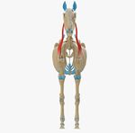
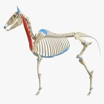
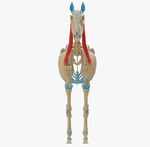
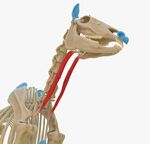
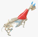
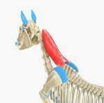
Muscles of Cervical spine[edit | edit source]
| Muscle | Origin | Insertion | Action | Innervation | |
|---|---|---|---|---|---|
| M. Omotransversarius | Fascia of shoulder | Scapular cartilage and transverse processes of C2-4 | Advances limb
Adducts limb Moves neck laterally |
Ventral branch of local cervical spinal nerve | |
| M. Brachiocephalicus | Mastoid process of temporal bone and first cervical vertebra | Deltoid tuberosity and crest of the humerus | Shoulder extension
Protraction Flexion of the neck towards the side of the protracting limb |
Accessory nerve | |
| M. Cleidobrachialis | Inscription of clavicle | Crest of the humerus | Advances limb
Adducts limb |
Axillary nerve | |
| M. Cleidomastoideus | Clavicular intersection | Mastoid process of temporal bone | Advances limb
Flexes neck Turns head |
Ventral branch of Accessory nerve
(cranial nerve XI) | |
| M. Sternocephalicus (Sternomandibularis) | Manubrium of the sternum | Caudal border of mandible | Turns head
Opens mouth | ||
| M. Omohyoideus | Subscapular fascia | Lingual process of basihyoid bone | Retracts basihyoid bone and tongue | Spinal nerve C1 | |
| M. Trapezius | Nuchal ligament and Supraspinous ligaments of C2-10 | Cervical part: Entire scapular spine | Advances thoracic limb
Abducts thoracic limb Elevates shoulder |
Dorsal branch of Accessory nerve
(cranial nerve XI) | |
| Thoracic part: Dorsal third of Scapular spine | |||||
| M. Rhomboideus
(cervicis and thoracis) |
Nuchal ligament and dorsoscapular ligaments of C2-T8 | Scapular cartilage | Elevates neck
Draws scapula cranially and dorsally |
Local thoracic nerve and Local cervical nerve | |
| M. Serratus ventralis (cervicis) | Transverse processes of C4-7 | Scapular cartilage and medial scapula | Supports trunk between forelimbs
Raises neck when the limb is fixed |
Ventral branch of local cervical nerve | |
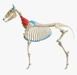
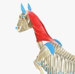
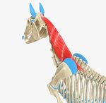
| M. Splenius (capitus and cervicis) | Nuchal ligament and spinous processes of T3-T5 | Nuchal crest and mastoid process of temporal bone | Extends neck
Elevates neck Bends neck laterally |
Dorsal branch of Accessory nerve and dorsal branch of local spinal nerve | |
| M. Longissimus (cervicis, capitus, atlantis) | Transverse processes of cervical and thoracic vertebrae | Wing of atlas and mastoid process of temporal bone | Elevates head and neck
Bends head and neck laterally Stabilizes and extends vertebral column |
Dorsal branch of local spinal nerve | |
| M. Semispinalis capitis | Articular processes of C2/3-7 and transverse processes of T1-6/7 | Occipital bone | Elevates head and neck
Bends head and neck laterally | ||
| M. Longus capitis | Transverse processes of C3-5 | Base of skull | Bends head and neck | Ventral branch of local spinal nerve | |
| M. Longus colli | Cervical part | Transverse processes of C3-7 | Ventral tubercle of atlas and bodies of cervical vertebrae | Flexes head | |
| Thoracic part | Bodies of T1-6 | Transverse processes of C6-7 | Flexes head
Bends head laterally | ||
| M. Obliqus capitis caudalis | Spinous process of the axis | Wing of the Atlas | Rotates atlas and Head | Dorsal branch of C2 | |
| M. Rectus capitis dorsalis | Major | Nuchal crest | Elevates head | Dorsal branch of C1 | |
| Minor | Dorsal arch of the atlas | Occipital bone | |||
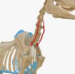
| M. Scalenes | Transverse processes of the last 4 cervical vertebrae | Anterior border and Outer surface of the first rib | Assists inspiration by drawing the first rib forward.
With the rib fixed, draws the neck downward and to one side. |
Cervical nerves | |
Muscles of Trunk[edit | edit source]
| Muscles | Origin | Insertion | Action | Innervation | |
|---|---|---|---|---|---|
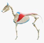
| M. Latissimus dorsi | Supraspinous ligaments from T3 and thoracolumbar fascia | Teres major tuberosity of humerus | Flexes shoulder and draws limb caudally.
Draws trunk cranially when the limb is flexed. |
Thoracodorsal nerve | |
| M. Serratus ventralis
(thoracis) |
Ribs 1-8/9 | Scapular cartilage and Medial scapula | Supports trunk between forelimbs.
Raises neck when the limb is flexed. |
Long thoracic nerve | |
| M. Serratus dorsalis | Cranialis | Supraspinous ligament | Cranial border of ribs 5-11 | Inspiration | Intercostal nerve |
| Caudalis | Thoracolumbar fascia | Caudal borders of ribs 11-18 | Expiration | ||
| M. External intercostal | Muscles run caudodorsally in the intercostal spaces | Inspiration | Intercostal nerve | ||
| M. Internal intercostal | Muscles run cranioventrally in the intercostal spaces | Expiration | |||
| M. External abdominal oblique | Thoracolumbar fascia and lateral aspect of ribs 4-18 | Linea alba
Prepubic tendon Pelvic tendon Coxal tendon Inguinal ligament |
Flexes the trunk | Ventral branch of lumbar nerve and local intercostal nerve | |
| M. Internal abdominal oblique | Coxal tuber and inguinal ligament | Linea alba
Prepubic tendon Last rib Cartilages of ribs 14-18 |
Flexes the trunk | Ventral branches of lumbar nerve and local intercostal nerve | |
| M. Transversus abdominis | Medial surface of Costal cartilage 7-18 and transverse processes of lumbar vertebrae | Linea alba | |||
| M. Rectus abdominis | Lateral surface of costal cartilages 4-9 | Prepubic tendon and the head of the femur | Flexes the trunk
Flexes lumbar spine and lumbosacral joint | ||
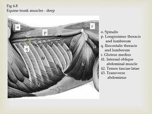
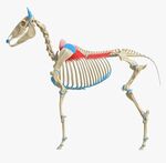
| M. Longissimus thoracis et lumborum | Spinous processes of thoracic, lumbar and sacral vertebrae and wing of ilium | Transverse processes of vertebrae and tubercles of ribs | Stabilizes and extends vertebral column | Dorsal branch of local spinal nerve | |
| M. Semispinalis thoracis and lumborum | Sacrum, the articular processes of the lumbar vertebrae and the transverse processes of the dorsa vertebrae | Spinous processes of third or fourth vertebra in front of the one from which it arises | Fixes the bone during the action of the large spinal muscle, and assists in spine extension | ||
| M. Iliocostalis thoracis and lumborum | Transverse processes of vertebrae | Bodies of the adjacent vertebrae and/or the tuberosities of the ribs | Expiration
Thoracolumbar extension | ||
| M. Cutaneous trunci | Superficial trunk fascia | Superficial shoulder fascia and medial surface of humerus | Moves the skin of the abdomen | Lateral thoracic nerve and intercostobrachial nerve | |
| M. Multifidus lumborum | Articular processes of each vertebra from C2 to sacrum | Spinous process of the preceding vertebrae | Stabilizes and rotates vertebral column | Dorsal branches of local spinal nerve | |
| M. Psoas major | Lumbar transverse processes and ventral surface of the last two ribs | Lesser trochanter of Femur | Rotates pelvic limb outward
Flexed hip Advances limb Stabilizes vertebral column when limb is fixed |
Ventral branches of lumbar and local intercostal nerve and lumbar plexus | |
References[edit | edit source]
- ↑ 1.00 1.01 1.02 1.03 1.04 1.05 1.06 1.07 1.08 1.09 1.10 1.11 1.12 1.13 1.14 1.15 1.16 1.17 1.18 1.19 1.20 1.21 Van der Walt A. Equine Spine and Head Presentation. Plus , 2021.
- ↑ King, Christine, BVSc, MACVSc, and Mansmann, Richard, VMD, PhD. "Equine Lameness." Equine Research, Inc. 1997.
- ↑ Riegal, Ronald J. DVM, and Susan E. Hakola RN. Illustrated Atlas of Clinical Equine Anatomy and Common Disorders of the Horse Vol. II. Equistar Publication, Limited. Marysville, OH. Copyright 2000.
- ↑ Budras, K. Sack, W.O., Anatomy of the Horse, 6th Edition (2012), Schlutersche Verlagsgesellschaft mbH & Co. KG
- ↑ Equine head. PeabodyDVM. Available from: https://www.youtube.com/watch?v=FhA-blWzC8Y[last accessed 01/05/2021]
- ↑ 6.0 6.1 Clayton HM, Townsend HG. Kinematics of the cervical spine of the adult horse. Equine Vet J. 1989;21(3):189-92.
- ↑ Baines EA, Grandage J, Herrtage ME, Baines SJ. Radiographic definition of the anticlinal vertebra in the dog. Veterinary Radiology & Ultrasound. 2009 Jan;50(1):69-73.
- ↑ Townsend HG, Leach DH, Fretz PB. Kinematics of the equine thoracolumbar spine. Equine Vet J. 1983;15(2):117-22.
- ↑ Degueurce C, Chateau H, Denoix JM. In vitro assessment of movements of the sacroiliac joint in the horse. Equine Vet J. 2004;36(8):694-8.
- ↑ Equine spine. Dani Jay - Equestrian. Available from: https://www.youtube.com/watch?v=ektcPGjKJj0 [last accessed 01/05/2021]
- ↑ 11.0 11.1 Aiello SE, Moses MA, Allen DG, editors. The Merck veterinary manual. Whitehouse Station, NJ, USA: Merck; 1998.
- ↑ Gilger BC, Stoppini R. Diseases of the eyelids, conjunctiva, and nasolacrimal system. GILGER BC. 2005.
- ↑ Brooks DE, Matthews AG. Equine ophthalmology. Veterinary ophthalmology. 1999;2:1108.
- ↑ Nicole Kitchener. Horse Sport. Available fromhttps://horsesport.com/magazine/health/horse-tongues-101/ [last accessed 01/05/2021]
- ↑ Norm G. Ducharme, Jonathan Cheetham, Equine Surgery (Fifth Edition), 2019
- ↑ Derksen FJ. Overview of upper airway function. InEquine Surgery 2012 Jan 1 (pp. 530-536). WB Saunders.
- ↑ Cornelisse CJ, Rosenstein DS, Derksen FJ, Holcombe SJ. Computed tomographic study of the effect of a tongue-tie on hyoid apparatus position and nasopharyngeal dimensions in anaesthetised horses. American journal of veterinary research. 2001 Dec 1;62(12):1865-9.
- ↑ Chalmers HJ, Cheetham J, Yeager AE, Ducharme NG. Ultrasonography of the equine larynx. Veterinary Radiology & Ultrasound. 2006 Sep;47(5):476-81.
- ↑ Kelly PG. Studies on serial resting and dynamic endoscopic examination of thoroughbred yearlings (Doctoral dissertation, University of Glasgow).
- ↑ Payne RC, Veenman P, Wilson AM. The role of the extrinsic thoracic limb muscles in equine locomotion. Journal of Anatomy. 2005 Feb;206(2):193-204.
- ↑ Wikivet. Online Veterinary Encyclopedia. Thoracic Limb Extrinsic Muscles - Horse Anatomy. Available from https://en.wikivet.net/Thoracic_Limb_Extrinsic_Muscles_-_Horse_Anatomy#:~:text=Innervation%3A%20Dorsal%20and%20ventral%20branches,cervical%20and%207th%20thoracic%20vertebrae [last accessed 01/05/2021]
- ↑ Superficial muscles. Jessica Blackwell. Available from: https://www.youtube.com/watch?v=2UtMocTNrWQ [last accessed 01/05/2021]
