Cytoskeleton Filaments: Difference between revisions
No edit summary |
No edit summary |
||
| Line 3: | Line 3: | ||
'''Top Contributors''' - {{Special:Contributors/{{FULLPAGENAME}}}} | '''Top Contributors''' - {{Special:Contributors/{{FULLPAGENAME}}}} | ||
== Introduction == | == Introduction == | ||
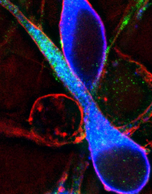
[[File:GnRH Neuron.png|thumb|Neurons (blue): | [[File:GnRH Neuron.png|thumb|Neurons (blue): cytoskeletons shown in red and green.|alt=|283x283px]] | ||
Within all eukaryotic cells there exists a network of filaments that play an important role in the functioning of the cell. They help cells maintain their shape and internal structure and mechanical support, enabling cells to carry out basic functions like division and movement. By means of this cytoskeletal network, the cell integrates numerous signals in order to coordinate its behavior, communicate with other cells, and adjust to its environment. The cytoskeleton is involved in virtually every cellular process, and when dysfunctional pathologies may evolve. | Within all eukaryotic cells there exists a network of filaments that play an important role in the functioning of the cell. They help cells maintain their shape and internal structure and mechanical support, enabling cells to carry out basic functions like division and movement. By means of this cytoskeletal network, the cell integrates numerous signals in order to coordinate its behavior, communicate with other cells, and adjust to its environment. The cytoskeleton is involved in virtually every cellular process, and when dysfunctional pathologies may evolve. | ||
== Filament Types == | == Filament Types == | ||
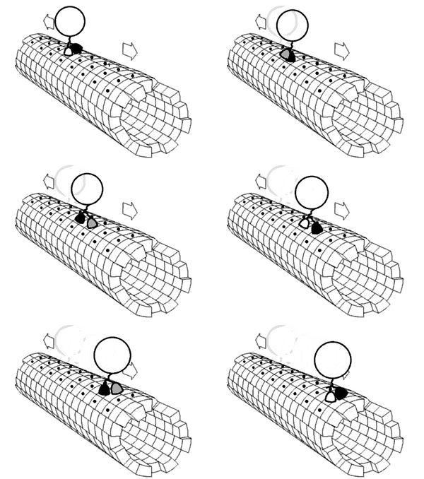
[[File:Kinesin walking.gif|thumb|A molecular motor “walking” on a MT]] | |||
There are three types of filaments make up the cytoskeleton: | There are three types of filaments make up the cytoskeleton: | ||
# Actin filaments occur in a cell in the form of meshworks or bundles of parallel fibres. They help in controlling the shape of the cell and also adhere the cell to the substrate. These filaments are constantly rearranging, helping move the cell and bring about specific activities within it eg cell cleavage during mitosis. Size, 6 nm in diameter and are composed of a protein called actin. | # Actin filaments occur in a cell in the form of meshworks or bundles of parallel fibres. They help in controlling the shape of the cell and also adhere the cell to the substrate. These filaments are constantly rearranging, helping move the cell and bring about specific activities within it eg cell cleavage during mitosis. Size, 6 nm in diameter and are composed of a protein called actin. | ||
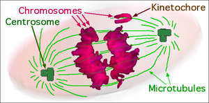
# Microtubules are longer filaments that are constantly assembling and disassembling. | # Microtubules (MT) are longer filaments that are constantly assembling and disassembling. MTs play a crucial role in eg.moving the daughter chromosomes to the newly forming daughter cells during mitosis. Bundles of MTs form the cilia and flagella found in protozoans and in the cells of some multicellular animals. Largest filaments, 24 nm in diameter, composed of a protein called tubulin. | ||
# Intermediate filaments, in contrast to actin filaments and microtubules, are very stable structures that form the true skeleton of the cell. They anchor the nucleus and position it within the cell, and they give the cell its elastic properties and its ability to withstand tension. Size, 10 nm in diameter, built from a number of different subunit proteins. | # Intermediate filaments, in contrast to actin filaments and microtubules, are very stable structures that form the true skeleton of the cell. They anchor the nucleus and position it within the cell, and they give the cell its elastic properties and its ability to withstand tension. Size, 10 nm in diameter, built from a number of different subunit proteins. | ||
| Line 23: | Line 24: | ||
== Striated Muscle Filaments == | == Striated Muscle Filaments == | ||
[[File:Sarcomere.gif|right|frameless]] | |||
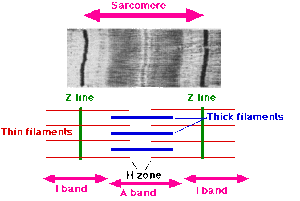
The cytoskeleton fibres in striated muscle have diverse roles as mentioned above, including as functioning as a structure for inter- and intracellular signaling. It does this by linking the complex functioning units of thee sarcomeres, enabling them to function effectively, and in turn provide a connection for these structures in turn to the sarcolemma, cell–cell junctions, the [[mitochondria]], and nucleus. When not functioning correctly pathologies occur as in eg myopathies. | The cytoskeleton fibres in striated muscle have diverse roles as mentioned above, including as functioning as a structure for inter- and intracellular signaling. It does this by linking the complex functioning units of thee sarcomeres, enabling them to function effectively, and in turn provide a connection for these structures in turn to the sarcolemma, cell–cell junctions, the [[mitochondria]], and nucleus. When not functioning correctly pathologies occur as in eg myopathies. | ||
Revision as of 01:48, 7 July 2022
Original Editor - Lucinda hampton
Top Contributors - Lucinda hampton and Vidya Acharya
Introduction[edit | edit source]
Within all eukaryotic cells there exists a network of filaments that play an important role in the functioning of the cell. They help cells maintain their shape and internal structure and mechanical support, enabling cells to carry out basic functions like division and movement. By means of this cytoskeletal network, the cell integrates numerous signals in order to coordinate its behavior, communicate with other cells, and adjust to its environment. The cytoskeleton is involved in virtually every cellular process, and when dysfunctional pathologies may evolve.
Filament Types[edit | edit source]
There are three types of filaments make up the cytoskeleton:
- Actin filaments occur in a cell in the form of meshworks or bundles of parallel fibres. They help in controlling the shape of the cell and also adhere the cell to the substrate. These filaments are constantly rearranging, helping move the cell and bring about specific activities within it eg cell cleavage during mitosis. Size, 6 nm in diameter and are composed of a protein called actin.
- Microtubules (MT) are longer filaments that are constantly assembling and disassembling. MTs play a crucial role in eg.moving the daughter chromosomes to the newly forming daughter cells during mitosis. Bundles of MTs form the cilia and flagella found in protozoans and in the cells of some multicellular animals. Largest filaments, 24 nm in diameter, composed of a protein called tubulin.
- Intermediate filaments, in contrast to actin filaments and microtubules, are very stable structures that form the true skeleton of the cell. They anchor the nucleus and position it within the cell, and they give the cell its elastic properties and its ability to withstand tension. Size, 10 nm in diameter, built from a number of different subunit proteins.
Filaments in Neurones[edit | edit source]
All the types of cytoskeleton filaments work together to guarantee correct formation of the nervous system during the embryonic development and to assure its function in adulthood. Both cytoskeletal filaments and motor proteins are required for axonal transport. Important neuronal events where the function of cytoskeletal filaments are required are
- During embryonic development: the cytoskeleton participating in the growth and guidance of axons
- Adult life: the cytoskeleton essential for maintaining neuronal homeostasis and neuronal plasticity
- Injury repair: peripheral axon needs to regenerate after being injured, peripheral neurons requiring a specific set of cytoskeletal proteins to ensure nerve regeneration upon damage.
Given the importance of cytoskeleton for neurons, it is easy to see how neurological disorders involve changes either in the expression, dynamics, and and/or the stability of cytoskeletal proteins or their mutations.
Striated Muscle Filaments[edit | edit source]
The cytoskeleton fibres in striated muscle have diverse roles as mentioned above, including as functioning as a structure for inter- and intracellular signaling. It does this by linking the complex functioning units of thee sarcomeres, enabling them to function effectively, and in turn provide a connection for these structures in turn to the sarcolemma, cell–cell junctions, the mitochondria, and nucleus. When not functioning correctly pathologies occur as in eg myopathies.
Resources[edit | edit source]
- bulleted list
- x
or
- numbered list
- x


