Myotendinous Junction: Difference between revisions
No edit summary |
No edit summary |
||
| (2 intermediate revisions by the same user not shown) | |||
| Line 5: | Line 5: | ||
</div> | </div> | ||
== Introduction == | == Introduction == | ||
[[File:Myotendinous junction structure.png|thumb| | [[File:Myotendinous junction structure.png|thumb|459x459px|MTJ structure|alt=]] | ||
Myotendinous junction (MTJ) is a part of the myotendinous unit | Myotendinous junction (MTJ) is a part of the myotendinous unit. The myotendinous unit consists usually of [[bone]], enthesis, [[Tendon Anatomy|tendon]], myotendinous junction and [[Muscle Cells (Myocyte)|muscle]], and is responsible for producing skeletal movement<ref>Radiopedia Myotendinous unit Available: https://radiopaedia.org/articles/myotendinous-unit?lang=us<nowiki/>(accessed 12.6.2022)</ref>. | ||
The MTJ has a | The MTJ has a distinctive form with the muscle membrane having many infolds which the [[collagen]] fibrils from the tendon join with (see image 1) . This unique structure creates an increased area for force transmission between muscle and tendon resulting in better force dispersal and less focal [[Stress Loading|stress]]<ref name=":0">Radiopedia Myotendinous junction Available:https://radiopaedia.org/articles/myotendinous-junction?lang=us (accessed 12.6.2022)</ref><ref name=":1">Jakobsen JR, Krogsgaard MR. The Myotendinous Junction—A Vulnerable Companion in Sports. A Narrative Review. Frontiers in physiology. 2021;12. Available;https://www.frontiersin.org/articles/10.3389/fphys.2021.635561/full (accessed 12.6.2022)</ref>. | ||
The MTJ transmits large forces from muscle to tendon in strenuous exercise, and hence is a common location for muscle strains. Most of these can be prevented by heavy eccentric exercise<ref name=":1" />. | |||
== Physiotherapy Implications == | == Physiotherapy Implications == | ||
[[File:Medial view of a cadaver dissection of the gastrocnemius–soleus junction.png|thumb|Dissection of the gastrocnemius–soleus MTJ|alt=|333x333px]] | [[File:Medial view of a cadaver dissection of the gastrocnemius–soleus junction.png|thumb|Dissection of the gastrocnemius–soleus MTJ|alt=|333x333px]] | ||
The myotendinous unit weakest region is the MTJ, and as such it is its most commonly injured part. | |||
* | * Large pennate muscle that are multi arthrodial and produce large tensile stresses are the most likely to suffer from MTJ injuries e.g. [[Biceps Femoris|biceps femoris]], [[Quadratus Femoris|quadratus femoris]], [[Biceps Brachii|biceps brachii]]<ref name=":0" />. | ||
* | * The interdigitations of the MTJ become shorter with aging, lessening the contact area for force transmission, and increase risk of injury.<ref>Wikimsk MTJ Available:https://wikimsk.org/wiki/Myotendinous_Junction (accessed 12.6.2022)</ref> | ||
== US and MRI == | == US and MRI == | ||
For correct diagnosis | For correct diagnosis a grading system of MTJ injuries exists based on [[MRI Scans|MRI]] or [[Ultrasound Scans|US]] scan results. | ||
# Mild [[Muscle Strain|strain]]: feathery interstitial edema and fluid/hemorrhage around the MTJ | # Mild [[Muscle Strain|strain]]: feathery interstitial edema and fluid/hemorrhage around the MTJ | ||
| Line 30: | Line 27: | ||
[[Scar Management|Scar tissue]], old [[Blood Physiology|blood]] products and atrophy/fatty degeneration of the muscle are indicative of an old strain<ref name=":0" />. | [[Scar Management|Scar tissue]], old [[Blood Physiology|blood]] products and atrophy/fatty degeneration of the muscle are indicative of an old strain<ref name=":0" />. | ||
[[File:Myotendinous junction and enthesis combined.png|center|thumb|826x826px|Myotendinous unit]] | |||
== References == | == References == | ||
Latest revision as of 02:33, 14 June 2022
Original Editor - Lucinda hampton
Top Contributors - Lucinda hampton
Introduction[edit | edit source]
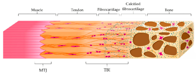
Myotendinous junction (MTJ) is a part of the myotendinous unit. The myotendinous unit consists usually of bone, enthesis, tendon, myotendinous junction and muscle, and is responsible for producing skeletal movement[1].
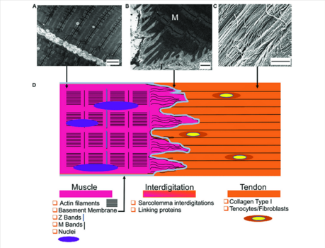
The MTJ has a distinctive form with the muscle membrane having many infolds which the collagen fibrils from the tendon join with (see image 1) . This unique structure creates an increased area for force transmission between muscle and tendon resulting in better force dispersal and less focal stress[2][3].
The MTJ transmits large forces from muscle to tendon in strenuous exercise, and hence is a common location for muscle strains. Most of these can be prevented by heavy eccentric exercise[3].
Physiotherapy Implications[edit | edit source]
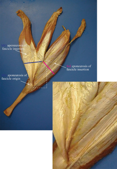
The myotendinous unit weakest region is the MTJ, and as such it is its most commonly injured part.
- Large pennate muscle that are multi arthrodial and produce large tensile stresses are the most likely to suffer from MTJ injuries e.g. biceps femoris, quadratus femoris, biceps brachii[2].
- The interdigitations of the MTJ become shorter with aging, lessening the contact area for force transmission, and increase risk of injury.[4]
US and MRI[edit | edit source]
For correct diagnosis a grading system of MTJ injuries exists based on MRI or US scan results.
- Mild strain: feathery interstitial edema and fluid/hemorrhage around the MTJ
- Moderate strain: intramuscular hematoma and perifascial fluid/hemorrhage
- Severe strain: MTJ tear with laxity/discontinuity of the tendon and muscle ends, sometimes with retraction
Scar tissue, old blood products and atrophy/fatty degeneration of the muscle are indicative of an old strain[2].
References[edit | edit source]
- ↑ Radiopedia Myotendinous unit Available: https://radiopaedia.org/articles/myotendinous-unit?lang=us(accessed 12.6.2022)
- ↑ 2.0 2.1 2.2 Radiopedia Myotendinous junction Available:https://radiopaedia.org/articles/myotendinous-junction?lang=us (accessed 12.6.2022)
- ↑ 3.0 3.1 Jakobsen JR, Krogsgaard MR. The Myotendinous Junction—A Vulnerable Companion in Sports. A Narrative Review. Frontiers in physiology. 2021;12. Available;https://www.frontiersin.org/articles/10.3389/fphys.2021.635561/full (accessed 12.6.2022)
- ↑ Wikimsk MTJ Available:https://wikimsk.org/wiki/Myotendinous_Junction (accessed 12.6.2022)


