Basic Anatomy of the Dancer's Ankle and Foot: Difference between revisions
Carin Hunter (talk | contribs) No edit summary |
Carin Hunter (talk | contribs) No edit summary |
||
| Line 3: | Line 3: | ||
The ankle and foot are complex and detailed structures that bear the weight of the whole body, and are designed to showcase a beautiful work of art. The ankle and foot is a very complex system.<ref>Brockett CL, Chapman GJ. [https://www.sciencedirect.com/science/article/pii/S1877132716300483 Biomechanics of the ankle.] Orthopaedics and trauma. 2016 Jun 1;30(3):232-8.</ref> This part of the body has to cope with high compressive and shearing forces and at the same time it has to offer a high degree of stability. The ankle is the kinetic link of the foot to higher up in the body and the foot is the body's interaction with the ground and has to control multi-axial motions occurring simultaneously.<ref>Houglum PA, Bertoti DB. [https://books.google.co.uk/books?hl=en&lr=&id=offuAwAAQBAJ&oi=fnd&pg=PR1&dq=Houglum+PA,+Bertoti+DB.+Brunnstrom%27s+clinical+kinesiology.+FA+Davis%3B+2012&ots=IxM_DDsDiu&sig=In1KgRYuAUxjjylNd2KuoISLCAw&redir_esc=y#v=onepage&q&f=false Brunnstrom's clinical kinesiology.] FA Davis; 2012</ref> | The ankle and foot are complex and detailed structures that bear the weight of the whole body, and are designed to showcase a beautiful work of art. The ankle and foot is a very complex system.<ref>Brockett CL, Chapman GJ. [https://www.sciencedirect.com/science/article/pii/S1877132716300483 Biomechanics of the ankle.] Orthopaedics and trauma. 2016 Jun 1;30(3):232-8.</ref> This part of the body has to cope with high compressive and shearing forces and at the same time it has to offer a high degree of stability. The ankle is the kinetic link of the foot to higher up in the body and the foot is the body's interaction with the ground and has to control multi-axial motions occurring simultaneously.<ref>Houglum PA, Bertoti DB. [https://books.google.co.uk/books?hl=en&lr=&id=offuAwAAQBAJ&oi=fnd&pg=PR1&dq=Houglum+PA,+Bertoti+DB.+Brunnstrom%27s+clinical+kinesiology.+FA+Davis%3B+2012&ots=IxM_DDsDiu&sig=In1KgRYuAUxjjylNd2KuoISLCAw&redir_esc=y#v=onepage&q&f=false Brunnstrom's clinical kinesiology.] FA Davis; 2012</ref> | ||
[[File:Transverse-tarsal-joint.jpg|thumb|Transverse-tarsal joint]] | [[File:Transverse-tarsal-joint.jpg|thumb|Transverse-tarsal joint]] | ||
Very simply, the ankle is made up of three bones, the [[Tibia]], [[Fibula]] and [[Talus]] and three joints, the '''transverse-tarsal joint''' which works together with the talocalcaneal joint, the '''talocalcaneal joint''' which controls inversion and eversion and the '''tibiotalar joint''' which helps with stability, flexion and extension. | |||
== Foot Summary == | == Foot Summary == | ||
| Line 9: | Line 9: | ||
# '''<u>Hind foot</u>''' | # '''<u>Hind foot</u>''' | ||
##'''Bones''' | ##'''Bones''' | ||
### '''Talus''' | ###'''Talus''' | ||
#### This is the highest foot bone | #### This is the highest foot bone | ||
#### There are no tendons attached to it, only the Deltoid ligament | |||
#### It is a mostly cartilage surface | |||
#### It has a poor blood supply and therefore relatively poor healing. | |||
### '''Calcaneous''' | ### '''Calcaneous''' | ||
### '''Lateral and medial malleolus''' | ### '''Lateral and medial malleolus''' | ||
| Line 58: | Line 61: | ||
### Inter-metatarsal ligament: This is found between the tarsal bones and keep the metatarsals moving in sync. If the nerve running between these joints gets irritated, this could result in a Morton's neuroma.[[File:On pointe good alignment.jpg|thumb|Pointe with Good Alignment]] | ### Inter-metatarsal ligament: This is found between the tarsal bones and keep the metatarsals moving in sync. If the nerve running between these joints gets irritated, this could result in a Morton's neuroma.[[File:On pointe good alignment.jpg|thumb|Pointe with Good Alignment]] | ||
# '''<u>Lateral Ligaments</u>''' | # '''<u>Lateral Ligaments</u>''' | ||
## | ## This is the most commonly injured side in dancers | ||
## | ## Anterior Talofibular Ligament | ||
## | ### Tightens in Plantar flexion | ||
## | ### Is the weakest ligament concluded by many authors | ||
### | ## Calcaneo-fibular Ligament | ||
### | ### Tightens in dorsiflexion | ||
### | ## The following ligaments contribute to ankle stability | ||
### Posterior Talofibular Ligament | |||
### Anterior-Inferior Talofibular Ligament | |||
### Posterior-Inferior Talofibular Ligament (Russel et al, 2008) | |||
==== '''Ballet Specific Ligament Anatomy''' ==== | |||
* In a Demi –plié, when the ankle is in dorsiflexion, the Anterior Talofibular Ligament will relax and the Calcaneo-fibular Ligament will be under tension | |||
* The opposite is expected when en pointe, when the ankle is in plantarflexion, although no studies have been done to examine extreme position en pointe | |||
* Strain in Anterior Talofibular Ligament increases with increasing Plantarflexion and is further accentuated during compressive loading through the ankle | |||
* Maximum plantarflexion en pointe places Anterior Talofibular Ligament parallel to fibula, thus functioning as a primary stabilizer of lateral ankle | |||
* This places Anterior Talofibular Ligament at particular risk at it's weakest and at it's longest at maximal tension force | |||
== Syndesmosis == | == Syndesmosis == | ||
Ligament formed by | Ligament formed by | ||
# | # Anterior-Inferior Talofibular Ligament | ||
# Interosseus membrane | # Interosseus membrane | ||
# | # Posterior-Inferior Talofibular Ligament | ||
# Transverse ligament | # Transverse ligament | ||
# Interosseus ligament | # Interosseus ligament | ||
| Line 104: | Line 112: | ||
## EDL | ## EDL | ||
## PT (eversion) | ## PT (eversion) | ||
==== 2. Intrinsic Foot Muscles ==== | ==== 2. Intrinsic Foot Muscles ==== | ||
| Line 157: | Line 127: | ||
==== Four Muscle Layers of the Plantar Foot ==== | ==== Four Muscle Layers of the Plantar Foot ==== | ||
[[File:Plantar muscles of the foot first layer Primal.png|thumb|First | [[File:Plantar muscles of the foot first layer Primal.png|thumb|First Layer]] | ||
'''Layer One''' | '''Layer One''' | ||
| Line 167: | Line 137: | ||
## Abductor digiti minimi | ## Abductor digiti minimi | ||
[[File:Plantar muscles of the foot second layer Primal.png|thumb|Second Layer]] | [[File:Plantar muscles of the foot second layer Primal.png|thumb|Second Layer]] | ||
'''Layer Two''' | '''Layer Two''' | ||
| Line 178: | Line 151: | ||
#* medial and lateral plantar arteries | #* medial and lateral plantar arteries | ||
[[File:Plantar muscles of the foot third layer Primal.png|thumb|Third Layer]]'''Layer Three''' | |||
# Muscles | |||
## Flexor hallucis brevis | ## Flexor hallucis brevis | ||
## Oblique and transverse heads of the adductor hallucis | ## Oblique and transverse heads of the adductor hallucis | ||
## Flexor digiti minimi brevis | ## Flexor digiti minimi brevis | ||
[[File:Plantar muscles of the foot fourth layer Primal.png|thumb|Fourth Layer]] | [[File:Plantar muscles of the foot fourth layer Primal.png|thumb|Fourth Layer]] | ||
'''Layer Four''' | '''Layer Four''' | ||
| Line 202: | Line 180: | ||
* Toe DF= tightens fascia, supports arch | * Toe DF= tightens fascia, supports arch | ||
* Windlass mechanism | * Windlass mechanism | ||
== Arches == | == Arches == | ||
| Line 216: | Line 195: | ||
Highest arch | Highest arch | ||
# '''Bone''' | # '''Bone''' | ||
## Shape of bones | ## Shape of bones | ||
## First 3 metatarsals | ## First 3 metatarsals | ||
| Line 222: | Line 201: | ||
## Navicular | ## Navicular | ||
## Talus | ## Talus | ||
## calcaneus | ## calcaneus[[File:Medial arch of the foot.gif|thumb|Medial arch of the foot]] | ||
# '''Ligament''' | # '''Ligament''' | ||
## Spring ligament | ## Spring ligament | ||
| Line 253: | Line 232: | ||
## Flexor digitorum longus | ## Flexor digitorum longus | ||
## Short muscles of the little toe | ## Short muscles of the little toe | ||
[[File:Demi pointe - correct, showing with shoe on&off.jpg|thumb|Demi Pointe with weight bearing surface on the Transverse Arch]] | |||
==== 3. Transverse Arch ==== | ==== 3. Transverse Arch ==== | ||
#'''Bone''' | |||
# '''Bone''' | |||
## Wedge shape of the lateral and intermediate cuniform | ## Wedge shape of the lateral and intermediate cuniform | ||
## Metatarsal bases | ## Metatarsal bases | ||
## Cuboid | ## Cuboid | ||
## 3 | ## 3 cuneiforms | ||
# '''Ligament''' | #'''Ligament''' | ||
## Deep transverse ligament | ## Deep transverse ligament | ||
## Dorsal and plantar ligament | ## Dorsal and plantar ligament | ||
# '''Muscle''' | #'''Muscle''' | ||
## Peroneus longus and brevis | ## Peroneus longus and brevis | ||
## Transverse head of adductor hallucis | ## Transverse head of adductor hallucis | ||
Revision as of 22:32, 21 December 2021
Top Contributors - Carin Hunter, Jess Bell, Kim Jackson, Wanda van Niekerk and Olajumoke Ogunleye
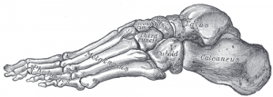
The ankle and foot are complex and detailed structures that bear the weight of the whole body, and are designed to showcase a beautiful work of art. The ankle and foot is a very complex system.[1] This part of the body has to cope with high compressive and shearing forces and at the same time it has to offer a high degree of stability. The ankle is the kinetic link of the foot to higher up in the body and the foot is the body's interaction with the ground and has to control multi-axial motions occurring simultaneously.[2]
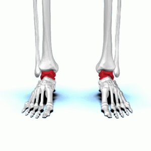
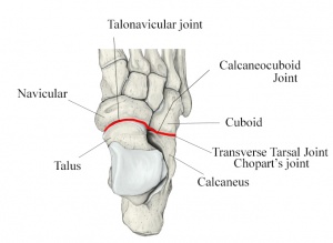
Very simply, the ankle is made up of three bones, the Tibia, Fibula and Talus and three joints, the transverse-tarsal joint which works together with the talocalcaneal joint, the talocalcaneal joint which controls inversion and eversion and the tibiotalar joint which helps with stability, flexion and extension.
Foot Summary[edit | edit source]
- Hind foot
- Bones
- Talus
- This is the highest foot bone
- There are no tendons attached to it, only the Deltoid ligament
- It is a mostly cartilage surface
- It has a poor blood supply and therefore relatively poor healing.
- Calcaneous
- Lateral and medial malleolus
- Talus
- Joints
- Tibiotalar Joint:
- The talus is at its widest anteriorly, meaning the joint is more stable in dorsiflexion.
- The conforming geometry of the tibiotalar joint is considered to contribute to the stability of the joint. In stance phase, the geometry of the joint alone is sufficient to provide resistance to eversion; otherwise stability is derived from the soft tissue structures.
- Subtalar joint:
- Absorbs rotational stress from higher up the body
- Transverse tarsal joint (combo of Talonavicular and CC joints) :
- Transitional link between the hindfoot and forefoot
- Tibiotalar Joint:
- Bones
- Mid Foot
- Bones
- Navicular
- The navicular has poor blood supply and the main attachment for posterior tibial tendon on medial side.
- Cuboid
- Three Cuneforms (Medial, Intermedius, Lateral).
- These are important for stability along with the plantar and dorsal ligaments.
- Navicular
- Joints
- Five Tarsal-metatarsal joints, also known as the Lisfranc joint.
- Ligaments, muscles and tendons
- Plantar fascia ligament
- This ligament is responsible for forming arches of feet and shock absorber when dancing.
- Plantar fascia ligament
- Bones
- Forefoot
- Bones
- 14 Phalanges
- 5 Metatarsals
- The heads are the main weightbearing surface in the following ballet positions: Releve, quarter pointe, demi-pointe, and three quarter pointe.
- 2 Sesmoid bones
- These are located inside Flexor Hallucis Brevis tendon and allows toe to move up and down
- Joints
- Metatarsophalangeal joints
- Ligaments, muscles and tendons
- 1st metatarsal bone is the location for the attachment of several tendons and is important for it's role in propulsion and weight bearing
- Bones
Ligaments of the Foot and Ankle[edit | edit source]
- Medial Ligaments
- Deltoid ligament is fan shaped comprising 4 ligament and resists eversion:
- ATTL (deep component)
- CTL
- TNL
- PTTL (deep component)
- Expansion of joint capsule
- Spring ligament: Cradles and supports the talar head
- Lisfranc ligaments: Series of ligaments that stabilize tarsometatarsal joints and provide stability to the arch of the foot. The Plantar ligament is stronger than the dorsal ligament.
- Inter-metatarsal ligament: This is found between the tarsal bones and keep the metatarsals moving in sync. If the nerve running between these joints gets irritated, this could result in a Morton's neuroma.
- Deltoid ligament is fan shaped comprising 4 ligament and resists eversion:
- Lateral Ligaments
- This is the most commonly injured side in dancers
- Anterior Talofibular Ligament
- Tightens in Plantar flexion
- Is the weakest ligament concluded by many authors
- Calcaneo-fibular Ligament
- Tightens in dorsiflexion
- The following ligaments contribute to ankle stability
- Posterior Talofibular Ligament
- Anterior-Inferior Talofibular Ligament
- Posterior-Inferior Talofibular Ligament (Russel et al, 2008)
Ballet Specific Ligament Anatomy[edit | edit source]
- In a Demi –plié, when the ankle is in dorsiflexion, the Anterior Talofibular Ligament will relax and the Calcaneo-fibular Ligament will be under tension
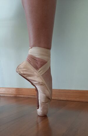
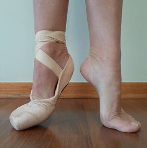
- The opposite is expected when en pointe, when the ankle is in plantarflexion, although no studies have been done to examine extreme position en pointe
- Strain in Anterior Talofibular Ligament increases with increasing Plantarflexion and is further accentuated during compressive loading through the ankle
- Maximum plantarflexion en pointe places Anterior Talofibular Ligament parallel to fibula, thus functioning as a primary stabilizer of lateral ankle
- This places Anterior Talofibular Ligament at particular risk at it's weakest and at it's longest at maximal tension force
Syndesmosis[edit | edit source]
Ligament formed by
- Anterior-Inferior Talofibular Ligament
- Interosseus membrane
- Posterior-Inferior Talofibular Ligament
- Transverse ligament
- Interosseus ligament
Their function is to hold the tibia and fibula together at the appropriate distance and form a mortise where talus sits.
Muscles of the Foot and Ankle[edit | edit source]
1. Extrinsic Foot Muscles[edit | edit source]
These muscles have contractile portions that lie outside the ankle, in the leg, and the tendons of those muscles insert onto the bones of the foot in such a way that ankle motion occurs when the muscles contract. There are four 4 compartments, separated by fascia. The Superficial Posterior compartment, Deep Posterior Compartment and the Lateral Compartment are all Plantarflexors and the Anterior compartment are the Dorsiflexors.
- Superficial Posterior compartment (Plantarflexors)
- Gastrocnemuis (TA)
- Soleus (TA)
- Plantaris
- Deep Posterior Compartment (Plantarflexors)
- FHL (inversion) (plantar surface of 1 toe)
- FDL (inversion) (plantar surface 2-5 toes)
- Tibialis Posterior (inversion) (navicular, medial cuneiform, 2-4 toes, other cuneiforms, cuboid)
- Lateral Compartment (Plantarflexors)
- PL (eversion, PF first metatarsal) (medial cuneiform and 1 toe)
- PB (eversion) (5 toe)
- Anterior compartment (Dorsiflexors)
- Tibialis anterior (inversion) (1 toe, medial cuneiform
- EHL (inversion)
- EDL
- PT (eversion)
2. Intrinsic Foot Muscles[edit | edit source]
These muscles all originate and insert within the foot. They are known to move the toes and stabilize the foot. Dancers refer to these muscles as the “core” muscles of the foot
19 muscles (2 on dorsum – EHB, EDB)
3 largest muscles are Abductor Hallucis, FDB, Quadratus Plantae
Provide support and stability of the arch
Most conditions require strength and control of these muscles
Dancer needs to learn to work with “straight” toes – provides counter stability to MT when pointing
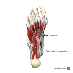
Four Muscle Layers of the Plantar Foot[edit | edit source]
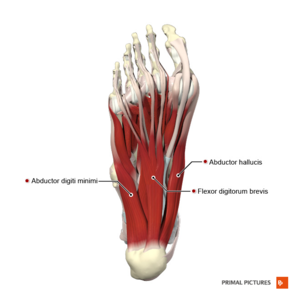
Layer One
Most superficial of all the layers
- Muscles
- Abductor hallucis
- Flexor digitorum brevis (FDB)
- Abductor digiti minimi
Layer Two
- Muscles
- quadratus plantae
- lumbrical muscles
- Tendons
- flexor digitorum longus (FDL)
- flexor hallucis longus (FHL)
- Neurovascular structures
- medial and lateral plantar arteries
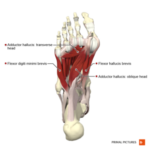
Layer Three
- Muscles
- Flexor hallucis brevis
- Oblique and transverse heads of the adductor hallucis
- Flexor digiti minimi brevis
Layer Four
Deepest layer and both tendons travel to their insertion point via fibro-osseus tunnels
- Muscles
- Dorsal interosseous
- Plantar interosseus
- Tendons
- Peroneus longus
- Tibialis posterior
Plantar fascia[edit | edit source]
- Strong fibrous tissue
- Originates deep within the plantar surface of the calcaneus and inserts on the base of each of the toes
- Toe DF= tightens fascia, supports arch
- Windlass mechanism
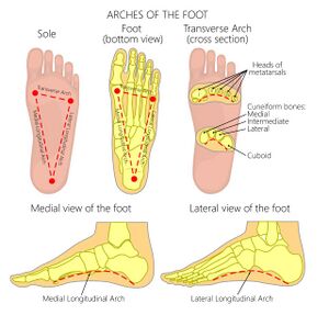
Arches[edit | edit source]
What makes up the arches of the foot?
Purpose:
- Spring
- Weight bearing
- Shock absorption
- Provides flexibility to the foot to facilitate function
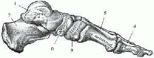
1. Medial longitudinal arch[edit | edit source]
Highest arch
- Bone
- Shape of bones
- First 3 metatarsals
- 3 cuneiforms
- Navicular
- Talus
- calcaneus
- Ligament
- Spring ligament
- Deltoid ligament
- Interosseus ligament
- Plantar aponeurosis
- Long and short plantar ligaments
- Muscle
- Tib posterior
- Tib anterior
- FHL
- FDL
- Short muscles of the big toe
2. Lateral longitudinal arch[edit | edit source]
Lies on ground in standing position
- Bone
- Shape of the bones
- Calcaneus
- Cuboid
- 4th and 5th metatarsals
- Ligament
- long and short plantar ligaments
- Interosseus ligament
- Plantar aponeurosis
- Muscle
- Peroneus longus and brevis
- Flexor digitorum longus
- Short muscles of the little toe
3. Transverse Arch[edit | edit source]
- Bone
- Wedge shape of the lateral and intermediate cuniform
- Metatarsal bases
- Cuboid
- 3 cuneiforms
- Ligament
- Deep transverse ligament
- Dorsal and plantar ligament
- Muscle
- Peroneus longus and brevis
- Transverse head of adductor hallucis
- Slips of tibial posterior
- ↑ Brockett CL, Chapman GJ. Biomechanics of the ankle. Orthopaedics and trauma. 2016 Jun 1;30(3):232-8.
- ↑ Houglum PA, Bertoti DB. Brunnstrom's clinical kinesiology. FA Davis; 2012
