Eversion Stress Test: Difference between revisions
Rucha Gadgil (talk | contribs) No edit summary |
Rucha Gadgil (talk | contribs) No edit summary |
||
| (3 intermediate revisions by the same user not shown) | |||
| Line 1: | Line 1: | ||
<div class="editorbox"> '''Original Editor '''- [[User: | <div class="editorbox"> '''Original Editor '''- [[User:Rucha Gadgil|Rucha Gadgil]]<br> | ||
'''Top Contributors''' - {{Special:Contributors/{{FULLPAGENAME}}}}</div> | '''Top Contributors''' - {{Special:Contributors/{{FULLPAGENAME}}}}</div> | ||
== Purpose == | == Purpose == | ||
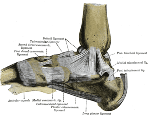
The Eversion Stress Test evaluates the integrity of the deltoid ligament and aids in determining the degree of instability after a medial ankle sprain<ref name=":0">Starkey C, Brown SD. Examination of orthopedic & athletic injuries. FA Davis; 2015 Feb | The Eversion Stress [[Stress tests for Ankle ligaments|Test]] evaluates the integrity of the deltoid ligament and aids in determining the degree of instability after a medial [[Ankle Sprain|ankle sprain]]<ref name=":0">Starkey C, Brown SD. Examination of orthopedic & athletic injuries. FA Davis; 2015 Feb.</ref><ref>de Vries JS, Kerkhoffs GM, Blankevoort L, van Dijk CN. Clinical evaluation of a dynamic test for lateral ankle ligament laxity. Knee Surg Sports Traumatol Arthrosc. 2010 May;18(5):628-33. </ref>.<br> | ||
[[File:Foot ligaments.png|alt=https://en.wikipedia.org/wiki/Deltoid_ligament#/media/File:Gray354.png|thumb|Medial Ankle Ligament]] | |||
== Technique == | == Technique == | ||
The patient is supine, side lying, or seated comfortable with the knee bent 90 degrees and gastrocnemius relaxed. The heel is held from below by one hand by the therapist while the other hand holds the lower leg. While maintaining the ankle in a neutral position, the clinician applies an abduction force to the calcaneus to tilt the talus<ref name=":0" />. | The patient is supine, side lying, or seated comfortable with the knee bent 90 degrees and gastrocnemius relaxed. The heel is held from below by one hand by the therapist while the other hand holds the lower leg. While maintaining the ankle in a neutral position, the clinician applies an abduction force to the [[calcaneus]] to tilt the talus<ref name=":0" />. | ||
Increased talar tilt or pain over the deltoid ligament, when compared bilaterally, indicates a positive test. A spongy or indefinite end feel is indicative of a complete tear<ref>Prentice W, Arnheim D. Principles of athletic training: A competency-based approach. McGraw-Hill Higher Education; 2013 Jan 25.</ref><ref>Manganaro D, Alsayouri K. Anatomy, Bony Pelvis and Lower Limb, Ankle Joint. In: StatPearls. StatPearls Publishing, Treasure Island (FL); 2020.</ref>. | Increased [[talar tilt]] or pain over the deltoid ligament, when compared bilaterally, indicates a positive test. A spongy or indefinite end feel is indicative of a complete tear<ref>Prentice W, Arnheim D. Principles of athletic training: A competency-based approach. McGraw-Hill Higher Education; 2013 Jan 25.</ref><ref>Manganaro D, Alsayouri K. Anatomy, Bony Pelvis and Lower Limb, Ankle Joint. In: StatPearls. StatPearls Publishing, Treasure Island (FL); 2020.</ref>. | ||
In the neutral position, a 2-degree or greater tilt during testing when compared bilaterally indicates a high probability of significant injury to the deltoid<ref>Leith JM, McConkey JP, Li D, Masri B. Valgus stress radiography in normal ankles. Foot & ankle international. 1997 Oct;18(10):654-7.</ref>. | |||
{{#ev:youtube|q6HYxNV28Vg|300}} | |||
== Evidence == | == Evidence == | ||
Studies show that a valgus tilt of the talus occurs when the superficial and deep fibers of the deltoid ligaments are involved. Neutral positioning of the ankle is suggested to test the superficial layer, while testing throughout available ankle range of motion assesses different deltoid fibers<ref>Magee DJ. Orthopedic physical assessment 5th ed. St. Louis, Mo, Saunders Elsevier. 2008.</ref>. | |||
Studies reporting the psychometric properties of this test are scarce and data related to it could not be identified. | |||
== References == | == References == | ||
<references /> | <references /> | ||
[[Category:Special Tests]] | |||
[[Category:Ankle - Special Tests]] | |||
[[Category:Foot - Special Tests]] | |||
[[Category:Ankle - Assessment and Examination]] | |||
Latest revision as of 15:10, 27 February 2021
Purpose[edit | edit source]
The Eversion Stress Test evaluates the integrity of the deltoid ligament and aids in determining the degree of instability after a medial ankle sprain[1][2].
Technique[edit | edit source]
The patient is supine, side lying, or seated comfortable with the knee bent 90 degrees and gastrocnemius relaxed. The heel is held from below by one hand by the therapist while the other hand holds the lower leg. While maintaining the ankle in a neutral position, the clinician applies an abduction force to the calcaneus to tilt the talus[1].
Increased talar tilt or pain over the deltoid ligament, when compared bilaterally, indicates a positive test. A spongy or indefinite end feel is indicative of a complete tear[3][4].
In the neutral position, a 2-degree or greater tilt during testing when compared bilaterally indicates a high probability of significant injury to the deltoid[5].
Evidence[edit | edit source]
Studies show that a valgus tilt of the talus occurs when the superficial and deep fibers of the deltoid ligaments are involved. Neutral positioning of the ankle is suggested to test the superficial layer, while testing throughout available ankle range of motion assesses different deltoid fibers[6].
Studies reporting the psychometric properties of this test are scarce and data related to it could not be identified.
References[edit | edit source]
- ↑ 1.0 1.1 Starkey C, Brown SD. Examination of orthopedic & athletic injuries. FA Davis; 2015 Feb.
- ↑ de Vries JS, Kerkhoffs GM, Blankevoort L, van Dijk CN. Clinical evaluation of a dynamic test for lateral ankle ligament laxity. Knee Surg Sports Traumatol Arthrosc. 2010 May;18(5):628-33.
- ↑ Prentice W, Arnheim D. Principles of athletic training: A competency-based approach. McGraw-Hill Higher Education; 2013 Jan 25.
- ↑ Manganaro D, Alsayouri K. Anatomy, Bony Pelvis and Lower Limb, Ankle Joint. In: StatPearls. StatPearls Publishing, Treasure Island (FL); 2020.
- ↑ Leith JM, McConkey JP, Li D, Masri B. Valgus stress radiography in normal ankles. Foot & ankle international. 1997 Oct;18(10):654-7.
- ↑ Magee DJ. Orthopedic physical assessment 5th ed. St. Louis, Mo, Saunders Elsevier. 2008.


