Gluteus Minimus: Difference between revisions
Kim Jackson (talk | contribs) m (Text replacement - "Category:Anatomy - Hip" to "") |
(removed the exisitng text as it was all copied, added description, anatomy, clinical significance and assessment.) |
||
| Line 6: | Line 6: | ||
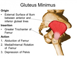
= Description[[Image:Gluteusminimus.jpg|thumb|right]] = | = Description[[Image:Gluteusminimus.jpg|thumb|right]] = | ||
Gluteus minimus is the smallest one of the three gluteal muscles, lying deep to the gluteus medius muscle. The gluteus minimus is smiliar to the gluteus medius in function, structure, nerve and blood supply. Gluteus minimus main function is hip stabilization and abduction.<ref name=":0">Greco AJ, Vilella RC. [https://www.ncbi.nlm.nih.gov/books/NBK556144/ Anatomy, Bony Pelvis and Lower Limb, Gluteus Minimus Muscle.] InStatPearls [Internet] 2020 Apr 11. StatPearls Publishing.</ref> | |||
= Anatomy = | = Anatomy = | ||
{{#ev:youtube|irU6Qmtw99I}} | {{#ev:youtube|irU6Qmtw99I}} | ||
=== Origin | === Origin === | ||
External surface of the ilium, between the anterior and inferior gluteal lines.<ref name=":1">Standring S, Ellis H, Healy J, Johnson D, Williams A, Collins P, Wigley C. Gray's anatomy: the anatomical basis of clinical practice. American Journal of Neuroradiology. 2005 Nov;26(10):2703.</ref> | |||
=== Insertion | === Insertion === | ||
Gluteus minimus muscle is fan-shaped, it attaches at the anterolateral aspect of the greater trochanter of the femur.<ref name=":1" /> | |||
=== Nerve and blood supply === | |||
<br>Superior gluteal nerve (L4, L5, S1) and superior gluteal artery<ref name=":0" /><ref name=":1" /> | |||
=== | === Action === | ||
Its main action is hip abduction. It stabilizes the pelvic during single limb support in gait, by engaging on the supported side, to keep the pelvic from dropping on the opposite swing side. Its anterior segment medially rotates the thigh.<ref name=":0" /> | |||
= Clinical Significance = | |||
Weakness in the gluteus minimus results in trendelenburg gait, where the pelvic drops on the unsupported side. | |||
Gluteus minimus tendinopathy results in Greater Trochanteric Pain Syndrome (GTPS). Which is characterized by lateral hip pain, tenderness at the greater trochanter and trendelenburg gait, it should be differentiated from trochanteric bursitis which is very rare. Anteroposterior radiograph of the pelvis is done to rule out hip osteoarthritis. | |||
Gluteus minimus trigger points referred pain starts at end of the lumbar spine and ends at the ankle, following a similar pain pathway of sciatic nerve but without neurological symptoms of the sciatic nerve.<ref name=":0" /> | |||
= Assessment = | = Assessment = | ||
Trendelenburg test is used to assess the strength of the hip abductors (gluteus medius and gluteus minimus). It is done by asking the patient to do single limb support on the tested leg, while observing the patient from behind to observe the pelvic alignment. If the pelvic drops or deviates from the midline it is indicative of hip abductors weakness.<ref>Whiler L, Fong M, Kim S, Ly A, Qin Y, Yeung E, Mathur S. [https://www.ncbi.nlm.nih.gov/pmc/articles/PMC5963550/#B18 Gluteus medius and minimus muscle structure, strength, and function in healthy adults: brief report.] Physiotherapy Canada. 2017;69(3):212-6.</ref> | |||
=== Palpation === | === Palpation === | ||
= Treatment = | = Treatment = | ||
| Line 49: | Line 42: | ||
| {{#ev:youtube|pRKvszxpSSs|412}} | | {{#ev:youtube|pRKvszxpSSs|412}} | ||
| {{#ev:youtube|sJhNTsMi0-U|412}} | | {{#ev:youtube|sJhNTsMi0-U|412}} | ||
|} | |} | ||
= References = | = References = | ||
| Line 68: | Line 49: | ||
<br> | <br> | ||
[[Category:Anatomy]] [[Category:Muscles]] [[Category:Hip]] [[Category:Musculoskeletal/Orthopaedics]] [[Category:Hip - Anatomy]] [[Category:Hip - Muscles]] | [[Category:Anatomy]] | ||
[[Category:Muscles]] | |||
[[Category:Hip]] | |||
[[Category:Musculoskeletal/Orthopaedics]] | |||
[[Category:Hip - Anatomy]] | |||
[[Category:Hip - Muscles]] | |||
Revision as of 19:51, 15 June 2020
Original Editor - George Prudden,
Top Contributors - Lucinda hampton, George Prudden, Elvira Muhic, Kim Jackson, Lilian Ashraf, Khloud Shreif, WikiSysop, Candace Goh, Evan Thomas and Joao Costa;
Description[edit | edit source]
Gluteus minimus is the smallest one of the three gluteal muscles, lying deep to the gluteus medius muscle. The gluteus minimus is smiliar to the gluteus medius in function, structure, nerve and blood supply. Gluteus minimus main function is hip stabilization and abduction.[1]
Anatomy[edit | edit source]
Origin[edit | edit source]
External surface of the ilium, between the anterior and inferior gluteal lines.[2]
Insertion[edit | edit source]
Gluteus minimus muscle is fan-shaped, it attaches at the anterolateral aspect of the greater trochanter of the femur.[2]
Nerve and blood supply[edit | edit source]
Superior gluteal nerve (L4, L5, S1) and superior gluteal artery[1][2]
Action[edit | edit source]
Its main action is hip abduction. It stabilizes the pelvic during single limb support in gait, by engaging on the supported side, to keep the pelvic from dropping on the opposite swing side. Its anterior segment medially rotates the thigh.[1]
Clinical Significance[edit | edit source]
Weakness in the gluteus minimus results in trendelenburg gait, where the pelvic drops on the unsupported side.
Gluteus minimus tendinopathy results in Greater Trochanteric Pain Syndrome (GTPS). Which is characterized by lateral hip pain, tenderness at the greater trochanter and trendelenburg gait, it should be differentiated from trochanteric bursitis which is very rare. Anteroposterior radiograph of the pelvis is done to rule out hip osteoarthritis.
Gluteus minimus trigger points referred pain starts at end of the lumbar spine and ends at the ankle, following a similar pain pathway of sciatic nerve but without neurological symptoms of the sciatic nerve.[1]
Assessment[edit | edit source]
Trendelenburg test is used to assess the strength of the hip abductors (gluteus medius and gluteus minimus). It is done by asking the patient to do single limb support on the tested leg, while observing the patient from behind to observe the pelvic alignment. If the pelvic drops or deviates from the midline it is indicative of hip abductors weakness.[3]
Palpation[edit | edit source]
Treatment[edit | edit source]
Trigger points[edit | edit source]
References[edit | edit source]
- ↑ 1.0 1.1 1.2 1.3 Greco AJ, Vilella RC. Anatomy, Bony Pelvis and Lower Limb, Gluteus Minimus Muscle. InStatPearls [Internet] 2020 Apr 11. StatPearls Publishing.
- ↑ 2.0 2.1 2.2 Standring S, Ellis H, Healy J, Johnson D, Williams A, Collins P, Wigley C. Gray's anatomy: the anatomical basis of clinical practice. American Journal of Neuroradiology. 2005 Nov;26(10):2703.
- ↑ Whiler L, Fong M, Kim S, Ly A, Qin Y, Yeung E, Mathur S. Gluteus medius and minimus muscle structure, strength, and function in healthy adults: brief report. Physiotherapy Canada. 2017;69(3):212-6.


