File:Lung and diaphragm.jpg: Difference between revisions
Marleen Moll (talk | contribs) No edit summary |
Marleen Moll (talk | contribs) mNo edit summary |
||
| (One intermediate revision by the same user not shown) | |||
| Line 1: | Line 1: | ||
<div class="pp-no-course-suggestions pp-no-article-suggestions"></div> | |||
== Summary == | |||
Anatomy of the respiratory system, showing the trachea and both lungs and their lobes and airways. Lymph nodes and the diaphragm are also shown. Oxygen is inhaled into the lungs and passes through the thin membranes of the alveoli and into the bloodstream (see inset). (1) Note: The text and images above are in the public domain and were reproduced or adapted from the websites of the National Cancer Institute (NCI): http://www.cancer.gov/ and http://visualsonline.cancer.gov/. | |||
== Licensing == | |||
{{Template:PD-USfg}} | |||
Latest revision as of 08:33, 15 April 2020
Summary[edit | edit source]
Anatomy of the respiratory system, showing the trachea and both lungs and their lobes and airways. Lymph nodes and the diaphragm are also shown. Oxygen is inhaled into the lungs and passes through the thin membranes of the alveoli and into the bloodstream (see inset). (1) Note: The text and images above are in the public domain and were reproduced or adapted from the websites of the National Cancer Institute (NCI): http://www.cancer.gov/ and http://visualsonline.cancer.gov/.
Licensing[edit | edit source]
This file is a work of an employee of the US federal government, taken or made during the course of the person's official duties. As a work of the U.S. federal government, the image is in the public domain.
File history
Click on a date/time to view the file as it appeared at that time.
| Date/Time | Thumbnail | Dimensions | User | Comment | |
|---|---|---|---|---|---|
| current | 08:27, 15 April 2020 | 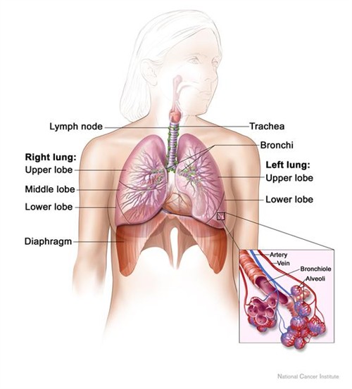 | 500 × 553 (46 KB) | Marleen Moll (talk | contribs) |
You cannot overwrite this file.
File usage
The following 2 pages use this file:

