When refering to evidence in academic writing, you should always try to reference the primary (original) source. That is usually the journal article where the information was first stated. In most cases Physiopedia articles are a secondary source and so should not be used as references. Physiopedia articles are best used to find the original sources of information (see the references list at the bottom of the article).
If you believe that this Physiopedia article is the primary source for the information you are refering to, you can use the button below to access a related citation statement.
Overview of the Pelvic Girdle Musculoskeletal Methodsm
The Pelvic Girdle Musculoskeletal Methodsm (PGM Methodsm) has been developed by Deborah Riczo, a physiotherapist and leading expert in the field of pelvic girdle pain. She has been teaching this method to physiotherapists and physiotherapy assistants since 2011.[1]
Components of the PGM Methodsm
The PGMsm method is a holistic approach to pelvic girdle pain and includes the following components[1]:
Interventions used to correct issues of innominate and sacral positional strain. These include:
Targeted strengthening exercises
Targeted stretching exercises
Core strengthening
Patient education
Consideration of sacroiliac belt
PGMsm Method Special Tests
This method includes two sets of special tests to assess dysfunction. For the purpose of this page and the Plus course, Introduction to the PGM method, dysfunction refers to a tendency for a positional strain caused by muscles that are overstrained versus a positional change. The two sets of special tests are:[1]
Innominate dysfunction cluster of special tests
Sacral dysfunction special tests
In the PGM method, it is important to screen the innominate first.
Innominate Imbalance Special Tests
These are a cluster of 4 special tests and 3 out of 4 of these tests must be positive.[1] The tests are:[1]
Asymmetrical hip internal rotation (IR) range of motion (>10 degrees between sides)
Asymmetry/ tenderness of the symphysis pubis
Unilateral tenderness of ASIS and/or the Iliac Crest
Unilateral tenderness of PSIS and/or long dorsal sacroiliac ligament
1. Special Test: Asymmetrical Hip Rotation Range of Motion (ROM)
Comparison of hip internal and external range of motion between sides
Optimal assessment position is prone
Can be assessed in other positions
Use an inclinometer or smartphone application to measure the range of motion
A difference of more than 10° between left and right internal hip rotation is regarded as a positive test result
Important to assess:
"Perceived" tightness unilaterally
Reproduction of symptoms unilaterally during the assessment
Evidence for the Relationship of Hip Range of Motion and Low Back Pain and Pelvic Girdle Dysfunction
Ellison et al. (1990)[3] reported differences in hip rotation in patients with low back pain compared to patients without low back pain (controls). The researchers reported that the total hip external rotation was greater than the total hip internal rotation in patients with low back pain. A significant result was a difference of more than 10°.[3]
Cibulka et al. (2002)[4] indicated that the presence of asymmetrical hip rotation range of motion may be an indication of sacroiliac region pain.[4]
Prather et al. (2017)[5] indicated an association between reduced hip internal rotation and low back pain.[5]
Hartigan et al. (2020)[6] investigated hip angles, joint moments and muscle activity during gait in women with self-reported stress urinary incontinence and found the following in this population[6]:
Weakness, poor endurance and timing of the pelvic floor muscles
Decreased ability of hip position control in the non-dominant leg
Decreased strength of the hip abductors and hip external rotators
Increased gluteus maximus activity of the non-dominant leg
Women with stress urinary incontinence present with altered hip biomechanics during the stance phase of gait. Although women with stress urinary incontinence seem to be using greater hip abduction and external rotation forces, motor control strategies seem to be lacking and unable to avoid greater hip adduction and internal rotation positions.[6]
Pizol et al. (2024)[7] concluded that the internal and external hip range of motion is lower in people with low back pain, compared to healthy individuals. Clinicians should assess and manage these changes in the treatment of people with low back pain or pelvic girdle pain.[7]
Test Reliability
Inter- and intra-rater reliability have been established for the measurement of hip internal and external rotation in prone.[3] Measurement of hip internal and external rotation in prone with a goniometer is performed as follows:
Hip internal rotation (IR) and external rotation (ER) are measured with the subject in prone, with the hip in a neutral position and the knees flexed to 90°. The goniometer is centred at the joint line, with the movement arm of the goniometer aligned along the midline of the tibia and the stationary arm aligned perpendicular to the floor.[8] The patient is asked to move the hip into internal rotation while keeping the hips stationary on the table. The patient is then asked to move the hip into external rotation while keeping the hips stationary on the table. Measures are assessed on the contralateral hip as well.
Measurement of hip rotation in prone with a smartphone application (there are various applications available) may be done as follows:[9][10]
The top of the smartphone is placed 3cm below the tibial tuberosity and the base of the smartphone is positioned towards the midline of the medial and lateral malleoli.[8] The patient is asked to move the hip into internal rotation (IR) and then into external rotation (ER). The clinician provides verbal cues to ensure that no compensatory movements occur during the motion.[11] Measures are also assessed on the contralateral limb.
Recent research demonstrated moderate to excellent intra-rater reliability for the measurement of active hip IR and ER when using the goniometer and a smartphone application (Clinometer).[9] Good to excellent interrater reliability was also reported with the measurement of active hip IR and ER with both goniometer and smartphone application. Additionally, it is suggested that the smartphone application may be a valid alternative to hand-held goniometry when measuring active hip IR and ER in clinical practice.[9]
Hip rotation range of motion can also be assessed in sitting as well as in supine in the PGM method.[1]
2. Special Test: Asymmetry/ Tenderness of the Symphysis Pubis
Palpation of the Symphysis Pubis
The presence of unilateral tenderness with palpation of the symphysis pubis is considered a positive result in the PGM method.[1] The sensitivity (66%) and specificity (99%) of palpation of the anterior surface of the symphysis pubis with the person in supine have been established. The examiner uses palpation to elicit pain that persists for more than 5 seconds after removal of the examiner’s hand.[14]
Anteroposterior or superoinferior displacement of the upper border of the pubic symphysis or pubic tubercle can also be felt with palpation.[15] This may sometimes be more difficult, for example, in the third trimester of pregnancy.[1]
A painful unilateral response on the side that the patient is complaining of the pain is a positive result in the PGM method.[1]
Garras et al. (2008)[17] investigated the range of movement at the symphysis pubis by using single-leg stance radiographs. Multiparous women had a significantly increased physiologic range of pubic translation compared to men and nulliparous women. Up to 5 mm of physiologic motion can occur at the pubic symphysis in asymptomatic individuals.[17]
3. Special Test: Unilateral Tenderness of ASIS and/or the Iliac Crest
As part of the PGM Method, only unilateral tenderness of the iliac crest and/or the anterior superior iliac spine (ASIS) is assessed and not asymmetry.[1] The standing ASIS asymmetry test has poor reliability and has no value during the examination.[18]
Test Reliability
Research has shown a good discriminative ability and inter-rater reliability when two criteria (localised tenderness and pain recognition) were combined.[19] Good interobserver agreement was reported for “localised tenderness” and “typical pain” criteria on iliac crest pain syndrome in patients with non-specific low back pain.[20] McCombe et al. (1989)[21] reported that palpation tests for bony tenderness were more reliable than palpation tests for soft tissue tenderness.[21]
4. Special Test: Unilateral Tenderness of PSIS and/or Long Dorsal Sacroiliac Ligament
In the PGM Method, tenderness with palpation of the posterior superior iliac spine (PSIS) and/or the long dorsal sacroiliac ligament on the symptomatic side is considered a positive test result.[1]
Palpation of the Long Dorsal Sacroiliac Ligament
The long dorsal sacroiliac ligament is known as a major pain generator for pelvic pain.[23][24] Tenderness with palpation of this ligament[25] does seem to indicate sacroiliac joint pain versus a low back problem. The sensitivity of this palpation test increases when it is combined with the active straight leg raise test and the thrust test.[26]
There are two reasons/indications for performing the sacral tests in the PGM Method:[1]
A negative cluster of PGM signs for the innominate (less than 3 out of 4 positive tests), but the patient has clinical history and symptoms consistent with sacroiliac pelvic girdle pain
After having completed the interventions for the innominate dysfunction to address the positional strain, the patient’s improve, innominate tests are negative, but the patient is still displaying some clinical signs and complaints of sacroiliac pain or pelvic girdle dysfunction
1. Special Test: Sacral Springing Test
This test is similar to the sacral thrust test and involves gross springing of the sacrum. The patient is in prone, the therapist uses the pisiform of their hand and applies a gentle springing of the sacrum.[28] If this relieves the symptoms of the patient, this can be used as a treatment technique. The test is positive if pain is elicited. If no pain is elicited with this test, but pelvic girdle dysfunction is still suspected, move on to assessing each individual pole of the sacrum.[1]
2. Special Test: Springing of Four Poles of Sacrum
This test involves the individual springing of the four poles of the sacrum in a similar way that the sacral springing test is performed and is considered positive if pain is present.[1] The four poles of the sacrum are:
The right and left base of the sacrum
The right and left inferior lower angles of the sacrum (ILA)
If the patient's symptoms are relieved with the individual springing on a specific pole, this can be used as a treatment technique.
When performing the individual springing on the four different poles it is important to take note of the pain provocation patterns.[1]
Pain Provocation Patterns
Diagonal
For example, painful with the springing of the right superior pole and the left inferior lateral angle (ILA) of the sacrum - this may indicate a torsion dysfunction[1]
Vertical
For example, pain provocation with the springing of the right superior pole and right inferior lateral angle of the sacrum - this may indicate the presence of an "up slip" or a "down slip." This is often best treated with postural type education and manual myofascial releases[1]
Horizontal
Pain provocation on both superior poles or inferior lateral angles of the sacrum (ILA) - this is not a torsion or "up slip/down slip" dysfunction and would more likely indicate a positional postural type dysfunction[1]
Virtual Assessment of Pelvic Girdle Dysfunction
The COVID-19pandemic has brought telehealth and virtual patient assessments to the forefront in physiotherapy.[30][31][32] Digital physiotherapy is recongnised as a helpful treatment for musculoskeletal issues such as pelvic girdle pain and lower back pain during and after pregnancy, especially for women who have limited access to a physiotherapist with expertise in women's health.[33] For more info on telehealth and rehabilitation, have a look at these pages:
Here are some recommendations on what and how to assess pelvic girdle dysfunction virtually:[1]
Initial screening
Rule out centralisation with the McKenzie approach (repeated back extensions or prone press-ups). If the patient's symptoms centralise, initial treatment should be focused on the lumbar spine before further assessment using the PGM Method.
PGM Method screening
Patient in standing (Special Tests 2-4):
Assess unilateral tenderness or pain with palpation of ASIS/iliac crest
Assess unilateral tenderness or pain with palpation of symphysis pubis
Assess unilateral tenderness or pain of the PSIS/long dorsal sacroiliac ligament
Patient in sitting (Special Test 1):
Asymmetrical hip rotation range of motion (ROM)
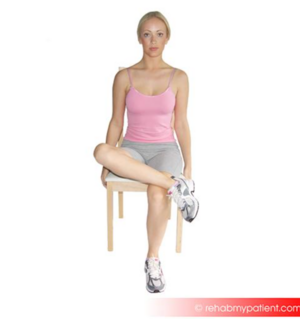
The patient can place the lateral malleoli of the ankle on the contralateral knee (figure of 4 - examining external rotation of the hip)
From the previous position, the patient can bring the leg across the body and assess pain with this movement (moving hip into internal rotation)
Knee to chest
Transitional movements should be assessed when the patient moves from sitting to supine
Patient in supine:
Asymmetrical Hip Rotation Range of Motion (ROM)
Figure of 4 (placing lateral malleoli of the ankle on the contralateral knee)
Piriformis stretch, knee pulled across body
Looking for pain, perceived tightness between sides
The patient can also palpate the anterior bony landmarks of the pelvis in supine
Other assessments as indicated (self palpation of muscles, other flexibility)
↑Chehrehrazi M, Nourbakhsh MR. The Effects of Pelvic Repositioning Exercise on Pelvic Floor Muscle Contractility in Continent Individuals With Asymmetric Pelvis: A Double-Blinded Randomized Control Study. The Journal of Women's & Pelvic Health Physical Therapy. 2024 Apr 1;48(2):75-83.
↑ 3.03.13.2Barbee Ellison J, Rose SJ, Sahrmann SA. Patterns of hip rotation range of motion: a comparison between healthy subjects and patients with low back pain. Physical therapy. 1990 Sep 1;70(9):537-41.
↑ 4.04.1Cibulka MT. Understanding sacroiliac joint movement as a guide to the management of a patient with unilateral low back pain. Manual Therapy. 2002 Nov 1;7(4):215-21.
↑ 5.05.1Prather H, Cheng A, Steger-May K, Maheshwari V, Van Dillen L. Hip and lumbar spine physical examination findings in people presenting with low back pain, with or without lower extremity pain. journal of orthopaedic & sports physical therapy. 2017 Mar;47(3):163-72.
↑ 6.06.16.2Hartigan E, McAuley JA, Lawrence M, Brucker W, King A, Tryon B, DeSilva M. Hip Angles, Joint Moments, and Muscle Activity During Gait in Women With and Without Self-reported Stress Urinary Incontinence. Journal of Women’s Health Physical Therapy. 2020 Jul 1;44(3):107-16.
↑ 8.08.1Charlton PC, Mentiplay BF, Pua YH, Clark RA. Reliability and concurrent validity of a Smartphone, bubble inclinometer and motion analysis system for measurement of hip joint range of motion. Journal of Science and Medicine in Sport. 2015 May 1;18(3):262-7.
↑ 9.09.19.2Miley E, May J, Albertin E, Takahashi E, Goodman C, Pettaway A. Reliability and Validity for Measuring Active Hip Rotation with the Clinometer Smartphone Application™. Journal of Rehabilitation Sciences & Research. 2019 Dec 1;6(4):193-9.
↑Cheatham S, Hanney WJ, Kolber MJ. Hip range of motion in recreational weight training participants: a descriptive report. International Journal of Sports Physical Therapy. 2017 Oct;12(5):764.
↑ATX Physical Therapy. Prone hip external and internal rotation assessment. Available from https://vimeo.com/399220225 (last accessed 3 January 2021)
↑Albert H, Godskesen M, Westergaard J. Evaluation of clinical tests used in classification procedures in pregnancy-related pelvic joint pain. European Spine Journal. 2000 Apr 1;9(2):161-6.
↑Jain S, Eedarapalli P, Jamjute P, Sawdy R. Symphysis pubis dysfunction: a practical approach to management. The Obstetrician & Gynaecologist. 2006 Jul;8(3):153-8.
↑ 17.017.1Garras DN, Carothers JT, Olson SA. Single-leg-stance (flamingo) radiographs to assess pelvic instability: how much motion is normal?. JBJS. 2008 Oct 1;90(10):2114-8.
↑Cook C, Hegedus E. Orthopedic Physical Examination Tests: An Evidence-Based Approach. 2nd ed. Prentice Hall; 2012
↑Njoo KH, Van der Does E. The occurrence and inter-rater reliability of myofascial trigger points in the quadratus lumborum and gluteus medius: a prospective study in non-specific low back pain patients and controls in general practice. Pain. 1994 Jan 1;58(3):317-23.
↑Njoo KH, Van der Does E, Stam HJ. Interobserver agreement on iliac crest pain syndrome in general practice. The Journal of rheumatology. 1995 Aug 1;22(8):1532-5.
↑ 21.021.1McCombe PF, Fairbank JC, Cockersole BC, Pynsent PB. 1989 Volvo Award in clinical sciences. Reproducibility of physical signs in low-back pain. Spine. 1989 Sep 1;14(9):908-18.
↑Vleeming A, Pool-Goudzwaard AL, Hammudoghlu D, Stoeckart R, Snijders CJ, Mens JM. The function of the long dorsal sacroiliac ligament: its implication for understanding low back pain. Spine. 1996 Mar 1;21(5):556-62.
↑Vleeming A, De Vries HJ, Mens JM, Wingerden JP. Possible role of the long dorsal sacroiliac ligament in women with peripartum pelvic pain. Acta obstetricia et gynecologica Scandinavica. 2002 Jan 1;81(5):430-6.
↑Saleh EA, Walton L, Said S, Raigangar V, Kim M. The association between Diastasis recti abdominis and pelvic floor dysfunction, sacroiliac joint dysfunction, and quality of life among postpartum women after Cesarean delivery. Physikalische Medizin, Rehabilitationsmedizin, Kurortmedizin. 2024 Feb;34(01):18-26.
↑Vleeming A, Albert HB, Östgaard HC, Sturesson B, Stuge B. European guidelines for the diagnosis and treatment of pelvic girdle pain. European Spine Journal. 2008 Jun 1;17(6):794-819.