When refering to evidence in academic writing, you should always try to reference the primary (original) source. That is usually the journal article where the information was first stated. In most cases Physiopedia articles are a secondary source and so should not be used as references. Physiopedia articles are best used to find the original sources of information (see the references list at the bottom of the article).
If you believe that this Physiopedia article is the primary source for the information you are refering to, you can use the button below to access a related citation statement.
The temporomandibular joint (TMJ) is a synovial joint that is made up of the articulating surface of the temporal bone and the head of the mandible (Figure 1 and 2).[1] Dysfunction of the TMJ is considered the most common cause of orofacial pain.[2] The joint itself is also associated with a number of important functions including eating[3], speaking,[4] breathing[5] and sleeping.[6]
Temporomandibular Joint Biomechanics
The TMJ is a ginglymoarthrodial joint[7] that allows for rotation and translation in the sagittal plane.[8][9]
Inferior and superior surfaces of the articular disc
The superior surface faces the fossa mandibularis
The inferior surface is in contact with the mandible condyle
The situation of the condyle inside the joint is a controversial subject. The so-called “central position” is a theoretical concept. The jaw is suspended, supported by the muscles and other stabilising elements, such as ligaments and the articular capsule.[8] For more information on the anatomy of the TMJ, please click here.
Maintenance of Jaw Position
Figure 2. TMJ - jaw open.
The maintenance of jaw position depends on jaw reflexes and the action of gravity. It is also affected by an individual’s position/posture and specific variations which allow functional jaw movements to occur.[8]
When the mandible is at rest, the mouth is slightly open, so that the teeth are not in contact.[10] This resting position is called physiological non-occlusion:[8][11]
In this position, the lips close the oral cavity without pressure - the teeth remain separated by a distance of around 2 mm - this distance is measured between the superior and inferior incisors[8]
This resting position is maintained by various reflexes (e.g. the jaw jerk reflex[12]), as well as active and passive mechanisms.
Periodontal mechanoreceptors and mechanoreceptors of the mucosa (i.e. gums, lips, tongue, palatal area)
Central control from the:
Cortico-visual system
Limbic system
Fusimotor-extrapyramidal afferences
The limbic and visual systems are not only actively involved in maintaining the position of the jaw, they also have an impact on the tone of the masticatory muscles. For instance, situations that cause emotional stress[13][14] or visual alterations[15] can change the tone of the jaw muscles and affect jaw position.[8]
Mastication marks the beginning of the digestive process. It is an essential step in the oral processing of food before deglutition (i.e. swallowing).[8][17]
The process of mastication, as well as other phases of swallowing, is controlled by the central pattern generator in the brainstem.[18] It occurs in the mouth with the help of the mandible and associated muscles.[8]
For mastication to occur, a range of information from sensory receptors (smell, taste and touch) is required,[3] as well as information from the tongue, palate, lips, masseter muscles and salivary glands.[8]
A change in one or more of these elements can cause issues with mastication.[8]
Huckabee and Daniels divide swallowing into four phases:[3]
Pre-oral (anticipatory) phase:
This phase “is the interaction of pre-oral motor, cognitive, psychosocial and somataesthetic elements which begin the swallowing process”[3]
Information about the food, which is obtained via the optic and olfactory nerves, is interpreted in the central nervous system and a swallowing plan is developed[3]
This information includes smell and specific routines that show the feeding act is about to begin[8][3]
The orofacial structures start to prepare to receive food - e.g. the salivary glands begin generating saliva[8]
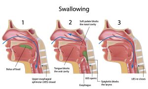
Figure 2. Swallowing.Oral phase:
The oral phase starts when food enters the mouth[8]
The lips close and the tongue forms a seal to prevent the food (which is being transformed into a bolus) from falling out of the mouth
The bolus is formed through the movement of the lips, jaw, cheeks and tongue[3] - i.e.the food is cut, split and ground up
Once the bolus is safe to be swallowed, it is pushed backwards by the tongue to the pharynx[3]
It has been found that the mastication process and the formation of the bolus is influenced by the physical characteristics of the food.[18][19] A study by Mishellany and colleagues found that individuals tend to achieve a similar bolus particle size, but that the amount of time and number of cycles to achieve this result varies between individuals. Thus, it appears that the size and distribution of the particles of the food influence the deglutition reflex.[20]
The oral phase can be affected by pathology of the TMJ. For some patients with TMJ dysfunction, it will be difficult for them to open their mouths. This will cause issues with mastication and, therefore, the overall digestive process.[8]
The pharyngeal phase refers to the movement of the bolus through the pharynx
During this phase, the airway is also protected from the bolus
The bolus moves from the base of the tongue to the wall of the posterior pharynx
Oesophageal phase:
This phase begins once the bolus passes through the upper oesophageal sphincter[8]
Peristalsis pushes the bolus down to the stomach via the lower oesophageal sphincter[3]
Speaking
Speaking is a complex, dynamic sensorimotor activity. It has been found that there is a connection between the intra-oral information and the oral and cervical muscles.[4]
Torisu and colleagues found, for instance, that intra-oral stimulation can inhibit neck muscle activity:[4]
This indicates that there is a neural connection between the trigeminal region and the cervical region
While this modulation might be largely due to nociceptive afferent nerves, non-nociceptive fibres may also be involved
As the authors note, this connection is significant because it may suggest that orofacial pain can affect head, neck and shoulder activity[4]
Breathing
Figure 3. Upper respiratory system.
Because breathing simultaneously occurs with all other oral activities, the respiratory pattern must be coordinated with the other functions occurring in the mouth / oral cavity (e.g. eating).[8]
The upper airways begin at the nasal cavity, before moving to the nasopharynx and oropharynx, down to the larynx and to the extra-thoracic trachea (see Figure 3).[21] When we breathe, air can enter through the nose or the mouth, but it always passes through the pharynx[5] - during swallowing, the pharynx is used as a passage for food. In healthy individuals, swallowing is dominant to respiration.[5] Breathing stops briefly when an individual is swallowing. This is caused by:[5]
The physical closure of the airway by the lifting of the soft palate and tilting of the epiglottis (see Figure 2)
Neural suppression of respiration by the brainstem
In the resting phase, air can enter via the nasal or oral cavity, generating two different respiratory pattern options. These patterns need to coordinate with the rest of the oral cavity's physiological functions. When the oral cavity is used for breathing, a degree of jaw opening is necessary. To achieve this, tone in the elevator muscles decreases, which allows air to circulate.[8]
Movements of the Temporomandibular Joint
Jaw Opening
Jaw opening is divided into the following phases:[8][22][23]
Open Jaw
Pure rotation of the condyles on their axis
Most of this movement happens in the infra-meniscal space of the condylo-discal complex
This is facilitated by the lateral pterygoid muscle (inferior part), mylohyoid, geniohyoid and digastric muscles
Translation of the condylo-disc complex forwards
This movement happens mainly in the superior compartment of the disc-temporal complex
The jaw opens around 40 to 50 mm
The temporomandibular ligament helps to maintain stability to prevent the jaw from dislocating forwards
The lateral pterygoid muscle is involved in this action
NB: The lateral pterygoid has opposite functions - while its superior fascicle relaxes during opening, stabilising the anterior displacement of the disc, the inferior fascicle contracts and allows movement of the condyle
The ligaments create stability at the end of the movement
The disc and condyles move medially and the collateral lateral ligaments on each side of the TMJ tighten
At a certain point, the condylo-discal complex is unable to move any further due to the tension in the ligaments and the joint capsule - at this point, it rotates on its axis
Jaw Closing
Jaw closure is associated with cervical extension. The elevator muscles of the jaw work against gravity. Closing of the jaw is divided into three phases:[8]
Closed Jaw
Condylar rotation in the inferior posterior meniscus area - this is similar to jaw opening, but in the opposite direction
This phase starts without any specific muscle action - rather it occurs due to the relaxation of the muscles involved in opening and the release of tension within the ligaments
Translation of the superior condylo-disc meniscal area
The complex made by the condyle and disc moves to the most posterior and superior part of the mandibular fossa
When the condyle has reached this point, there is a rotation in the posterior direction of the condyle in the intra-meniscal space - this ends with occlusal contact (NB occlusion refers to the relationship between the upper and lower teeth when the jaw closes[24])
In normal conditions, a slight lateral displacement of condyles can be observed in a sagittal view.[8]
The jaw is slightly opened to avoid any interference from the teeth (i.e. occlusion) - this opening causes anterior rotation in the sagittal plane
The condyle translates forwards and downwards - this is due to the disposition of the fossa mandibularis, which makes the condyle move down
This movement occurs due to the coordinated action of both fascicles of the lateral pterygoid muscle. As jaw opening does not progress in protraction, the jaw is stabilised by the contraction of the temporalis muscles.[8]
The condyle translates backwards and upwards inside the fossa mandibularis - this movement is activated by the temporalis muscle and the posterior belly of the digastric muscle
Finally there is a posterior rotation of the condyle at the intra-meniscal level (i.e. the condylo-disc complex)[8]
The condyles work together to achieve lateral movements of the jaw. When assessing lateral movement, it is necessary to differentiate one condyle from the other:
The "working side" is the side that moves laterally when taking the chin as a reference
The "non-working side" is the side that moves towards the midline
On the working side, there is a rotation of the condyle over its vertical axis and a transversal displacement of about 0.9 mm. This movement is caused by the deep masseter muscle and the medial and posterior fascicles of the temporalis muscle.[8]
On the non working side, the condyle moves to the midline, going forward and moving closer to the midline. It also moves transversally around 0.4 mm. In this case, the muscles activated are the lateral pterygoid (inferior fascicle) and the medial pterygoid.[8]
References
↑Maini K, Dua A. Temporomandibular Joint Syndrome. [Updated 2021 Apr 25]. In: StatPearls [Internet]. Treasure Island (FL): StatPearls Publishing; 2021 Jan-. Available from: https://www.ncbi.nlm.nih.gov/books/NBK551612/
↑Hollis JH. The effect of mastication on food intake, satiety and body weight. Physiol Behav. 2018;193(Pt B):242-245.
↑ 18.018.1Hwang J, Kim DK, Bae JH, Kang SH, Seo KM, Kim BK, et al. The effect of rheological properties of foods on bolus characteristics after mastication. Ann Rehabil Med. 2012;36(6):776-84.
↑van der Bilt A, Abbink JH. The influence of food consistency on chewing rate and muscular work. Arch Oral Biol. 2017;83:105-10.
↑Mishellany A, Woda A, Labas R, Peyron MA. The challenge of mastication: preparing a bolus suitable for deglutition. Dysphagia. 2006;21(2):87-94.
↑Bordoni B, Varacallo M. Anatomy, Head and Neck, Temporomandibular Joint. [Updated 2021 Feb 7]. In: StatPearls [Internet]. Treasure Island (FL): StatPearls Publishing; 2021 Jan-. Available from: https://www.ncbi.nlm.nih.gov/books/NBK538486/
↑Pai SA, Poojari SR, Ramachandra K, Patel RKV, Jyothi M. Temporomandibular joint - an anatomical view. Journal of Advanced Clinical & Research Insights. 2019;6:1-5.
↑Alila Medical Media. Temporomandibular Joint (TMJ) Anatomy and Disc Displacement Animation. Available from: https://www.youtube.com/watch?v=mB468Jh9aAY [last accessed 29/6/2021]