When refering to evidence in academic writing, you should always try to reference the primary (original) source. That is usually the journal article where the information was first stated. In most cases Physiopedia articles are a secondary source and so should not be used as references. Physiopedia articles are best used to find the original sources of information (see the references list at the bottom of the article).
If you believe that this Physiopedia article is the primary source for the information you are refering to, you can use the button below to access a related citation statement.
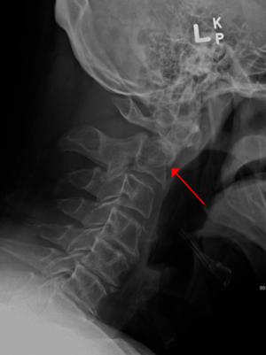
Cervical fractures refer to fractures in the vertebrae of the cervical spine (C1 to C7), often caused by high-impact trauma. These fractures can range from mild to severe, and if the spinal cord is affected, they may lead to neurological impairments, including paralysis. Cervical fractures are considered a medical emergency due to the proximity of the spinal cord to the vertebral bones.
Clinically Relevant Anatomy
The cervical spine comprises of seven vertebrae (C1-C7). The first two vertebrae (C1, C2) are unique and referred to as the upper C-Spine. The C1 (atlas) supports the skull, while the C2 (axis) allows rotation of the head. Stability is primarily obtained from ligaments, especially the transverse ligaments of the altas. The lower or sub axial C-spine which is C3 to C7 which in return gets stability equally from bony structures and ligaments.The vertebral column protects the spinal cord, which passes through the vertebral foramen. Damage to the cervical spine can result in nerve root compression or spinal cord injury, leading to severe functional impairments, such as quadriplegia. [1]
Etiology
Primary causes:
Motor vehicle accidents (most common - 29.3% of cases)[2]
Falls (especially in elderly)
Spinal cord injury is reported to occur in 10 to 50% of cases.[3]
A CT scan of the spine with thin slices and multi-planar reconstructions is recommended. If a CT scan is unavailable or cannot be obtained, full cervical spine plain radiographs, including adequate anteroposterior (AP), lateral, and odontoid views, should be performed.[4]
Although Cervical imaging evaluations are not necessary for all suspected cervical spine injuries. Clinical decision tools have been developed, they consist of a combination of patient history, physical examinations and injury mechanism.[5]
Occipital Condyle Fractures (Anderson and Montesano Classification).
The classification of occipital condyle fractures (OCFs) is based on mechanisms of injury and stability of the fracture.
Type I:
Mechanism: Impact fracture due to axial load.
Stability: Stable fracture because the tectorial membrane and contralateral alar ligaments remain intact, helping to preserve stability
Type II:
Mechanism: Part of a cranial base fracture, often from direct regional trauma (such as a blow to the head), with a fracture line extending toward the foramen magnum.
Stability: Stable, as the tectorial membrane and alar ligaments remain intact.
Type III:
Mechanism: Avulsion fracture of the occipital condyle by the alar ligament, often due to rotational or lateral movements of the head.
Stability: Potentially unstable due to disruption of the alar ligaments and/or tectorial membrane.
Mechanism: Burst fracture of the atlas due to axial compression. This causes splitting of the lateral masses of the C1 vertebra. The force drives the skull downward onto the atlas, causing it to compress and rupture at its weakest points (the anterior and posterior arches).
Type 1:Isolated anterior OR posterior arch fracture
Type 2: Fracture of the anterior AND posterior arch
Type 3:Fracture of lateral mass with or without fracture of the posterior arch
These injuries usually result from high-energy trauma, such as motor vehicle accidents or falls from height, and may be associated with significant neurological damage.
Rheumatoid arthritis is a common cause of subluxation due to existing atlantoaxial instability.
Grisel's Syndrome is a rare cause of C1-C2 rotatory subluxation, typically associated with inflammatory processes (like a throat or ear infection) that cause ligament laxity and instability.
Odontoid Dens (C2) Fractures (Anderson and D'Alonzo Classification)
Type I: Involves the upper part of the odontoid (the tip of the dens).Type II:At the base of the odontoid (the junction of the dens and the axis body).
Type III: Through the body of the axis.
Hangman’s Fracture (Levine and Edwards Classification)[2]
Mechanism: This is typically a hyperextension-distraction injury, often resulting from a high-impact motor vehicle accident or hangings. Normally with this fracture it rarely leads to injury to the spinal cord because the canal widens and not narrows.[2]
Type I: Fracture of the pars interarticularis without significant angular deviation or displacement (<3.5mm).
Type II: Significant displacement and angular deviation of the vertebrae.
Type IIa: A subtype of Type II with less displacement but a wide angulation.
Type III: Large displacement with facet dislocation, often requiring surgical intervention..
Lower Cervical Spine Fractures (C3-C7)
Can be classified using either:The AO classification system is widely used and focuses on the mechanism of injury, providing a classification based on the fracture pattern.
Type A:Compression fractures
Mechanism: These fractures result from axial compression forces. The vertebral body is compressed, often leading to a wedge shape. For example: Simple anterior compression fracture, where the posterior elements remain intact.
Type B: Distraction fractures
Mechanism: These fractures occur from a distraction force, typically resulting from hyperextension or flexion with axial loading. This type of fracture often involves a failure of the posterior ligamentous complex, leading to instability. Example: Teardrop fractures, spinous process fractures or fractures involving the disc-ligamentous complex.
Type C: Rotation fracture
Mechanism: These fractures are caused by a rotational force that causes either facet dislocation, fractures of the facet or disruption of the posterior elements. For example: Facet dislocation, rotary subluxation or fractures through the facet joints.
AO Classification
Type A: Compression
Type B: Distraction
Type C: Rotation
SLIC (Subaxial Injury Classification) Scale: Scores based on:[7]
The SLIC was created to by systemic reviewing surgical treatment of the lower spine and a treatment algorithm was created. The classification takes into account the following: Morphology, Status of the disco-ligamentous complex and Neurological assessment.
Morphogy (fracture pattern):Describes the fracture type (e.g., compression, distraction, rotation, or others). This is similar to the AO classification.
Disco-ligamentous complex: Assesses the integrity of the posterior ligamentous complex, which is critical for maintaining stability.
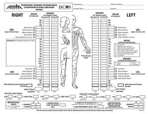
Neurological assessment: Assesses the presence of any neurological deficits , such as paralysis or sensory loss.
Subaxial Injury Classification (SLIC) scale
Points
Morphology
No abnormality
0
Compression & Burst
1+1=2
Distraction
3
Rotation or translation
4
Disc-ligamentous complex
Intact
0
Indeterminate
1
Disrupted
2
Neurological status
Intact
0
Root injury
1
Complete cord injury
2
Incomplete cord injury
3
Continuous cord compression
+1
A score is given as shown above to each injury. Treatment is decided on the total score received:[2]
Score < 4 : The patient do not need surgical treatment
Score =4: Either surgical or conservative treatment. Normally the deciding decision is made based upon the surgeons personal experience.
Score >4: Surgical treatment is usually required.
Management / Intervention
Pre-Hospital Management:
Immobilisation: Patients with suspected C-spine fractures are initially managed in the pre-hospital setting with a rigid cervical collar (hard collar) applied in a neutral position to prevent unnecessary movement of the cervical spine and protect against further injury.[1]
Initial Assessment in the Emergency Department:[1]
C-Spine Protection: On arrival at the emergency department, C-spine injury should be assumed until proven otherwise. A detailed history from the patient or emergency crew is essential, focusing on the type of trauma (high vs. low energy), mechanism of injury, and the presence of neurological or physical symptoms.
Advanced Trauma Life Support (ATLS): The ATLS protocol is followed to guide the prioritisation of initial management. This begins with:
Airway Management: Ensuring the airway is patent and securing it.
C-Spine Immobilisation: Triple immobilisation (hard collar, sandbags, head blocks, and tape) is applied to prevent movement and protect the spinal cord.
Breathing: Assess breathing for adequate oxygenation.
Circulation: Assess circulation for sufficient perfusion and address any hypotension due to haemorrhagic or neurogenic shock with aggressive fluid resuscitation.
Clinical Examination Limitations: Clinical examination alone is not sufficient to identify all potential C-spine injuries. Imaging is required for a definitive diagnosis.
Imaging Protocol: For unconscious patients, a CT trauma series (including C-spine, chest, abdomen, and pelvis) is typically performed. For conscious patients, imaging is guided by CCR or NEXUS.
SCI must be quickly identified as it can result in long-term disability. Prompt intervention and management, including appropriate imaging and possible surgical intervention, are essential to minimize neurological sequel.
Following below is a general guidelines to follow after cervical surgery:[14]
Neck and Shoulder Muscle Strengthening
Start with non-resistance and isometric exercises to strengthen deep neck and shoulder muscles and progress to resistance exercises as tolerated. These exercises not only target muscle hypertrophy but also improve local blood circulation, reduce pain, and facilitate recovery.[14]
Hand Dexterity and Movement Exercises
Use task-specific exercises to mimic daily activities—such as pinching, cutting, and handling objects with utensils. These exercises are essential for restoring the dexterity needed for ADLs.
Neural Mobilisations (NM)
Do mobilisation techniques for nerves like the median, radial, and ulnar nerves to reduce pain, improve circulation, and enhance mobility. NM has shown effectiveness in reducing upper limb pain and improving quality of life.
Exercises should focus on improving lower limb strength and postural control. Activities may include standing on unstable surfaces, reacting to treadmill speed changes, and using game-based exercises for center of gravity control. Balance rehabilitation also lowers fall risks, which are particularly high for patients with cervical spinal cord involvement.
VR creates an immersive environment where patients practice controlled movements, which has shown to positively impact muscle strength, gait, and balance, especially in those with spinal cord injuries. VR can also stimulate neuroplasticity, making it a promising adjunct to traditional therapy.
In summary, a comprehensive approach to cervical spine postoperative rehabilitation should include progressive strengthening, hand dexterity improvement, neural mobilisation, balance training, and, where feasible, VR to aid in motor function restoration. Such an approach is designed to enhance recovery outcomes by addressing the unique needs of cervical surgery patients.
For a detailed management of a spinal cord injury management please see the following pages:
↑ 2.02.12.22.32.42.5Marcon R, Cristante A, Teixeira W, Narasaki D, Oliveira R, Barros Filho T. Fractures of the cervical spine. Clinics [Internet]. 2013 Nov 1;68(11):1455–61. Available from: https://www.ncbi.nlm.nih.gov/pmc/articles/PMC3812556/