Vastus Medialis Oblique: Difference between revisions
No edit summary |
No edit summary |
||
| (21 intermediate revisions by 5 users not shown) | |||
| Line 1: | Line 1: | ||
</div> | |||
<div class="editorbox"> | <div class="editorbox"> | ||
'''Original Editor '''- [[User:Rafet Irmak|Rafet Irmak]] | '''Original Editor '''- [[User:Rafet Irmak|Rafet Irmak]] | ||
| Line 5: | Line 7: | ||
</div> | </div> | ||
== Description == | |||
The Vastus Medialis Oblique (VMO) muscle is the muscle that makes up the distal most fibres of the [[Vastus Medialis|Vastus Medialis (VM)]] muscle of the [[Quadriceps Muscle|Quadriceps]].<ref name=":2">El Sawy MM, Mikkawy DM, El-Sayed SM, Desouky AM. [https://www.ncbi.nlm.nih.gov/pmc/articles/PMC8017455/ Morphometric analysis of vastus medialis oblique muscle and its influence on anterior knee pain.] Anatomy & cell biology. 2021 Mar 1;54(1):1-9.</ref> The VMO and VM muscles are functionally different due to different orientation of muscle fibres. VMO runs obliquely and helps with medial translation of the [[Patella]] where VM runs more longitudinally and contributes more to knee extension. [[Patellofemoral Joint|Patellofemoral joint]] load is decreased by advanced and adequate medially directed force by the VMO.<ref name=":0">Peng YL, Tenan MS, Griffin L. [https://journals.physiology.org/doi/pdf/10.1152/japplphysiol.00702.2017 Hip position and sex differences in motor unit firing patterns of the vastus medialis and vastus medialis oblique in healthy individuals]. Journal of Applied Physiology. 2018 Jun 1;124(6):1438-46.</ref> | |||
[[File:VMO1.png|alt=|center|572x572px]] | |||
== | === Origin === | ||
The VMO has multiple points of origin. The muscle fibres largely originate from the pubic points of the [[Adductor Magnus]] tendon. The other points of origin include the medial lip of linea aspera and the medial supracondylar line.<ref name=":2" /><ref name=":1">Rajput HB, Rajani SJ, Vaniya VH. [https://www.ncbi.nlm.nih.gov/pmc/articles/PMC5713709/pdf/jcdr-11-AC01.pdf Variation in morphometry of vastus medialis muscle]. Journal of Clinical and Diagnostic Research: JCDR. 2017 Sep;11(9):AC01.</ref> | |||
=== | === Insertion === | ||
VMO inserts onto the medial border of the patella and the knee joint capsule. It also has a small area where it directly continues with the patella tendon<ref name=":2" />. | |||
== | === Action === | ||
The primary function the VMO muscle is to pull the patella medially. The alignment of the muscle fibres, which is horizontal, makes it the primary medial stabilizer of the patella. The VMO along with [[Vastus Lateralis|Vastus Lateralis (VL)]] helps align the patella within the trochlear groove during knee flexion and extension.<ref>Miao P, Xu Y, Pan C, Liu H, Wang C. [https://bmcmusculoskeletdisord.biomedcentral.com/articles/10.1186/s12891-015-0736-6 Vastus medialis oblique and vastus lateralis activity during a double-leg semisquat with or without hip adduction in patients with patellofemoral pain syndrome.] BMC musculoskeletal disorders. 2015 Dec;16(1):1-8.</ref> The VMO also participates in the last phase of knee extension. <ref name=":2" /> | |||
== | === Nerve Supply === | ||
VMO receives its nerve supply from the branches of the Femoral Nerve. <ref name=":1" /> | |||
=== Bood Supply === | |||
The femoral artery provides the blood supply for the VMO muscle. <ref>Mirjalili SA. Anatomy of the lumbar plexus. InNerves and Nerve Injuries 2015 Jan 1 (pp. 609-617). Academic Press.</ref> | |||
== Assessment == | |||
The VMO muscle can often be evaluated using surface electromyography (EMG). <ref>Abdelraouf OR, Abdel-Aziem AA, Ahmed AA, Nassif NS, Matar AG. [https://www.ncbi.nlm.nih.gov/pmc/articles/PMC6706826/ Backward walking alters vastus medialis oblique/vastus lateralis muscle activity ratio in females with patellofemoral pain syndrome.] Turkish journal of physical medicine and rehabilitation. 2019 Jun;65(2):169.</ref> | |||
== Clinical relevance == | |||
Due to VMO's primary function, its clinical relevance is with respect to knee function and stability. | |||
* | * '''Patellar stability:''' VMO muscle weakness or any alteration in its muscle activity can result in mal-tracking of patella resulting in instability and subsequently causing damage to surround structures and pain in the area.<ref name=":2" /> | ||
* '''Patellofemoral Pain Syndrome (PFPS):''' People with PFPS on evaluation are found to have lower volume of VMO and often benefit from strengthening exercises. <ref>Kasitinon D, Li WX, Wang EX, Fredericson M. [https://www.ncbi.nlm.nih.gov/pmc/articles/PMC8733121/ Physical examination and patellofemoral pain syndrome: an updated review]. Current reviews in musculoskeletal medicine. 2021 Dec 1:1-7.</ref> | |||
== Exercises == | |||
The video below demonstrates few exercises that can be incorporated into rehabilitation programmes for people with reduced VMO strength: | |||
{{#ev:youtube|ialuYCEdje4}}<ref>''VMO strengthening exercises - ask doctor jo'' (2016) ''YouTube''. Available at: <nowiki>https://www.youtube.com/watch?v=ialuYCEdje4</nowiki> </ref> | |||
== References == | == References == | ||
| Line 58: | Line 43: | ||
<references /> | <references /> | ||
[[Category:Anatomy]] [[Category:Knee]] [[Category:Muscles]] [[Category:Musculoskeletal/Orthopaedics|Orthopaedics]] | [[Category:Anatomy]] | ||
[[Category:Knee - Anatomy]] | |||
[[Category:Knee]] | |||
[[Category:Muscles]] | |||
[[Category:Knee - Muscles]] | |||
[[Category:Musculoskeletal/Orthopaedics|Orthopaedics]] | |||
Latest revision as of 18:46, 24 January 2024
Original Editor - Rafet Irmak
Top Contributors - Admin, Ananya Bunglae Sudindar, Rafet Irmak, George Prudden, Wendy Snyders, Richard Benes, Kim Jackson, Evan Thomas, WikiSysop and Claudia Karina
Description[edit | edit source]
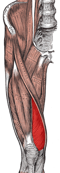
The Vastus Medialis Oblique (VMO) muscle is the muscle that makes up the distal most fibres of the Vastus Medialis (VM) muscle of the Quadriceps.[1] The VMO and VM muscles are functionally different due to different orientation of muscle fibres. VMO runs obliquely and helps with medial translation of the Patella where VM runs more longitudinally and contributes more to knee extension. Patellofemoral joint load is decreased by advanced and adequate medially directed force by the VMO.[2]
Origin[edit | edit source]
The VMO has multiple points of origin. The muscle fibres largely originate from the pubic points of the Adductor Magnus tendon. The other points of origin include the medial lip of linea aspera and the medial supracondylar line.[1][3]
Insertion[edit | edit source]
VMO inserts onto the medial border of the patella and the knee joint capsule. It also has a small area where it directly continues with the patella tendon[1].
Action[edit | edit source]
The primary function the VMO muscle is to pull the patella medially. The alignment of the muscle fibres, which is horizontal, makes it the primary medial stabilizer of the patella. The VMO along with Vastus Lateralis (VL) helps align the patella within the trochlear groove during knee flexion and extension.[4] The VMO also participates in the last phase of knee extension. [1]
Nerve Supply[edit | edit source]
VMO receives its nerve supply from the branches of the Femoral Nerve. [3]
Bood Supply[edit | edit source]
The femoral artery provides the blood supply for the VMO muscle. [5]
Assessment[edit | edit source]
The VMO muscle can often be evaluated using surface electromyography (EMG). [6]
Clinical relevance[edit | edit source]
Due to VMO's primary function, its clinical relevance is with respect to knee function and stability.
- Patellar stability: VMO muscle weakness or any alteration in its muscle activity can result in mal-tracking of patella resulting in instability and subsequently causing damage to surround structures and pain in the area.[1]
- Patellofemoral Pain Syndrome (PFPS): People with PFPS on evaluation are found to have lower volume of VMO and often benefit from strengthening exercises. [7]
Exercises[edit | edit source]
The video below demonstrates few exercises that can be incorporated into rehabilitation programmes for people with reduced VMO strength:
References[edit | edit source]
- ↑ 1.0 1.1 1.2 1.3 1.4 El Sawy MM, Mikkawy DM, El-Sayed SM, Desouky AM. Morphometric analysis of vastus medialis oblique muscle and its influence on anterior knee pain. Anatomy & cell biology. 2021 Mar 1;54(1):1-9.
- ↑ Peng YL, Tenan MS, Griffin L. Hip position and sex differences in motor unit firing patterns of the vastus medialis and vastus medialis oblique in healthy individuals. Journal of Applied Physiology. 2018 Jun 1;124(6):1438-46.
- ↑ 3.0 3.1 Rajput HB, Rajani SJ, Vaniya VH. Variation in morphometry of vastus medialis muscle. Journal of Clinical and Diagnostic Research: JCDR. 2017 Sep;11(9):AC01.
- ↑ Miao P, Xu Y, Pan C, Liu H, Wang C. Vastus medialis oblique and vastus lateralis activity during a double-leg semisquat with or without hip adduction in patients with patellofemoral pain syndrome. BMC musculoskeletal disorders. 2015 Dec;16(1):1-8.
- ↑ Mirjalili SA. Anatomy of the lumbar plexus. InNerves and Nerve Injuries 2015 Jan 1 (pp. 609-617). Academic Press.
- ↑ Abdelraouf OR, Abdel-Aziem AA, Ahmed AA, Nassif NS, Matar AG. Backward walking alters vastus medialis oblique/vastus lateralis muscle activity ratio in females with patellofemoral pain syndrome. Turkish journal of physical medicine and rehabilitation. 2019 Jun;65(2):169.
- ↑ Kasitinon D, Li WX, Wang EX, Fredericson M. Physical examination and patellofemoral pain syndrome: an updated review. Current reviews in musculoskeletal medicine. 2021 Dec 1:1-7.
- ↑ VMO strengthening exercises - ask doctor jo (2016) YouTube. Available at: https://www.youtube.com/watch?v=ialuYCEdje4


