Vastus Medialis: Difference between revisions
Evan Thomas (talk | contribs) No edit summary |
Joao Costa (talk | contribs) No edit summary |
||
| (14 intermediate revisions by 7 users not shown) | |||
| Line 1: | Line 1: | ||
<div class="editorbox"> | <div class="editorbox"> | ||
'''Original Editor '''- | '''Original Editor '''- [[User:Evan Thomas|Evan Thomas]] | ||
'''Top Contributors''' - {{Special:Contributors/{{FULLPAGENAME}}}} | '''Top Contributors''' - {{Special:Contributors/{{FULLPAGENAME}}}} | ||
</div> | </div> | ||
== Description == | == Description == | ||
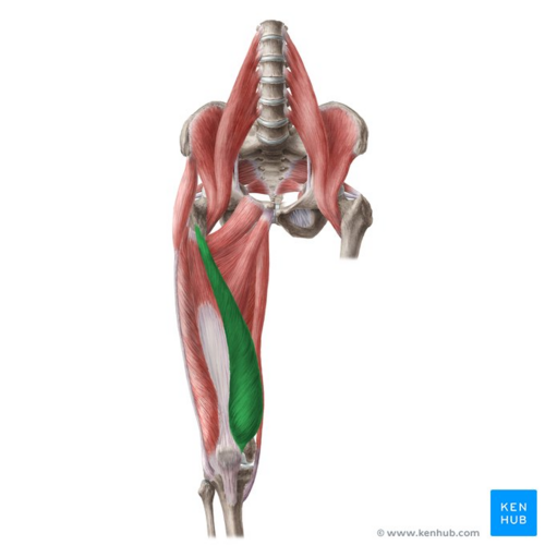
[[File:Vastus medialis muscle - Kenhub.png|alt=Vastus medialis muscle (highlighted in green) - anterior view|right|frameless|500x500px|Vastus medialis muscle (highlighted in green) - anterior view]] | |||
Vastus medialis is one of the four muscles that make up the [[Quadriceps Muscle|quadriceps]] group of muscles. It originates from the upper part of the [[Femur|femoral shaft]] and inserts as a flattened tendon into the quadriceps femoris tendon, which inserts into the upper border of the [[patella]].<ref name=":0">Anatomy.tv | 3D Human Anatomy | Primal Pictures [Internet]. Anatomy.tv. 2018 [cited 16 March 2018]. Available from: <nowiki>http://www.anatomy.tv/</nowiki></ref> | |||
Image: Vastus medialis muscle (highlighted in green) - anterior view<ref >Vastus medialis muscle (highlighted in green) - anterior view image - © Kenhub https://www.kenhub.com/en/study/main-muscles-of-lower-limb</ref> | |||
=== Origin === | |||
= | Lower part of the intertrochanteric line, along the spiral line to the medial lip of the linea aspera, the medial intermuscular septum and the aponeurosis of adductor magnus.<ref name=":0" /> | ||
=== Insertion === | === Insertion === | ||
= | Into the medial side of the quadriceps tendon, joining with [[Rectus Femoris|rectus femoris]] and the other quadriceps muscles, enveloping the [[patella]], then by the patellar ligament into the tibial tuberosity.<ref name=":0" /> | ||
=== | === Nerve === | ||
A branch from the posterior division of the femoral nerve, derived from L2, 3 and 4.<ref name=":0" /> | |||
== | === Artery === | ||
Femoral artery and branches from the profunda femoris artery.<ref name=":0" /> | |||
== | == Function == | ||
Vastus medialis, together with the other muscles that make up quadriceps femoris, extends the [[Knee|knee joint]] <ref name=":0" />and it also contributes to correct tracking of the patella.<ref>Vastus Medialis. Available from: <nowiki>https://en.wikipedia.org/wiki/Vastus_medialis</nowiki> (Accessed, 21/07/2021).</ref> | |||
=== | === Clinical relevance === | ||
Weakness of the vastus medials is associated with patellar maltracking and [[Patellofemoral Pain Syndrome|patellofemoral pain]]. An approach to treatment attempts to restore balance between vastus medialis and [[Vastus Lateralis|lateralis]], which requires strengthening of the oblique fibres of medialis, as well as assessment of the degree of dynamic supination and pronation of the foot.<ref>Werner S. [https://www.ncbi.nlm.nih.gov/pubmed/24997734 Anterior knee pain: an update of physical therapy.] Knee Surgery, Sports Traumatology, Arthroscopy. 2014 Oct 1;22(10):2286-94.</ref><ref name=":0" /> | |||
VMO strengthening has become less popular approach to the treatment of anterior knee pain as the evidence supporting isolated exercises has been criticised for its poor quality.<ref>Crossley K, Bennell K, Green S, McConnell J. [https://www.ncbi.nlm.nih.gov/pubmed/11403109 A systematic review of physical interventions for patellofemoral pain syndrome.] Clinical Journal of Sport Medicine. 2001 Apr 1;11(2):103-10.</ref><ref>Heintjes EM, Berger MY, Bierma-Zeinstra SM, Bernsen RM, Verhaar JA, Koes BW. [https://www.ncbi.nlm.nih.gov/pubmed/14583980 Exercise therapy for patellofemoral pain syndrome]. Cochrane Database Syst Rev. 2003 Jan 1;4.</ref><ref>Rodriguez-Merchan EC. [https://www.ncbi.nlm.nih.gov/pmc/articles/PMC4151435/ Evidence based conservative management of patello-femoral syndrome]. Archives of Bone and Joint Surgery. 2014 Mar;2(1):4.</ref><ref>Seeley MK, Park J, King D, Hopkins JT. [https://www.ncbi.nlm.nih.gov/pmc/articles/PMC3655747/ A novel experimental knee-pain model affects perceived pain and movement biomechanics.] Journal of athletic training. 2013 May;48(3):337-45.</ref> Furthermore researchers doubt the existence of VMO<ref>Smith TO, Nichols R, Harle D, Donell ST. [https://www.ncbi.nlm.nih.gov/pubmed/19090000 Do the vastus medialis obliquus and vastus medialis longus really exist?] A systematic review. Clinical anatomy. 2009 Mar 1;22(2):183-99.</ref> and have found that any quadricep exercise will similarly activate the vastus muscles.<ref>Smith TO, Bowyer D, Dixon J, Stephenson R, Chester R, Donell ST. [https://www.ncbi.nlm.nih.gov/pubmed/19212898 Can vastus medialis oblique be preferentially activated? A systematic review of electromyographic studies]. Physiotherapy theory and practice. 2009 Jan 1;25(2):69-98.</ref> Strengthening further up the kinetic chain has been suggested as more effective approach, Khayambashi et al. found that hip strengthening was more effective for improving patellofemoral pain than knee strengthening.<ref>Khayambashi K, Fallah A, Movahedi A, Bagwell J, Powers C. [https://www.ncbi.nlm.nih.gov/pubmed/24440362 Posterolateral hip muscle strengthening versus quadriceps strengthening for patellofemoral pain: a comparative control trial.] Archives of physical medicine and rehabilitation. 2014 May 1;95(5):900-7.</ref> | |||
== | == Assessment == | ||
=== | === Palpation === | ||
It can be palpated along its entire length. Distally, the quadriceps tendon can be palpated attaching to the proximal border (base) of the patella. | |||
== | == Treatment == | ||
== Resources == | == Resources == | ||
{| width="100%" cellspacing="1" cellpadding="1" | |||
|- | |||
| {{#ev:youtube|3RafHu6ir_c|412}} | |||
| {{#ev:youtube|8trLEtQYDGw|412}} | |||
|} | |||
== References == | == References == | ||
<references /> | <references /> | ||
[[Category:Anatomy]] [[Category: | [[Category:Anatomy]] | ||
[[Category:Knee]] | |||
[[Category:Knee - Anatomy]] | |||
[[Category:Muscles]] | |||
[[Category:Knee - Muscles]] | |||
[[Category:Musculoskeletal/Orthopaedics|Orthopaedics]] | |||
Latest revision as of 05:17, 1 April 2022
Original Editor - Evan Thomas
Top Contributors - George Prudden, Evan Thomas, Kim Jackson, Joao Costa, WikiSysop, Abbey Wright, Lucinda hampton and Kirenga Bamurange Liliane
Description[edit | edit source]
Vastus medialis is one of the four muscles that make up the quadriceps group of muscles. It originates from the upper part of the femoral shaft and inserts as a flattened tendon into the quadriceps femoris tendon, which inserts into the upper border of the patella.[1]
Image: Vastus medialis muscle (highlighted in green) - anterior view[2]
Origin[edit | edit source]
Lower part of the intertrochanteric line, along the spiral line to the medial lip of the linea aspera, the medial intermuscular septum and the aponeurosis of adductor magnus.[1]
Insertion[edit | edit source]
Into the medial side of the quadriceps tendon, joining with rectus femoris and the other quadriceps muscles, enveloping the patella, then by the patellar ligament into the tibial tuberosity.[1]
Nerve[edit | edit source]
A branch from the posterior division of the femoral nerve, derived from L2, 3 and 4.[1]
Artery[edit | edit source]
Femoral artery and branches from the profunda femoris artery.[1]
Function[edit | edit source]
Vastus medialis, together with the other muscles that make up quadriceps femoris, extends the knee joint [1]and it also contributes to correct tracking of the patella.[3]
Clinical relevance[edit | edit source]
Weakness of the vastus medials is associated with patellar maltracking and patellofemoral pain. An approach to treatment attempts to restore balance between vastus medialis and lateralis, which requires strengthening of the oblique fibres of medialis, as well as assessment of the degree of dynamic supination and pronation of the foot.[4][1]
VMO strengthening has become less popular approach to the treatment of anterior knee pain as the evidence supporting isolated exercises has been criticised for its poor quality.[5][6][7][8] Furthermore researchers doubt the existence of VMO[9] and have found that any quadricep exercise will similarly activate the vastus muscles.[10] Strengthening further up the kinetic chain has been suggested as more effective approach, Khayambashi et al. found that hip strengthening was more effective for improving patellofemoral pain than knee strengthening.[11]
Assessment[edit | edit source]
Palpation[edit | edit source]
It can be palpated along its entire length. Distally, the quadriceps tendon can be palpated attaching to the proximal border (base) of the patella.
Treatment[edit | edit source]
Resources[edit | edit source]
References[edit | edit source]
- ↑ 1.0 1.1 1.2 1.3 1.4 1.5 1.6 Anatomy.tv | 3D Human Anatomy | Primal Pictures [Internet]. Anatomy.tv. 2018 [cited 16 March 2018]. Available from: http://www.anatomy.tv/
- ↑ Vastus medialis muscle (highlighted in green) - anterior view image - © Kenhub https://www.kenhub.com/en/study/main-muscles-of-lower-limb
- ↑ Vastus Medialis. Available from: https://en.wikipedia.org/wiki/Vastus_medialis (Accessed, 21/07/2021).
- ↑ Werner S. Anterior knee pain: an update of physical therapy. Knee Surgery, Sports Traumatology, Arthroscopy. 2014 Oct 1;22(10):2286-94.
- ↑ Crossley K, Bennell K, Green S, McConnell J. A systematic review of physical interventions for patellofemoral pain syndrome. Clinical Journal of Sport Medicine. 2001 Apr 1;11(2):103-10.
- ↑ Heintjes EM, Berger MY, Bierma-Zeinstra SM, Bernsen RM, Verhaar JA, Koes BW. Exercise therapy for patellofemoral pain syndrome. Cochrane Database Syst Rev. 2003 Jan 1;4.
- ↑ Rodriguez-Merchan EC. Evidence based conservative management of patello-femoral syndrome. Archives of Bone and Joint Surgery. 2014 Mar;2(1):4.
- ↑ Seeley MK, Park J, King D, Hopkins JT. A novel experimental knee-pain model affects perceived pain and movement biomechanics. Journal of athletic training. 2013 May;48(3):337-45.
- ↑ Smith TO, Nichols R, Harle D, Donell ST. Do the vastus medialis obliquus and vastus medialis longus really exist? A systematic review. Clinical anatomy. 2009 Mar 1;22(2):183-99.
- ↑ Smith TO, Bowyer D, Dixon J, Stephenson R, Chester R, Donell ST. Can vastus medialis oblique be preferentially activated? A systematic review of electromyographic studies. Physiotherapy theory and practice. 2009 Jan 1;25(2):69-98.
- ↑ Khayambashi K, Fallah A, Movahedi A, Bagwell J, Powers C. Posterolateral hip muscle strengthening versus quadriceps strengthening for patellofemoral pain: a comparative control trial. Archives of physical medicine and rehabilitation. 2014 May 1;95(5):900-7.


