Trapezius
Original Editor - Daphne Jackson
Lead Editors - Lucinda hampton, Andeela Hafeez, Kim Jackson, Kai A. Sigel, Daphne Jackson, WikiSysop, Joao Costa, Kirenga Bamurange Liliane, Rachael Lowe, Evan Thomas and Tarina van der Stockt
Description[edit | edit source]
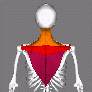
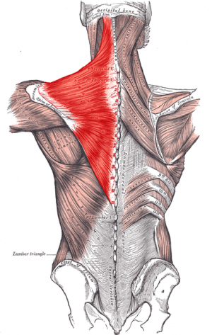
The trapezius is a broad, flat, superficial muscle extending from the cervical to thoracic region on the posterior aspect of the neck and trunk. The muscle is divided into three parts: descending (superior), ascending (inferior), and middle[1]. The muscle contributes to scapulohumeral rhythm through attachments on the clavicle and scapula, and to head balance through muscular control of the cervical spine.
In people who are chronically stressed or anxious the trapezius suffers. Hunching up the shoulders and holding tension in the shoulders and neck, puts constant strain on the trapezius. The trapezius is a major source of headache pain, usually a tension headache. [2]
Origin[edit | edit source]
The muscle attaches to the medial third of superior nuchal line; external occipital protruberance, nuchal ligament, and spinous processes of C7 - T12 vertebrae[3].
Insertion[edit | edit source]
The muscle inserts on the lateral third of clavicle, acromion, and spine of scapula[3].
Nerve Supply[edit | edit source]
- Spinal root of accessory nerve (CN XI) (motor)[3]
- Cervical nerves (C3 and C4) (pain and proprioception)[3]
Blood Supply[edit | edit source]
Transverse cervical artery (cervicodorsal trunk)[3][1]
Function[edit | edit source]
The trapezius is a muscle made up of particularly long muscle fibers that spanning a large width of the upper back. Functionally, this enables the trapezius to assist in mainly postural attributes, allowing and supporting the spinal column to remain erect when the person is standing.[4]
Shoulder pain and dysfunction are among the most common orthopedic problems managed by physiotherapists.
- Appropriate kinematics of the scapula is crucial for optimal shoulder joint function, eg enabling repetitive movements of the hand over the head.
- Trapezius and Serratus anterior are the main muscles that optimize scapula position and scapulohumeral rhythm.
- If the scapula stabilizing muscles are weakened eg trapezius, the normal position and kinematics of the scapula become altered[5].
The trapezius muscle is also used for active movements eg side bending and turning the head, elevating and depressing the shoulders, and internally rotating the arm[4].
Action[edit | edit source]
- Descending part: elevates pectoral girdle[1]
- Middle part: retracts scapula[1]
- Ascending part: depresses shoulders[1]
- Descending and ascending together: rotates scapula upwards[1]
- Bilateral contraction: extends neck[6]
- Unilateral contraction:
Video[edit | edit source]
Techniques[edit | edit source]
Palpation[edit | edit source]
superficial, can palpate upper, middle and lower
Upper is frequently involved in neck injuries
Hold the sloping superior lateral portion between fingers and thumb and palpate from
origin (O) toward the clavicle/acromion and its insertion (I)
With patient (pt.) standing: abduct shoulders to 90 degrees and retract shoulder girdle.
Slightly bend the trunk forward so antigravity.
Upper can also be seen with elevation and lower with depression [7]
Length Tension Testing[edit | edit source]
Patient: Positioned in supine lying with the arms resting by the side and the knees flexed.
Therapist: Standing at the head of the bed.
Action: The therapist supports the posterior aspect of the head with both hands and then passively flexes the craniovertebral joint. The right
hand stabilizes the lateral one third of the patient’s right clavicle and acromion palpating the muscle. The therapist then gently and slowly flexes, left side flexes and right rotates the mid and lower cervical spine with the left hand. The hand stabilizing and the hand moving the body part senses the tension in the muscle and barrier. The amount of range and the end feel is assessed and the reproduction of any symptoms is noted. This test is then repeated on the contralateral side and compared. [8]
Strength Testing[edit | edit source]
Treatment[edit | edit source]
Below are some treatment ideas.
Exercise
Stretching
Sit upright in a chair and make sure that your posture is correct. These stretches can be done in repetitions of 15-20 every hour to decrease trapezius muscle pain. Begin by rolling the shoulders back so that the shoulder blades feel like they are being pinched together. Then raise your shoulders up towards the ceiling and lower them down gently. You can then bend your neck from side to side by tilting your head towards your shoulder and counting to 3, then repeating in each direction.
Massage
Pain in the shoulder and neck can be prevented or reduced with massage.It is possible to reduce trapezius muscle pain through self-massage. Reach back with one hand and find your trapezius muscle. Beginning at the base of the neck, try to knead the trapezius muscles.
Trigger Points
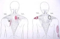
There are six main trigger points of the trapezius, two in each portion of the muscle, plus one unusual one which causes only autonomic phenomena.[2]See Trigger Points
References[edit | edit source]
- ↑ 1.0 1.1 1.2 1.3 1.4 1.5 Moore KL, et al. Essential clinical anatomy. 4th ed. Baltimore, MD: Lippincott Williams and Wilkins, 2011.
- ↑ 2.0 2.1 Weebly The Trapezius Muscle Available: https://trapeziusmuscle.weebly.com/interesting-facts.html(accessed 6.1.2022)
- ↑ 3.0 3.1 3.2 3.3 3.4 Department of Radiology, UW Medicine. Trapezius. www.rad.washington.edu/academics/academic-sections/msk/muscle-atlas/upper-body/trapezius (accessed 27 January 2014).
- ↑ 4.0 4.1 Ourieff J, Scheckel B, Agarwal A. Anatomy, Back, Trapezius. StatPearls [Internet]. 2020 Apr 17.Available: https://www.ncbi.nlm.nih.gov/books/NBK518994/(accessed 6.1.2022)
- ↑ Hwang M, Lee S, Lim C. Effects of the Proprioceptive Neuromuscular Facilitation Technique on Scapula Function in Office Workers with Scapula Dyskinesis. Medicina. 2021 Apr;57(4):332. Available:https://www.ncbi.nlm.nih.gov/pmc/articles/PMC8067054/ (accessed 6.1.2022)
- ↑ 6.0 6.1 6.2 Neumann DA. Kinesiology of the musculoskletal system: foundations for rehabilitation. 2nd ed. St. Louis, MO: Mosby Elsevier, 2010.
- ↑ https://www.fgc.edu/wp-content/uploads/2011/12/palpation-of-muscles.pdf
- ↑ http://activepotentialrehab.com/docs/pdf/sample_upperextremity11_21.pdf
- ↑ Physiotutors. Trapezius Strength Test. Available from: https://www.youtube.com/watch?v=9tfVeIhlY-o
- ↑ Physiotutors. Tight Upper Traps? Try These Exercises!. Available from: https://www.youtube.com/watch?v=4D6_sK6hxLQ


