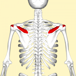
Supraspinatus
Original Editor - Wendy Walker
Lead Editors - Andeela Hafeez, Wendy Walker, Kim Jackson, Kai A. Sigel, Joao Costa, George Prudden, Aya Alhindi, Admin, Rachael Lowe, Naomi O'Reilly and WikiSysop
Description[edit | edit source]
Supraspinatus is the smallest of the 4 muscles which comprise the Rotator Cuff of the shoulder joint.
It travels underneath the acromion.
Origin[edit | edit source]
Supraspinatus fossa of the scapula.
Insertion[edit | edit source]
Greater tuberosity of the humerus, superior facet.
Nerve Supply[edit | edit source]
Suprascapular Nerve, C5 & 6, superior trunk of the brachial plexus.
Blood Supply[edit | edit source]
Suprascapular Artery.
Action[edit | edit source]
It abducts the arm from 0 to 15 degrees, when it is the main agonist, then assists deltoid to produce abduction beyond this range up to 90 degrees.
Function[edit | edit source]
Shoulder Stability[edit | edit source]
As part of the Rotator Cuff, supraspinatus helps to resist the gravitational forces which act on the shoulder joint to pull from the weight of the upper limb downward.
It also helps to stabilize the shoulder joint by keeping the head of the humerus firmly pressed medially against the glenoid fossa of the scapula.
Active Movement[edit | edit source]
Supraspinatus is commonly thought to be instrumental in the initiation of shoulder abduction.
A study in 2011 used electromyography to study the levels of activity in the shoulder muscles during flexion, and found that supraspinatus was consistently recruited prior to movement of the limb at all loads; the authors concluded that "Posterior rotator cuff muscles appear to be counterbalancing anterior translational forces produced during flexion and it would appear that supraspinatus is one of the muscles that consistently 'initiates' flexion."[1]
Resources[edit | edit source]
Recent Related Research (from Pubmed)[edit | edit source]
Extension:RSS -- Error: Not a valid URL: Feed goes here!!|charset=UTF-8|short|max=10
References[edit | edit source]
References will automatically be added here, see adding references tutorial.
- ↑ A comprehensive analysis of muscle recruitment patterns during shoulder flexion: an electromyographic study. Wattanaprakornkul D1, Halaki M, Boettcher C, Cathers I, Ginn KA Clin Anat. 2011 Jul;24(5):619-26


