Spondylolisthesis: Difference between revisions
No edit summary |
(text, sections, refs,) |
||
| Line 8: | Line 8: | ||
== Definition/Description: == | == Definition/Description: == | ||
Spondylolisthesis is the slippage of one vertebral body with respect to the adjacent vertebral body causing mechanical or radicular symptoms or pain. It can be due to congenital, acquired, or idiopathic causes. Spondylolisthesis is graded based on the degree of slippage of one vertebral body on the adjacent vertebral body.<ref name=":1">Tenny S, Gillis CC. [https://www.ncbi.nlm.nih.gov/books/NBK430767/ Spondylolisthesis]. InStatPearls [Internet] 2019 Mar 27. StatPearls Publishing. Available from:https://www.ncbi.nlm.nih.gov/books/NBK430767/ (last accessed 26.1.2020)</ref><u></u><sub></sub><sup></sup><br> | Spondylolisthesis is the slippage of one vertebral body with respect to the adjacent vertebral body causing mechanical or radicular symptoms or pain. It can be due to congenital, acquired, or idiopathic causes. Spondylolisthesis is graded based on the degree of slippage of one vertebral body on the adjacent vertebral body.<ref name=":1">Tenny S, Gillis CC. [https://www.ncbi.nlm.nih.gov/books/NBK430767/ Spondylolisthesis]. InStatPearls [Internet] 2019 Mar 27. StatPearls Publishing. Available from:https://www.ncbi.nlm.nih.gov/books/NBK430767/ (last accessed 26.1.2020)</ref><u></u><sub></sub><sup></sup><br>Figure 2: Highest stress during various lumbar motions is found at the pars interarticularis, as shown in a threedimensional finite element model <ref name="Mays">Mays, S. (2006). Spondylolysis, spondylolisthesis, and lumbo-sacral morphology in a medieval English skeletal population. American Journal of Physical Anthropolgy, 131, 352–62. (Level of evidence: 2B)</ref> [[File:Stress_lumbar_vertebra.png|right|391x391px]] | ||
http://www.spine-health.com/video/degenerative-spondylolisthesis-video | http://www.spine-health.com/video/degenerative-spondylolisthesis-video | ||
| Line 31: | Line 31: | ||
== Etiology: == | == Etiology: == | ||
There are different classification systems regarding the etiology, terminology, subtypes of spondylolysis and spondylolisthesis, and treatment.<br> | There are different classification systems regarding the etiology, terminology, subtypes of spondylolysis and spondylolisthesis, and treatment.<br>'''A'''. '''Wiltse Classification''': It is one of the most commonly used classification systems to convey the etiology of spondylolisthesis (see table below). It has five major etiologies: degenerative, isthmic, traumatic, dysplastic, or pathologic. | ||
# Degenerative spondylolisthesis occurs from degenerative changes in the spine without any defect in the pars interarticularis. It is usually related to combined facet joint and disc degeneration leading to instability and forward movement of one vertebral body relative to the adjacent vertebral body. | |||
'''A'''. '''Wiltse Classification''': It is one of the most commonly used classification systems to convey the etiology of spondylolisthesis. | # Isthmic spondylolisthesis results from defects in the pars interarticularis. The cause of isthmic spondylolisthesis is undetermined, but a possible etiology includes microtrauma in adolescence related to sports such as wrestling, football, and gymnastics, where repeated lumbar extension occurs. | ||
# Traumatic spondylolisthesis occurs after fractures of the pars interarticularis or the facet joint structure and is most common after trauma. | |||
# Dysplastic spondylolisthesis is congenital and secondary to variation in the orientation of the facet joints to an abnormal alignment. In dysplastic spondylolisthesis, the facet joints are more sagittally oriented than the typical coronal orientation. | |||
# Pathologic spondylolisthesis can be from systemic causes such as bone or connective tissue disorders or a focal process, including infection, neoplasm, or iatrogenic origin. | |||
Additional risk factors for spondylolisthesis include a first-degree relative with spondylolisthesis, scoliosis, or occult spina bifida at the S1 level<ref name=":1" /> | |||
Degenerative spondylolisthesis occurs from degenerative changes in the spine without any defect in the pars interarticularis. It is usually related to combined facet joint and disc degeneration leading to instability and forward movement of one vertebral body relative to the adjacent vertebral body. | |||
Isthmic spondylolisthesis results from defects in the pars interarticularis. The cause of isthmic spondylolisthesis is undetermined, but a possible etiology includes microtrauma in adolescence related to sports such as wrestling, football, and gymnastics, where repeated lumbar extension occurs. | |||
Traumatic spondylolisthesis occurs after fractures of the pars interarticularis or the facet joint structure and is most common after trauma. | |||
Dysplastic spondylolisthesis is congenital and secondary to variation in the orientation of the facet joints to an abnormal alignment. In dysplastic spondylolisthesis, the facet joints are more sagittally oriented than the typical coronal orientation. | |||
Pathologic spondylolisthesis can be from systemic causes such as bone or connective tissue disorders or a focal process, including infection, neoplasm, or iatrogenic origin. | |||
Additional risk factors for spondylolisthesis include a first-degree relative with spondylolisthesis, scoliosis, or occult spina bifida at the S1 level | |||
[[Image:Wlitse.jpg]] <u></u><sub></sub><sup></sup> | |||
== Characteristics/Clinical Presentation: == | == Characteristics/Clinical Presentation: == | ||
'''Symptoms and findings in spondylolisthesis are:'''<ref>Antony Wicker et al; Spondylolysis and spondylolisthesis in sports; International SportMed Journal, Vol. 9 No.2, 2008 pp.74-7 (level of evidence 2B) | '''Symptoms and findings in spondylolisthesis are:'''<ref>Antony Wicker et al; Spondylolysis and spondylolisthesis in sports; International SportMed Journal, Vol. 9 No.2, 2008 pp.74-7 (level of evidence 2B) | ||
</ref>: | </ref>: | ||
*Low-back pain | *Low-back pain | ||
*Pain radiating down the leg | *Pain radiating down the leg | ||
| Line 104: | Line 77: | ||
== Diagnostic Procedures: == | == Diagnostic Procedures: == | ||
# [[X-Rays|X Ray]]<nowiki/>s - Anteroposterior and lateral plain films, as well as lateral flexion-extension plain films, are the standard for the initial diagnosis of spondylolisthesis. One is looking for the abnormal alignment of one vertebral body to the next as well as possible motion with flexion and extension, which would indicate instability. In isthmic spondylolisthesis, there may be a pars defect, which is termed the "Scotty dog collar." The "Scotty dog collar" shows a hyperdensity where the collar would be on the cartoon dog, which represents the fracture of the pars interarticularis. | |||
# Computed tomography ([[CT Scans|CT]]) of the spine - provides the highest sensitivity and specificity for diagnosing spondylolisthesis. Spondylolisthesis can be better appreciated on sagittal reconstructions as compared to axial CT imaging. | |||
# [[MRI Scans|MRI]] of the spine can show associated soft tissue and disc abnormalities, but it is relatively more challenging to appreciate bony detail and a potential pars defect on MRI.<ref name=":1" /> | |||
=== Outcome Measures === | |||
* Disability: <font color="#0066cc">Oswestry Disability Index, the SF-36 Physical Functioning scale, the Quebec Back Pain Disability Scale</font><ref>Davidson, M. & Keating, J.L., A comparison of five low back disability questionnaires: reliability and responsiveness. Phys Ther. 2002 Jan;82(1):8-24 (Level of evidence 2B) | * Disability: <font color="#0066cc">Oswestry Disability Index, the SF-36 Physical Functioning scale, the Quebec Back Pain Disability Scale</font><ref>Davidson, M. & Keating, J.L., A comparison of five low back disability questionnaires: reliability and responsiveness. Phys Ther. 2002 Jan;82(1):8-24 (Level of evidence 2B) | ||
</ref> | </ref> | ||
| Line 133: | Line 90: | ||
* Kinesiophobia and Catastrophising: <font color="#0066cc">Tampa Scale for Kinesiophobia, Pain Catastrophising Scale</font><ref name=":14" /> | * Kinesiophobia and Catastrophising: <font color="#0066cc">Tampa Scale for Kinesiophobia, Pain Catastrophising Scale</font><ref name=":14" /> | ||
== Examination | == Examination == | ||
* History: Specific questions referring to pain, location, severity, duration, quality- tingling, burning sensations, exacerbating factors, alleviating factors, leisure activities , occupational risks and pain changes throughout day- difference morning compared to evening/night? | * History: Specific questions referring to pain, location, severity, duration, quality- tingling, burning sensations, exacerbating factors, alleviating factors, leisure activities , occupational risks and pain changes throughout day- difference morning compared to evening/night? | ||
* General<ref name=":2" /><br> | * General<ref name=":2" /><br> | ||
== Medical Management | == Medical Management == | ||
For grade I and II spondylolisthesis, treatment typically begins with conservative therapy, including nonsteroidal anti-inflammatory drugs (NSAIDs), heat, light exercise, traction, bracing, and/or bed rest. | |||
'''Conservative''' <ref name="Kalichman" /> <ref name="Weinstein">James N. Weinstein, Surgical versus Nonsurgical Treatment for Lumbar Degenerative Spondylolisthesis,New England Journal of Medicine. May 2007; 356:2257-2270. (Level of evidence 2B)</ref> | |||
*Initially resting and avoiding movements like lifting, bending, and sports. | *Initially resting and avoiding movements like lifting, bending, and sports. | ||
*Analgesics and NSAIDs reduce musculoskeletal pain and have an anti-inflammatory effect on nerve root and joint irritation. | *Analgesics and NSAIDs reduce musculoskeletal pain and have an anti-inflammatory effect on nerve root and joint irritation. | ||
| Line 146: | Line 103: | ||
*A brace may be useful to decrease segmental spinal instability and pain. <ref name="Funao">Funao H, Tsuji T, Hosogane N, Watanabe K, Ishii K, Nakamura M, Chiba K, Toyama Y, Matsumoto M. Comparative study of spinopelvic sagittal alignment between patients with and without degenerative spondylolisthesis. European Spine Journal. 2012 Nov 1;21(11):2181-7.</ref> | *A brace may be useful to decrease segmental spinal instability and pain. <ref name="Funao">Funao H, Tsuji T, Hosogane N, Watanabe K, Ishii K, Nakamura M, Chiba K, Toyama Y, Matsumoto M. Comparative study of spinopelvic sagittal alignment between patients with and without degenerative spondylolisthesis. European Spine Journal. 2012 Nov 1;21(11):2181-7.</ref> | ||
<br>'''Surgical | <br>'''Surgical''' | ||
Approximately 10% to 15% of younger patients with low-grade spondylolisthesis will fail conservative treatment and need surgical treatment. | |||
* No definitive standards exist for surgical treatment. | |||
* Surgical treatment includes a varying combination of decompression, fusion with or without instrumentation, or interbody fusion. | |||
* Patients with instability are more likely to require operative intervention. | |||
* Some surgeons recommend a reduction of the spondylolisthesis if able as this not only decreases foraminal narrowing but also can improve spinopelvic sagittal alignment and decrease the risk for further degenerative spinal changes in the future. The reduction can be more difficult and riskier in higher grades and impacted spondylolisthesis<ref name=":1" /> | |||
Indications for Surgery<ref name=":8">Kalichman L. et al, Diagnosis and conservative management of degenerative lumbar spondylolisthesis, Eur Spine J, 2008; 17:327-335 (Level of evidence 2B) | |||
</ref><ref>Sairyo K., Decompression Surgery For Lumbar Spondylolysis Without Fusion: A Review Article, The Internet Journal of Spine Surgery, 2005. (Level of evidence 1A) | |||
</ref> | </ref> | ||
| Line 164: | Line 124: | ||
*Postural deformity and gait abnormality | *Postural deformity and gait abnormality | ||
'''Contra-indications to Surgery | '''Contra-indications to Surgery'''<ref name=":0">Amir Vokshoor et al., Spondylolisthesis, Spondylolysis, and Spondylosis. Medscape, updated Sep 10, 2014, Consulted on Oct 20, 2014 (Level of evidence 2A)</ref> | ||
*Poor medical health | *Poor medical health | ||
*High operative risk (higher risk than potential benefits) | *High operative risk (higher risk than potential benefits) | ||
*High risk of hemorrhage: Anticoagulation with warfarin, or antiplatelet therapy | *High risk of hemorrhage: Anticoagulation with warfarin, or antiplatelet therapy | ||
*Smoking | *Smoking<u></u><sub></sub><sup></sup> | ||
== Physical Therapy Management: == | == Physical Therapy Management: == | ||
| Line 183: | Line 140: | ||
There is strong evidence for exercise therapy, which consists of strengthening exercises of the deep abdominal musculature.. In addition, isometric and isotonic exercises may be beneficial for strengthening of the main muscle groups of the trunk, which stabilize the spine. These techniques may also play a role in pain reduction.. In order to improve the patient’s mobility, physical therapy includes stretching exercises of the hamstrings, hip flexors and lumbar paraspinal muscles<ref>Jerrad MD. Et al., Bony Healing in a Patient with Bilateral L5 Spondylolysis. Current Sports Medicine Reports, (2005) 35-37. (Level of evidence 5) | There is strong evidence for exercise therapy, which consists of strengthening exercises of the deep abdominal musculature.. In addition, isometric and isotonic exercises may be beneficial for strengthening of the main muscle groups of the trunk, which stabilize the spine. These techniques may also play a role in pain reduction.. In order to improve the patient’s mobility, physical therapy includes stretching exercises of the hamstrings, hip flexors and lumbar paraspinal muscles<ref>Jerrad MD. Et al., Bony Healing in a Patient with Bilateral L5 Spondylolysis. Current Sports Medicine Reports, (2005) 35-37. (Level of evidence 5) | ||
</ref><ref name=":16">Van Tulder M.W. et al, Conservative treatment of acute and chronic nonspecific low back pain. A systematic review of randomized controlled trials of the most common interventions, Spine, 1997; 22(18): 2128-2156 f(Level of evidence 1A) </ref><ref name=":13" /> | </ref><ref name=":16">Van Tulder M.W. et al, Conservative treatment of acute and chronic nonspecific low back pain. A systematic review of randomized controlled trials of the most common interventions, Spine, 1997; 22(18): 2128-2156 f(Level of evidence 1A) </ref><ref name=":13">Sinaki M. et al. Lumbar spondylolisthesis: retrospective comparison and three year follow-up of two conservative treatment programs. Arch Phys Med Rehabil 1989 70:594-598. (level of evidence 1B) | ||
</ref> | |||
Furthermore, endurance training is effective for chronic low back pain.<ref name=":16" /><ref>Margaret L. McNeely et al.; A systematic review of physiotherapy for spondylolysis and spondylolisthesis; Manual Therapy (2003) 8(2), 80–91. ( level of evidence 1B) | Furthermore, endurance training is effective for chronic low back pain.<ref name=":16" /><ref>Margaret L. McNeely et al.; A systematic review of physiotherapy for spondylolysis and spondylolisthesis; Manual Therapy (2003) 8(2), 80–91. ( level of evidence 1B) | ||
| Line 250: | Line 208: | ||
• Steven, S.A. et al, (20mc10). Contemporary management of isthmic spondylolisthesis: pediatric and adult. The Spine Journal, 2010, 530-543. (Level of evidence: 1A)<br>• Van Tulder M.W. et al, Conservative treatment of acute and chronic nonspecific low back pain. A systematic review of randomized controlled trials of the most common interventions, Spine, 1997; 22(18): 2128-2156 f(Level of evidence 1A)<br>• Paulo H Ferreira et al.; Specific stabilisation exercise for spinal and pelvic pain: A systematic review; Australian Journal of Physiotherapy 2006 Vol. 52 ( level of evidence 1A)<br> | • Steven, S.A. et al, (20mc10). Contemporary management of isthmic spondylolisthesis: pediatric and adult. The Spine Journal, 2010, 530-543. (Level of evidence: 1A)<br>• Van Tulder M.W. et al, Conservative treatment of acute and chronic nonspecific low back pain. A systematic review of randomized controlled trials of the most common interventions, Spine, 1997; 22(18): 2128-2156 f(Level of evidence 1A)<br>• Paulo H Ferreira et al.; Specific stabilisation exercise for spinal and pelvic pain: A systematic review; Australian Journal of Physiotherapy 2006 Vol. 52 ( level of evidence 1A)<br> | ||
== Clinical bottom line == | == Clinical bottom line == | ||
* An interprofessional team consisting of a specialty-trained orthopedic nurse, physical therapist, and an orthopedic surgeon or neurosurgeon will provide the best outcome and long-term care of patients with degenerative spondylolisthesis. | |||
* Treating clinician will decide on the management plan, and then have the other team members engaged - surgical cases with include the nursing staff in pre-, intra-, and post-operative care, and coordinating with PT for rehabilitation. | |||
* In non-operative cases (majority), the PT keeps the rest of the team informed of progress (or lack of). | |||
* The team should encourage weight loss as weight reduction may reduce symptoms and increase the quality of life. | |||
* Interprofessional collaboration, as above, will drive patient outcomes to their best results.<ref name=":1" /> | |||
== References == | == References == | ||
Revision as of 23:39, 25 January 2020
Original Editors - Margo De Mesmaeker
Top Contributors - Maëlle Cormond, Simisola Ajeyalemi, Evi Peeters, Margo De Mesmaeker, Kim Jackson, Admin, Chrysolite Jyothi Kommu, Kenneth de Becker, Shaimaa Eldib, Lucinda hampton, Rachael Lowe, Mike Myracle, Evan Thomas, Carlos De Coster, Johnathan Fahrner, Aminat Abolade, Mariam Hashem, Shreya Pavaskar, Camille Linussio, Claire Knott, Rucha Gadgil, Laura Ritchie, Jess Bell, Kirenga Bamurange Liliane and Scott Buxton
Definition/Description:[edit | edit source]
Spondylolisthesis is the slippage of one vertebral body with respect to the adjacent vertebral body causing mechanical or radicular symptoms or pain. It can be due to congenital, acquired, or idiopathic causes. Spondylolisthesis is graded based on the degree of slippage of one vertebral body on the adjacent vertebral body.[1]
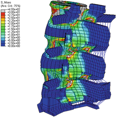
Figure 2: Highest stress during various lumbar motions is found at the pars interarticularis, as shown in a threedimensional finite element model [2]
http://www.spine-health.com/video/degenerative-spondylolisthesis-video
Clinically Relevant Anatomy:[edit | edit source]
Spondylolisthesis most commonly occurs in the lower lumbar spine but can also occur in the cervical spine and rarely, except for trauma, in the thoracic spine.[1]
Spondylolysthesis, regardless of the type, is mostly common preceded by spondylolysis. This pathology involves
- a fractured pars interarticularis of the lumbar vertebrae, also called the isthmus.
- this affects the supporting structural integrity of the vertebrae, which could lead to slippage of the corpus of the vertebrae, called spondylolysthesis.
- in turn, leads to one of the most obvious manifestations of lumbar instability.
- slippage can occur in 2 directions: most commonly in anterior translation, called anterolisthesis, or a backward translation, called retrolisthesis.[4]
A study of Dai L.Y. analysed the correlation between disc degeneration and the age, duration and severity of clinical symptoms and grade of vertebral slip. The disc degeneration on subsegmental level was significantly related to age and duration of clinical symptoms, although it was not related to the severity of clinical symptoms or the grade of vertebral slip[5].
Epidemiology[edit | edit source]
- Degenerative spondylolisthesis predominately occurs in adults and is more common in females than males with increased risk in the obese.
- Isthmic spondylolisthesis is more common in the adolescent and young adult population but may go unrecognized until symptoms develop in adulthood. There is a higher prevalence of isthmic spondylolisthesis in males.
- Dysplastic spondylolisthesis is more common in the pediatric population, with females more commonly affected than males. Current estimates for prevalence are 6 to 7% for isthmic spondylolisthesis by the age of 18 years, and up to 18% of adult patients undergoing MRI of the lumbar spine.
- Grade I spondylolisthesis accounts for 75% of all cases.
- Spondylolisthesis most commonly occurs at the L5-S1 level with anterior translation of the L5 vertebral body on the S1 vertebral body.
- The L4-5 level is the second most common location for spondylolisthesis.[1]
Etiology:[edit | edit source]
There are different classification systems regarding the etiology, terminology, subtypes of spondylolysis and spondylolisthesis, and treatment.
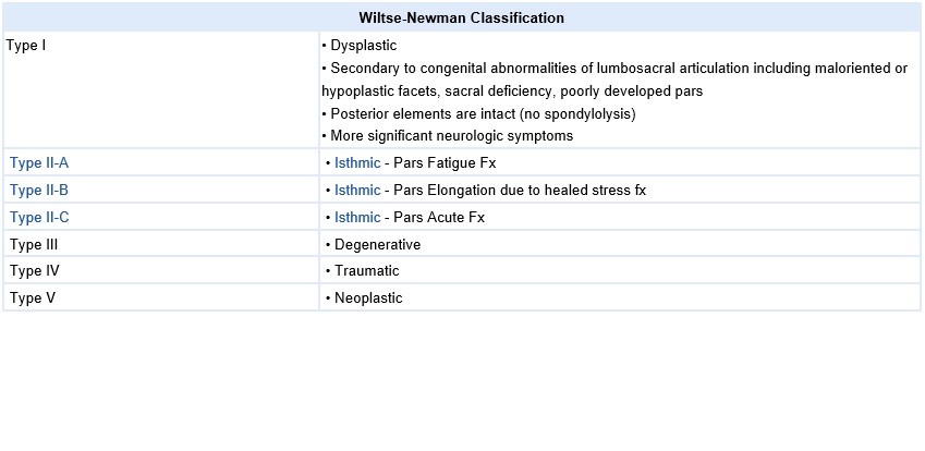
A. Wiltse Classification: It is one of the most commonly used classification systems to convey the etiology of spondylolisthesis (see table below). It has five major etiologies: degenerative, isthmic, traumatic, dysplastic, or pathologic.
- Degenerative spondylolisthesis occurs from degenerative changes in the spine without any defect in the pars interarticularis. It is usually related to combined facet joint and disc degeneration leading to instability and forward movement of one vertebral body relative to the adjacent vertebral body.
- Isthmic spondylolisthesis results from defects in the pars interarticularis. The cause of isthmic spondylolisthesis is undetermined, but a possible etiology includes microtrauma in adolescence related to sports such as wrestling, football, and gymnastics, where repeated lumbar extension occurs.
- Traumatic spondylolisthesis occurs after fractures of the pars interarticularis or the facet joint structure and is most common after trauma.
- Dysplastic spondylolisthesis is congenital and secondary to variation in the orientation of the facet joints to an abnormal alignment. In dysplastic spondylolisthesis, the facet joints are more sagittally oriented than the typical coronal orientation.
- Pathologic spondylolisthesis can be from systemic causes such as bone or connective tissue disorders or a focal process, including infection, neoplasm, or iatrogenic origin.
Additional risk factors for spondylolisthesis include a first-degree relative with spondylolisthesis, scoliosis, or occult spina bifida at the S1 level[1]
Characteristics/Clinical Presentation:[edit | edit source]
Symptoms and findings in spondylolisthesis are:[6]:
- Low-back pain
- Pain radiating down the leg
- Neurological symptoms (possible evolution towards cauda equine syndrome)
- Atrophy of the muscles, muscle weakness
- Tense hamstrings, hamstrings spasms
- Diminished ROM (spine)
- Disturbances in coordination and balance
Patients usually report that their symptoms vary as a function of mechanical loads (such as in going from supine to erect position) and pain frequently worsens over the course of the day. Radiation into the posterolateral thighs is also common and is independent of neurological signs and symptoms. The pain could be diffuse in the lower extremities, involving the L5 and/or L4 roots unilaterally or bilaterally, but generally bilaterally [7]
Symptoms decrease with sitting or standing with lumbar flexion and with lying. As symptoms worsen patients are more and more limited in their activities and walking distance. This relationship is known as neurogenic intermittent claudication[8]
Spondylolisthesis can occur with other disorders and seems to have a link with some of them:
- Spina Bifida Occulta [2] [9] [10]; Several studies support a positive association between spina bifida occulta and spondylolysthesis [11]. This high association may not be due to mechanical factors but to genetic factors [9].
- Cerebral Palsy [12]; A number of studies proved the association between cerebral palsy and spondylolysthesis, certainly in athetoid cerebral palsy (60%)
- Scheuermanns Disease also known as Schuermann's Kyphosis[13][14]; Ogilvie and Sherman reported a 50% incidence of spondylolIsthesis in 18 patients with Scheuermann’s disease [13]. Greene et al. found spondylolIsthesis (grade I or II) at L5-S1 in 32% of patients with Scheuermann’s disease [14].
- Scoliosis[11]: Fisk et al. reported that the incidence in 539 patients with idiopathic scoliosis was 6.2%, which corresponded to that found in the general population [15]. But the relation between scoliosis and spondylolisthesis has not been clarified[11]
- Spinal stenosis[16]
Differential Diagnosis:[edit | edit source]
- Spondylolysis [17] [18]
- Metastatic disease [19]
- Low back pain [20]
- Osteoarthritis [20]
- Neuroforaminal stenosis [20]
- Spinal Stenosis [20]
Diagnostic Procedures:[edit | edit source]
- X Rays - Anteroposterior and lateral plain films, as well as lateral flexion-extension plain films, are the standard for the initial diagnosis of spondylolisthesis. One is looking for the abnormal alignment of one vertebral body to the next as well as possible motion with flexion and extension, which would indicate instability. In isthmic spondylolisthesis, there may be a pars defect, which is termed the "Scotty dog collar." The "Scotty dog collar" shows a hyperdensity where the collar would be on the cartoon dog, which represents the fracture of the pars interarticularis.
- Computed tomography (CT) of the spine - provides the highest sensitivity and specificity for diagnosing spondylolisthesis. Spondylolisthesis can be better appreciated on sagittal reconstructions as compared to axial CT imaging.
- MRI of the spine can show associated soft tissue and disc abnormalities, but it is relatively more challenging to appreciate bony detail and a potential pars defect on MRI.[1]
Outcome Measures[edit | edit source]
- Disability: Oswestry Disability Index, the SF-36 Physical Functioning scale, the Quebec Back Pain Disability Scale[21]
- Dysfunctional thoughts: Short Form of the Medical Outcomes Study (SF-36)[22]
- Pain: Pain Numerical Rating Scale, VAS.
- Quality of life: Short-Form Health Survey [22]
- Kinesiophobia and Catastrophising: Tampa Scale for Kinesiophobia, Pain Catastrophising Scale[22]
Examination[edit | edit source]
- History: Specific questions referring to pain, location, severity, duration, quality- tingling, burning sensations, exacerbating factors, alleviating factors, leisure activities , occupational risks and pain changes throughout day- difference morning compared to evening/night?
- General[4]
Medical Management[edit | edit source]
For grade I and II spondylolisthesis, treatment typically begins with conservative therapy, including nonsteroidal anti-inflammatory drugs (NSAIDs), heat, light exercise, traction, bracing, and/or bed rest.
- Initially resting and avoiding movements like lifting, bending, and sports.
- Analgesics and NSAIDs reduce musculoskeletal pain and have an anti-inflammatory effect on nerve root and joint irritation.
- Epidural steroid injections can be used to relieve low back pain, lower extremity pain related to radiculopathy and neurogenic claudication.
- A brace may be useful to decrease segmental spinal instability and pain. [24]
Surgical
Approximately 10% to 15% of younger patients with low-grade spondylolisthesis will fail conservative treatment and need surgical treatment.
- No definitive standards exist for surgical treatment.
- Surgical treatment includes a varying combination of decompression, fusion with or without instrumentation, or interbody fusion.
- Patients with instability are more likely to require operative intervention.
- Some surgeons recommend a reduction of the spondylolisthesis if able as this not only decreases foraminal narrowing but also can improve spinopelvic sagittal alignment and decrease the risk for further degenerative spinal changes in the future. The reduction can be more difficult and riskier in higher grades and impacted spondylolisthesis[1]
Indications for Surgery[25][26]
- Neurologic signs- neurogenic claudication / radiculopathy
- Myelopathy
- High-grade slip (>50%)
- Type 1 and 2 slips with evidence of instability, progression of spondylolisthesis
- Type 3 spondylolisthesis with gross instability and incapacitating pain
- Bladder or bowel symptoms (especially in type 3)
- Traumatic spondylolisthesis
- Iatrogenic spondylolisthesis
- Postural deformity and gait abnormality
Contra-indications to Surgery[27]
- Poor medical health
- High operative risk (higher risk than potential benefits)
- High risk of hemorrhage: Anticoagulation with warfarin, or antiplatelet therapy
- Smoking
Physical Therapy Management:[edit | edit source]
A systematic review including 10 articles from which 5 were RCT, concluded that no consensus could be reached on the role of nonoperative care vs. surgery due to the heterogeneity of the different studies reported. Spondylolisthesis should be treated first with conservative therapy, which includes physical therapy, rest, medication and braces.[28][29]
- Exercise therapy
There is strong evidence for exercise therapy, which consists of strengthening exercises of the deep abdominal musculature.. In addition, isometric and isotonic exercises may be beneficial for strengthening of the main muscle groups of the trunk, which stabilize the spine. These techniques may also play a role in pain reduction.. In order to improve the patient’s mobility, physical therapy includes stretching exercises of the hamstrings, hip flexors and lumbar paraspinal muscles[30][31][32]
Furthermore, endurance training is effective for chronic low back pain.[31][33]The objective of stretching and strengthening is to decrease the extension forces on the lumbar spine, due to agonist muscle tightness, antagonist weakness, or both, which may result in decreased lumbar lordosis.[31] Rehabilitation programs should be designed to improve muscle balance rather than muscle strength alone. [34]
There is evidence that suggests that specific stabilization exercises and core stability exercises can be useful in reducing pain and disability in chronic low back pain in patient with spondylolisthesis.[35][34]
- Movements in closed-chain-kinetics
- Renewing of the motion-pattern
- Antilordotic movement patterns of the spine
- Elastic band exercises in the lying position
- Gait training
- Brace-gymnastics
- Stretching exercises
- Sensomotoric training on unstable devices
- Functional electric stimulation
- Walking in all variations
- Underwater therapy
- Balance training
- Coordinative skills
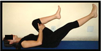
Figure 3: Strengthening of the deep abdominal muscles.
Alternating legs, with leg extension while exhaling, maintaining contraction of transverse abdominis, paravertebral and pelvic floor muscles [36]
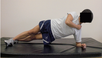
Figure 4: Horizontal side support exercise for core stability [37] .
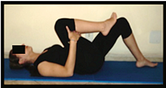
Figure 5: Stretching of the erector spine muscles. [36]
- Lumbosacral braces or corset
In healthy subjects, it has been found that the lumbosacral brace can improve the sitting position of the patient. The fact that there was wear of the brace, indicates that the brace has an important function in the sitting position.[38] According to Prateepavanich et al., a lumbosacral corset can be used to improve walking distance and to reduce pain in daily activities[39], but it does not reduce the shift of the vertebra. It is a good aid during the painful periods but should be discontinued when the patients' complaints are reduced.
- Posture and lifting techniques
Special attention has to be given to posture and proper lifting techniques[40] wherein the physiotherapist has an important educational role. Lifting techniques is effective for chronic low back pain.[41][31]
- Management of catastrophising and kinesiophobia
Physical therapy treatment in combination with management of catastrophising and kinesiophobia gave good results. The disability, pain, dysfunctional thoughts were significant reduced.[42]
- Alternative cardiovascular exercise
Athletes with spondylolysis and first-degree spondylolisthesis can take part in all sports activities. However, attention should be given to those kinds of sport where recurring trauma resulting from repeated flexion, hyperextension and twisting is usually undertaken (e.g. gymnastics, aerobics, swimming in the dolphin technique). Athletes with a grade 2, 3 or 4 can also participate in all the sport activities but have to do this with a special and individually adapted directive.[43] Low aerobic impact sports are highly recommended.[40] Sports that certainly can be practiced are walking, swimming and cross-training. Although these activities will not improve the shift, these sports are a good alternative for cardiovascular exercises.[25] Impact sports like running should not be done in order to avoid wear. The adolescent athlete or manual laborer should avoid hyperextension and/or contact sports.[40]
- Massage therapy
A case report showed that the onset of low back pain was delayed during walking/standing over the course of treatment, hyperlordosis decreased, and hypertonicity of iliopsoas and quadratus lumborum muscles decreased. Bilateral net reduction of illial rotation was achieved, but with irregular changes. These results were inconclusive but bring into question the role of hip flexor and spinal extensor muscles in normalizing postural misalignment associated with spondylolisthesis.[44]
- Pulsed radiofrequency (PRF)
Results of a cohort study demonstrated that the application of PRF might be more effective than steroid and bupivacaine injection in decreasing back pain due to degenerative facet pain and improvement in function of patients with degenerative spondylolisthesis.[45]
Key Research[edit | edit source]
• Steven, S.A. et al, (20mc10). Contemporary management of isthmic spondylolisthesis: pediatric and adult. The Spine Journal, 2010, 530-543. (Level of evidence: 1A)
• Van Tulder M.W. et al, Conservative treatment of acute and chronic nonspecific low back pain. A systematic review of randomized controlled trials of the most common interventions, Spine, 1997; 22(18): 2128-2156 f(Level of evidence 1A)
• Paulo H Ferreira et al.; Specific stabilisation exercise for spinal and pelvic pain: A systematic review; Australian Journal of Physiotherapy 2006 Vol. 52 ( level of evidence 1A)
Clinical bottom line[edit | edit source]
- An interprofessional team consisting of a specialty-trained orthopedic nurse, physical therapist, and an orthopedic surgeon or neurosurgeon will provide the best outcome and long-term care of patients with degenerative spondylolisthesis.
- Treating clinician will decide on the management plan, and then have the other team members engaged - surgical cases with include the nursing staff in pre-, intra-, and post-operative care, and coordinating with PT for rehabilitation.
- In non-operative cases (majority), the PT keeps the rest of the team informed of progress (or lack of).
- The team should encourage weight loss as weight reduction may reduce symptoms and increase the quality of life.
- Interprofessional collaboration, as above, will drive patient outcomes to their best results.[1]
References[edit | edit source]
- ↑ 1.0 1.1 1.2 1.3 1.4 1.5 1.6 Tenny S, Gillis CC. Spondylolisthesis. InStatPearls [Internet] 2019 Mar 27. StatPearls Publishing. Available from:https://www.ncbi.nlm.nih.gov/books/NBK430767/ (last accessed 26.1.2020)
- ↑ 2.0 2.1 Mays, S. (2006). Spondylolysis, spondylolisthesis, and lumbo-sacral morphology in a medieval English skeletal population. American Journal of Physical Anthropolgy, 131, 352–62. (Level of evidence: 2B)
- ↑ Foreman P. et al, L5 spondylolysis/spondylolisthesis: a comprehensive review with an anatomic focus, Childs Nerv Syst. 2013;29(2):209-16 (Level of evidence 1B)
- ↑ 4.0 4.1 O’sullivan RCT + Iguchi T. et al., Lumbar multilevel degenerative spondylolisthesis: radiological evaluation and factors related to anterolisthesis and retrolisthesis, J Spinal Disord Tech. 2002. Apr;15(2):93-9 (Level of evidence 1B)
- ↑ Iguchi T. et al., Lumbar multilevel degenerative spondylolisthesis: radiological evaluation and factors related to anterolisthesis and retrolisthesis, J Spinal Disord Tech. 2002. Apr;15(2):93-9 (Level of evidence 2B)
- ↑ Antony Wicker et al; Spondylolysis and spondylolisthesis in sports; International SportMed Journal, Vol. 9 No.2, 2008 pp.74-7 (level of evidence 2B)
- ↑ Frymoyer, J.W.,Degenerative spondylolisthesis. In: Andersson GBJ, McNeill TW (eds) Lumbar spinal stenosis. Mosby Year Book, St Louis, 1992 (Level of Evidence: 5)
- ↑ Phalen GS. et al, Spondylolisthesis and tight hamstrings. J Bone Joint Surg, 1961, 43:505–512 (Level of evidence 1B)
- ↑ 9.0 9.1 Sairyo, K., Goel, V.K., Vadapalli, S., Vishnubhotla, S.L., Biyani, A., Ebraheim, N. et al. (2006). Biomechanical comparison of lumbar spine with or without spina bifida occulta: a finite element analysis. Spinal Cord, 44, 440–4. (Level of evidence: 2C)
- ↑ Burkus, J.K. (1990). Unilateral spondylolysis associated with spina bifida occulta and nerve root compression. Spine (Phila Pa 1976), 15, 555–9. (Level of evidence: 3B)
- ↑ 11.0 11.1 11.2 Toshinori, S, Koichi, S., Naoto, S., Hirofumi, K., Natsuo, Y. (2010). Incidence and etiology of lumbar spondylolysis: review of the literature. Journal of Orthopaedic Science, 15, 281-288. (Level of evidence: 2A)
- ↑ Sakai, T., Yamada, H., Nakamura, T., Nanamori, K., Kawasaki, Y., Hanaoka, N. et al. (2006). Lumbar spinal disorders in patients with athetoid cerebral palsy: a clinical and biomechanical study. Spine (Phila Pa 1976), 31, E66–70. (Level of evidence: 2B)
- ↑ 13.0 13.1 Ogilvie, J.W., Sherman, J. (1987). Spondylolysis in Scheuermann’s disease. Spine (Phila Pa 1976), 12, 251–3. (Level of evidence: 2B)
- ↑ 14.0 14.1 Greene, T.L., Hensinger, R.N., Hunter, L.Y. (1985). Back pain and vertebral changes simulating Scheuermann’s disease. Journal of Pediatrics Orthopedics, 5, 1–7. (Level of evidence: 2B)
- ↑ Fisk JR, Moe JH, Winter RB. Scoliosis, spondylolysis, and spondylolisthesis: their relationship as reviewed in 539 patients. Spine (Phila Pa 1976) 1978;3:234–45. (Level of evidence 2B)
- ↑ Anderson T. et al, Degenerative spondylolisthesis is associated with low spinal bone density: a comparative study between spinal stenosis and degenerative spondylolisthesis, Biomed Res Int. 2013; 123847 (Level of evidence: 2A)
- ↑ Tsirikos AI, Garrido EG. Spondylolysis and spondylolisthesis in children and adolescents. J Bone Joint Surg Br. 2010 Jun;92(6):751-9. doi: 10.1302/0301-620X.92B6.23014 (Level of evidence 1A)
- ↑ Thein-Nissenbaum J, Boissonnault WG. Differential diagnosis of spondylolysis in a patient with chronic low back pain. J Orthop Sports Phys Ther. 2005 May;35(5):319-26. (Level of evidence 4)
- ↑ 19.0 19.1 Kalichman, L., Kim, D., Li, L., Guermazi, A., Berkin, V., & Hunter, D. (2009). Spondylolysis and spondylolisthesis: Prevalence and association with low back pain in the adult community‐based population. Spine, 34(2), 99–205.
- ↑ 20.0 20.1 20.2 20.3 Metzger R, Chaney S. Spondylolysis and spondylolisthesis: what the primary care provider should know. J Am Assoc Nurse Pract. 2014 Jan;26(1):5-12. doi: 10.1002/2327-6924.12083. (Level of evidence 1C)
- ↑ Davidson, M. & Keating, J.L., A comparison of five low back disability questionnaires: reliability and responsiveness. Phys Ther. 2002 Jan;82(1):8-24 (Level of evidence 2B)
- ↑ 22.0 22.1 22.2 Monticone M. et al., Management of catastrophising and kinesiophobia improves rehabilitation after fusion for lumbar spondylolisthesis and stenosis. A randomised controlled trial. Eur Spine J. (2014) Jan 23(1):87-95. (LE 1B)
- ↑ James N. Weinstein, Surgical versus Nonsurgical Treatment for Lumbar Degenerative Spondylolisthesis,New England Journal of Medicine. May 2007; 356:2257-2270. (Level of evidence 2B)
- ↑ Funao H, Tsuji T, Hosogane N, Watanabe K, Ishii K, Nakamura M, Chiba K, Toyama Y, Matsumoto M. Comparative study of spinopelvic sagittal alignment between patients with and without degenerative spondylolisthesis. European Spine Journal. 2012 Nov 1;21(11):2181-7.
- ↑ 25.0 25.1 Kalichman L. et al, Diagnosis and conservative management of degenerative lumbar spondylolisthesis, Eur Spine J, 2008; 17:327-335 (Level of evidence 2B)
- ↑ Sairyo K., Decompression Surgery For Lumbar Spondylolysis Without Fusion: A Review Article, The Internet Journal of Spine Surgery, 2005. (Level of evidence 1A)
- ↑ Amir Vokshoor et al., Spondylolisthesis, Spondylolysis, and Spondylosis. Medscape, updated Sep 10, 2014, Consulted on Oct 20, 2014 (Level of evidence 2A)
- ↑ Serena S. Hu et al., Spondylolisthesis and Spondylolysis, J Bone Joint Surg Am, 2008 Mar 01;90(3):656-671 (Level of evidence 3A)
- ↑ Kalpakcioglua B. et al; Determination of spondylolisthesis in low back pain by clinical evaluation; Journal of Back and Musculoskeletal Rehabilitation 22 (2009) 27–32 ( level of evidence 2B)
- ↑ Jerrad MD. Et al., Bony Healing in a Patient with Bilateral L5 Spondylolysis. Current Sports Medicine Reports, (2005) 35-37. (Level of evidence 5)
- ↑ 31.0 31.1 31.2 31.3 Van Tulder M.W. et al, Conservative treatment of acute and chronic nonspecific low back pain. A systematic review of randomized controlled trials of the most common interventions, Spine, 1997; 22(18): 2128-2156 f(Level of evidence 1A)
- ↑ Sinaki M. et al. Lumbar spondylolisthesis: retrospective comparison and three year follow-up of two conservative treatment programs. Arch Phys Med Rehabil 1989 70:594-598. (level of evidence 1B)
- ↑ Margaret L. McNeely et al.; A systematic review of physiotherapy for spondylolysis and spondylolisthesis; Manual Therapy (2003) 8(2), 80–91. ( level of evidence 1B)
- ↑ 34.0 34.1 Nava-Bringasa T.I. et al; Association of strength, muscle balance, and atrophy with pain and function in patients with degenerative spondylolisthesis; Journal of Back and Musculoskeletal Rehabilitation 27 (2014) 371–376 (level of evidence 2B)
- ↑ Paulo H Ferreira et al.; Specific stabilisation exercise for spinal and pelvic pain: A systematic review; Australian Journal of Physiotherapy 2006 Vol. 52 ( level of evidence 1A)
- ↑ 36.0 36.1 Garcia A.N. et al., Effectiveness of back school versus McKenzie exercises in patients with chronic nonspecific low back pain : a randomized controlled trial, Phys Ther, 2013; 93(6):729-47. (Level of evidence 1B)
- ↑ Childs J.D. et al, Effects of traditional sit-up training versus core stabilization exercises on short-term musculoskeletal injuries in US Army soldiers: a cluster randomized trial, Phys Ther, 2010; 90 (10): 1404-12. (Level of evidence 1B)
- ↑ Mathias M. et al, In healthy subjects, the sitting position can be used to validate the postural effects induced by wearing a lumbar lordosis brace. , Ann Phys Rehabil Med. 2010 Oct (level of evidence 2B)
- ↑ Prateepavanich P. et al., The effectiveness of lumbosacral corset in symptomatic degenerative lumbar spinal stenosis, J Med Assoc Thai., 2001; 84(4):572-6. (Level of evidence 2B)
- ↑ 40.0 40.1 40.2 Agabegi SS, Fischgrund JS. Contemporary management of isthmic spondylolisthesis: pediatric and adult. The Spine Journal, 2010, 530-543. (Level of evidence: 1A)
- ↑ Margaret L. McNeely et al.; A systematic review of physiotherapy for spondylolysis and spondylolisthesis; Manual Therapy (2003) 8(2), 80–91. ( level of evidence 1B)
- ↑ Monticone M. et al, Management of catastrophising and kinesiophobia improves rehabilitation after fusion for lumbar spondylolisthesis and stenosis. A randomised controlled trial , Eur Spine J. 2014 Jan (level of evidence 1B)
- ↑ Antony Wicker et al; Spondylolysis and spondylolisthesis in sports; International SportMed Journal, Vol. 9 No.2, 2008 pp.74-7 (level of evidence 2B)
- ↑ Halpin, Shannon. "Case report: The effects of massage therapy on lumbar spondylolisthesis." Journal of bodywork and movement therapies 16.1 (2012): 115-123. (level of evidence: 4)
- ↑ Hashemi, Masoud, et al. "Effect of pulsed radiofrequency in treatment of facet-joint origin back pain in patients with degenerative spondylolisthesis." European Spine Journal 23.9 (2014): 1927-1932. (level of evidence 2B)
![Scottie dog .png[3]](/images/a/a4/Scottie_dog.png)
