Plantar Heel Pain: Difference between revisions
No edit summary |
No edit summary |
||
| (43 intermediate revisions by 6 users not shown) | |||
| Line 3: | Line 3: | ||
'''Top Contributors''' - {{Special:Contributors/{{FULLPAGENAME}}}} | '''Top Contributors''' - {{Special:Contributors/{{FULLPAGENAME}}}} | ||
</div> | </div> | ||
== Introduction == | == Introduction == | ||
The term | [[File:Arch tendonitis.jpg|right|frameless]] | ||
Many terms have been used to describe pain on the plantar aspect of the heel, including plantar fasciitis, plantar fasciopathy, plantar fasciosis, jogger’s heel and [[Introduction to Plantar Heel Pain|more]]. The term "plantar heel pain" is now considered a more appropriate term than [https://physio-pedia.com/Plantar_Fasciitis plantar fasciitis or fasciopathy]<ref>Riel H, Cotchett M, Delahunt E, Rathleff MS, Vicenzino B, Weir A, Landorf KB. Is 'plantar heel pain' a more appropriate term than 'plantar fasciitis'? Time to move on. Br J Sports Med. 2017;51(22):1576-7.</ref> because pain in this area can be caused by several conditions, including those of bony or soft tissue origins:<ref name=":0">Alshami AM, Souvlis T, Coppieters MW. A review of plantar heel pain of neural origin: differential diagnosis and management. Manual therapy. 2008 Apr 1;13(2):103-11.</ref> | |||
Skeletal problems include: | '''Skeletal problems include:''' | ||
* [https://physio-pedia.com/Calcaneal_Fractures | * [https://physio-pedia.com/Calcaneal_Fractures Calcaneal stress fracture] | ||
* [https://physio-pedia.com/Sever's_disease Apophysitis of the calcaneus (Sever’s disease)] | * [https://physio-pedia.com/Sever's_disease Apophysitis of the calcaneus (Sever’s disease)] | ||
* [https://physio-pedia.com/Ankle_and_Foot_Arthropathies Osteomyelitis, or inflammatory arthropathy | * [https://physio-pedia.com/Ankle_and_Foot_Arthropathies Osteomyelitis, or inflammatory arthropathy] | ||
Soft tissue pathology includes: | '''Soft tissue pathology includes:''' | ||
* Fat pad atrophy (FPA) or contusion | * Fat pad atrophy (FPA) or contusion | ||
* Plantar fascia rupture | * Plantar fascia rupture | ||
* [https://physio-pedia.com/Plantar_Fasciitis Plantar fasciitis (PF).] | * [https://physio-pedia.com/Plantar_Fasciitis Plantar fasciitis (PF).] | ||
* Nerve compression/pathology: | *[[File:Anatomy ankle and foot 6.png|500x500px|right|frameless]]Nerve compression/pathology: eg | ||
** The first branch of the lateral plantar nerve ([[Baxter's Nerve Entrapment|Baxter’s nerve]]) | |||
** Medial calcaneal branch of posterior [[Tibial Nerve|tibial nerve]] or nerve to [[Abductor Digiti Minimi (Foot)|abductor digiti quinti muscle]] (see image). | |||
** S1 [[radiculopathy]] | |||
** [[Tarsal Tunnel Syndrome|Tarsal tunnel]] syndrome | |||
** [[Neuropathies|Peripheral neuropathy]] | |||
Plantar heel pain is most common in middle-aged women<ref name=":1">Simpson H. Plantar Heel Pain Course. | Plantar heel pain is one of the most common lower limb musculoskeletal conditions that affects both sedentary and physically active people.<ref>Thomas MJ, Whittle R, Menz HB, Rathod-Mistry T, Marshall M, Roddy E. Plantar heel pain in middle-aged and older adults: population prevalence, associations with health status and lifestyle factors, and frequency of healthcare use. BMC Musculoskelet Disord. 2019;20(1):337. </ref><ref>Harvey HD, Game C, Walsh TP, Wearing SC, Platt SR. [https://www.sciencedirect.com/science/article/abs/pii/S0972978X22001350 Are models of plantar heel pain suitable for competitive runners? A narrative review]. J Orthop. 2022;33:9-14. </ref> It can have a significant impact on health-related quality of life,<ref>Cotchett M, Rathleff MS, Dilnot M, Landorf KB, Morrissey D, Barton C. [https://link.springer.com/article/10.1186/s13047-020-0377-3 Lived experience and attitudes of people with plantar heel pain: a qualitative exploration]. J Foot Ankle Res. 2020;13(1):12. </ref> including work and activities.<ref name=":3">Sullivan J, Pappas E, Adams R, Crosbie J, Burns J. Determinants of footwear difficulties in people with plantar heel pain. Journal of Foot and Ankle Research. 2015 Dec;8(1):1-7.</ref> | ||
* It accounted for approximately one million physician consultations per year in the United States of America between 1995 and 2000.<ref>Riddle DL, Schappert SM. Volume of ambulatory care visits and patterns of care for patients diagnosed with plantar fasciitis: a national study of medical doctors. Foot & ankle international. 2004 May;25(5):303-10.</ref> | |||
'''There | * 11-18 % of people continue to report symptoms beyond 1 year following conservative management.<ref name=":3" /> | ||
* Plantar heel pain was found to be most common in middle-aged women.<ref name=":1">Simpson H. Plantar Heel Pain Course. Plus2020 </ref> | |||
* The reasons for the high incidence in women are not known, however, it has been linked to early [[menopause]]. The hormonal changes are believed to attribute to a weakening of the [[fascia]] and maybe increased stress. | |||
[[File:Plantar pain obese.jpg|right|frameless|390x390px]] | |||
'''There are a number of risk factors found to be associated with plantar heel pain, including:'''<ref name=":2">Im Yi T, Lee GE, Seo IS, Huh WS, Yoon TH, Kim BR. Clinical characteristics of the causes of plantar heel pain. Annals of rehabilitation medicine. 2011 Aug;35(4):507.</ref><ref name=":0" /> | |||
* Prolonged standing | * Prolonged standing | ||
* Recent changing of shoe wear | * Recent changing of shoe wear | ||
* Stress | * [https://physio-pedia.com/Stress_and_Health Stress] | ||
* Excessive running or suddenly increasing running distance | * Excessive [[Running Errors|running]] or suddenly increasing running distance | ||
* Pes planus | * [https://physio-pedia.com/Pes_Planus Pes planus] | ||
* | * Limited ankle dorsiflexion and foot strength deficits<ref>Sullivan J, Pappas E, Burns J. [https://www.sciencedirect.com/science/article/pii/S0958259219300835 Role of mechanical factors in the clinical presentation of plantar heel pain: Implications for management]. Foot (Edinb). 2020;42:101636.</ref> | ||
* [https://physio-pedia.com/Obesity Obesity] | |||
Patients often use the internet for education on plantar heel pain, advice and remedies for their symptoms. | |||
* Some patients we see at the clinic may have tried strapping, rolling the foot on the ice bottle, rolling it on golf balls, and doing various stretches. | |||
* Often they would say these treatments have been particularly painful, but they haven't made a difference to the pain. | |||
* This drives patients to look for professional advice. | |||
If we look at the literature, there are various studies showing that physiotherapy can have positive benefits on plantar heel pain.<ref>Grim C, Kramer R, Engelhardt M, John SM, Hotfiel T, Hoppe MW. Effectiveness of Manual Therapy, Customised Foot Orthoses and Combined Therapy in the Management of Plantar Fasciitis—A RCT. Sports. 2019 Jun;7(6):128.</ref> However, when it comes to clinical practice, different measures should be considered to apply research findings properly and see positive results. | |||
* Often, the treatment that works for a woman might differ from the treatment that works for a man. | |||
* The level of activity is an important factor, as well as the patient's history. | |||
* Considering individual factors and using clinical reasoning skills are mandatory when it comes to the treatment of plantar heel pain and other muscuoloskeletal issues. | |||
== Nerve Entrapment == | |||
#[[File:Gray833.png|right|frameless|600x600px]]Entrapment of the lateral plantar nerve (see image), a branch of the [https://physio-pedia.com/Tibial_Nerve Tibial nerve], represents 15–20% of the chronic plantar heel pain presentations.<ref>Watson TS, Anderson RB, Davis WH, Kiebzak GM. Distal tarsal tunnel release with partial plantar fasciotomy for chronic heel pain: an outcome analysis. Foot & ankle international. 2002 Jun;23(6):530-7.</ref> This can result from compression between the abductor hallucis and quadratus plantae muscles.<ref>May TJ, Judy TA, Conti M, Cowan JE. Current treatment of plantar fasciitis. Current sports medicine reports. 2002 Oct;1(5):278-84.</ref> | |||
# The close proximity of this nerve to the [[Calcaneus|calcaneal tuberosity]] suggests the possibility of entrapment, resulting in plantar heel pain.<ref name=":0" /> | |||
3. The medial calcaneal nerve (MCN) provides sensory innervation to most of the heel fat pad and to the superficial tissues overlying the inferior part of the calcaneus. MCN entrapment only occurred in 5 out of 200 surgical cases in one study.<ref>Schon LC, Glennon TP, Baxter DE. Heel pain syndrome: electrodiagnostic support for nerve entrapment. Foot & ankle. 1993 Mar;14(3):129-35.</ref> | |||
[https://physio-pedia.com/Tarsal_Tunnel_Syndrome Tarsal tunnel syndrome (TTS)] | 4. [https://physio-pedia.com/Tarsal_Tunnel_Syndrome Tarsal tunnel syndrome (TTS)] can also contribute to plantar heel pain (the tunnel lies posterior to the medial malleolus of the ankle, beneath the flexor retinaculum).<ref>Lau JT, Stavrou P. Posterior tibial nerve--primary. Foot and ankle clinics. 2004 Jun;9(2):271-85.</ref> | ||
== Presentation == | == Presentation == | ||
# '''Pain:''' usually burning, sharp, shooting, shock-like, electric, and occasionally as dull aching. Pain can be local or radiating proximally or distally. Symptoms tend to be worse during or after activities involving weight-bearing but this pain improves with rest. Progressively, the pain can occur with rest and in non weight-bearing positions. If the symptoms are caused by nerve entrapment, patients might experience night pain as a result of venostasis (slowing of venous outflow) and venous engorgement (local congestion an distension with blood)<ref name=":0" /> | # '''[[Pain Behaviours|Pain]]'''<ref name=":4">Lim AT, How CH, Tan B. Management of plantar fasciitis in the outpatient setting. Singapore medical journal. 2016 Apr;57(4):168.</ref>''':''' usually burning, sharp, shooting, shock-like, electric, and occasionally described as a dull aching. Pain can be local or radiating proximally or distally. Symptoms tend to be worse during or after activities involving weight-bearing, but this pain improves with rest. Progressively, the pain can occur with rest and in non weight-bearing positions. If the symptoms are caused by nerve entrapment, patients might experience night pain as a result of venostasis (slowing of venous outflow) and venous engorgement (local congestion an distension with blood).<ref name=":0" /> | ||
# '''Post-static dyskinesia:''' pain when a patient first stands after periods of rest. It is most commonly associated with nerve-related plantar heel pain. Patients report severe pain in the morning after rising from the bed. If the pain is due to neural compression it tends to ease with ambulation and movement | # '''Post-static dyskinesia:''' pain when a patient first stands after periods of rest. It is most commonly associated with nerve-related plantar heel pain. Patients report severe pain in the morning after rising from the bed. If the pain is due to neural compression it tends to ease with ambulation and movement. However, it tends to worsen if the pain is caused by plantar fasciitis.<ref name=":0" /> | ||
# '''Paraesthesiae and neurological changes''' such as numbness, sensory changes, pins and needles around the medial and plantar | # '''Paraesthesiae and neurological changes''' such as numbness, sensory changes, pins and needles around the medial and plantar aspect of the heel.<ref name=":0" /> | ||
== Assessment == | == Assessment == | ||
=== Subjective Assessment === | === Subjective Assessment === | ||
'''Pain Location.''' Physiotherapists are | '''Pain Location.''' Physiotherapists are skilled at figuring out the source of the pain - i.e. the pain driver. | ||
'''Pain Behaviour''': It is important to understand the nature of the pain to get an idea of the main contributing factor. The pain can be neural or mechanical | '''Pain Behaviour''': It is important to understand the nature of the pain to get an idea of the main contributing factor. | ||
* The pain can be neural or mechanical. | |||
Allowing patients to talk about their pain can be very insightful and | * Ask questions to investigate the pain pattern across the day and the aggravating and easing factors. | ||
* Allowing patients to talk about their pain can be very insightful and can help us recognise catastrophising behavior, which is common in chronic pain patients. | |||
One | * One useful motivational interviewing tip is to give your patients time to reflect on what they said. Try to summarise what they told you to understand the impact on the [[Quality of Life|quality of their life]].<ref>Roscher M. Motivational Interviewing course. Plus2019</ref> | ||
=== Objective Assessment === | === Objective Assessment === | ||
'''Observe and Assess:''' | '''Observe and Assess:''' | ||
# Foot, lower leg and the entire kinetic chain. | # Foot, lower leg and the entire [[Kinetic Chain|kinetic chain.]] | ||
# Posture-related problems such as leaning backward and shifting the weight to the ankles during gait or if the weight is mostly on the toes | # Posture-related problems such as leaning backward and shifting the weight to the ankles during gait or if the weight is mostly on the toes. [[File:Pronated-Kids-Feet-400x301 (1).jpg|right|frameless]]The shape of the foot e.g. collapsed arch on one side. You can link the history of trauma from the subjective assessment to the findings on observation. Also, compare the foot shape with the other foot; bilateral collapsed arches will be treated differently from somebody who comes in with steep thick high arched feet. The [[Pes Planus|pes planus]] (s<nowiki/>ee image) foot is not going to respond to the same treatment strategies as a very rigid [[pes cavus]] foot. A study by Im Yi et al.<ref name=":2" /> found excessive pronation increased the stress on plantar fascia resulting in collapsed arch. The pronatory effect is believed to increase with age and is related to limited ankle dorsiflexion. | ||
# [https://physio-pedia.com/Foot_Orthoses Orthotics] and footwear. The shape of footwear and the level of comfort they provide are an important assessment measure. One study reported that people with chronic plantar heel pain have greater difficulties with footwear comfort, fit and choice.<ref>Irving DB, Cook JL, Young MA, Menz HB. Impact of chronic plantar heel pain on health-related quality of life. Journal of the American Podiatric Medical Association. 2008 Jul 1;98(4):283-9.</ref> Another found a link between toe flexor weakness and foot-wear difficulties in people with plantar heel pain.<ref>Sullivan J, Pappas E, Adams R, Crosbie J, Burns J. Determinants of footwear difficulties in people with plantar heel pain. Journal of Foot and Ankle Research. 2015 Dec;8(1):1-7.</ref> | |||
# | # Neural examination is also recommended. Some patients might describe various "bizarre" pain sensations which might be in different locations and are often disregarded because the main concern is the foot pain. Therefore, [[Neurodynamics|neural dynamics]] should be included in your assessment.<ref name=":1" /><ref name=":0" /> | ||
# Neural examination is also recommended. Some patients might describe | # [https://physio-pedia.com/Functional_Gait_Assessment Gait assessment] | ||
# Gait Assessment | # See also [[Biomechanical Assessment of Foot and Ankle]] | ||
'''Things to observe during gait:''' | '''Things to observe during gait:''' | ||
* Overstriding | * Overstriding | ||
| Line 83: | Line 85: | ||
=== Clinical Tests: === | === Clinical Tests: === | ||
[[File:Intrinsic foot muscles.png| | [[File:Intrinsic foot muscles.png|right|frameless|668x668px]] | ||
'''Palpation:''' over the abductor hallucis and/or on the medial calcaneal tuberosity reproduced symptoms | '''Palpation:''' over the abductor hallucis and/or on the medial calcaneal tuberosity for reproduced symptoms. | ||
The presence of maximal tenderness over the | The presence of maximal tenderness over the lateral plantar nerve suggests that it is entrapped. | ||
To diagnose medial calcaneal nerve entrapment, look for the following findings: | |||
# Maximal tenderness over the medial anterior part of the heel fat pad and abductor hallucis | # Maximal tenderness over the medial anterior part of the heel fat pad and abductor hallucis | ||
# Distal radiating pain with pressure on the nerve | # Distal radiating pain with pressure on the nerve | ||
# Minimal tenderness over the plantar fascia origin | # Minimal tenderness over the plantar fascia origin | ||
In | In medial plantar nerve entrapment, the tenderness is typically located over the plantar aspect of the medial arch around the navicular tuberosity. | ||
'''Dorsiflexion-eversion | '''Dorsiflexion-eversion neurodynamic test:''' With this test, all metatarsophalangeal joints are passively extended while the ankle is held in dorsiflexion and eversion. This test is aimed mainly at reproducing symptoms in tarsal tunnel syndrome patients. By extending the metatarsophalangeal joints, we increase strain on the medial plantar and tibial nerves. Adding hip flexion with knee extension to ankle dorsiflexion can further increase the strain on the tibial and plantar nerves at the ankle and foot without increasing the tension in the plantar fascia.<ref>Coppieters MW, Alshami AM, Babri AS, Souvlis T, Kippers V, Hodges PW. Strain and excursion of the sciatic, tibial, and plantar nerves during a modified straight leg raising test. Journal of Orthopaedic Research. 2006 Sep;24(9):1883-9.</ref> | ||
'''Tinel’s test:''' tapping along the course of a nerve. If | '''[[Tinel’s Test|Tinel’s test]]:''' tapping along the course of a nerve. If tapping produces tingling along the nerve distribution, the test is considered positive.<ref name=":0" /> | ||
{{#ev:youtube|Z2LsxI8JWBE|300}}<ref>Dorsiflexion Eversion Test . Available from:https://www.youtube.com/watch?v=Z2LsxI8JWBE[last accessed 30/06/2020]</ref> | {{#ev:youtube|Z2LsxI8JWBE|300}}<ref>Dorsiflexion Eversion Test . Available from:https://www.youtube.com/watch?v=Z2LsxI8JWBE[last accessed 30/06/2020]</ref> | ||
{{#ev:youtube|xWi1tX7yrOA|300}}<ref>Tinel's test (sign) for Tarsal Tunnel Syndrome . Available from:https://www.youtube.com/watch?v=xWi1tX7yrOA[last accessed 30/06/2020]</ref> | {{#ev:youtube|xWi1tX7yrOA|300}}<ref>Tinel's test (sign) for Tarsal Tunnel Syndrome . Available from:https://www.youtube.com/watch?v=xWi1tX7yrOA[last accessed 30/06/2020]</ref> | ||
== | === '''Differential Diagnosis''' === | ||
[[File:Plantar Fat Pad.PNG|264x264px|right|frameless]] | |||
* [[Tibial Nerve|Tibial nerve]] | |||
*Fat pad: fat pad atrophy and limited shock absorption capacity was found in patients older than 40 years. Wearing hard-soled shoes and walking on hard surfaces usually aggravate pain in people with a soft and thin heel fat pad. On palpation, tenderness tends to be localised to the fat pad and does not radiate to the medial calceneal tuberosity and plantar fascia.<ref name=":0" /> | |||
* [[Kinetic Chain|Kinetic chain]] problems | |||
== Case Study == | |||
In this section, we discuss a case presentation of a patient who had plantar heel pain and the successful management plan that she followed. | |||
Our patient is a 50-year-old professional woman who travels for work from her city to another city on a weekly basis. She wears high heels and regularly runs in her high heels from one meeting to the next. She is a competitive cyclist and previously, she would train at the weekend when she was home. | |||
However, she has recently decided to take up marathon running as well. In order to achieve her training goals, she has add treadmill running during the week to her exercise regime. She continues to cycle long distances at the weekend. [[File:Calceneal Spur.jpg|thumb]] | |||
At one conference, she decided to wear flat shoes because she knew she would be walking a lot. Two days later, she started to get pain in her feet. She reported a burning pain in her heels that felt like electric shocks when she put her feet on the floor in the mornings. | |||
Eight weeks later, she was still | She went to see a physiotherapist who did some shockwave therapy which she didn't find helpful. She was then referred to an orthopaedic specialist who took some x-rays and found a heel spur. The specialist felt that this spur explained the patient's pain. She was, therefore, put in a boot during waking hours for six to eight weeks and asked to stop exercising. At night, a splint was advised. In addition, it was recommended that she roll her foot on a PVC pipe to ease the pain. | ||
Eight weeks later, she was still unable to walk. She was frustrated and felt unfit. So she presented for a second opinion to a physiotherapist.<ref name=":1" /> | |||
<br> | <br> | ||
<blockquote>Remember: The spur is not the cause of the symptoms! </blockquote> | <blockquote>Remember: The spur is not the cause of the symptoms! </blockquote> | ||
The spur is only an indication that there are some traction forces on the | The spur is only an indication that there are some traction forces on the plantar fascia. Identifying these traction forces and the reason these forces generated a spur should guide our thinking to help address the symptoms.<ref name=":1" /> | ||
Also remember the | Also, remember the plantar fascia is integral to the [[Achilles Tendon|Achilles tendon]] complex. By unloading and immobilising the fascia, the tendons are also unloaded and thus, they are no longer able to assist the plantar fascia. Thus, more stress is applied to the plantar fascia, which results in more pain. Typically, the Achilles and [[Tibialis Posterior|tibialis posterior tendons]] are responsible for unloading the foot when it is in contact with the ground. Unloading these tendons will make these tissues weaker and, consequently, further increase the stress on the plantar fascia.<ref name=":1" /> | ||
=== Addressing the pain === | === Addressing the pain === | ||
In this patient's case, it was | In this patient's case, it was essential to try and address her morning pain as this was her main complaint. Starting the day with pain affected the quality of her whole day. | ||
Stretching the Plantar Fascia in a non-weight bearing position | '''[[Stretching]] the Plantar Fascia''' in a non-weight bearing position is recommended by Lim et al.<ref name=":4" /> and is a great way to warm up and ease fascial pain at the start of the day. | ||
{{#ev:youtube|pKx7swh47Uc}}<ref>Bay Podiatry. Plantar Fascia Stretch for Plantar Fasciitis (aka the DiGiovanni stretch). Available from: https://www.youtube.com/watch?v=pKx7swh47Uc [last accessed 6/9/2022]</ref> | |||
'''[[Load Management|Managing load]]:'''<ref name=":4" /> The patient still has to be mobile and active at work and it was important to help her to return to running in the evenings on the treadmill. However, while treadmills are great in the short term, they can be problematic because they increase the tension and load on the Achilles tendon. By running thirty minutes to an hour on a treadmill, the load on the soft tissue structures is significantly increased. The patient was, therefore, advised to switch to stationary cycling while away for work during the week and to run during the weekends. | |||
This patient also had rigid feet from wearing high heels for years. Manual | Instead of running on the treadmill, the patient was encouraged to do trail running, across undulating, different terrains, which provide more cushioning. It was also advised that she complete shorter runs twice a day. All these strategies were introduced to manage her load over a 24 hour period. In cases of tendon issues, a response to the training load can be seen over 24 hours. So in this case, the patient was asked to monitor her response to the load over 24 hours.<ref name=":1" /> | ||
This patient also had rigid feet from wearing high heels for years. '''Manual mobilisation''' for stiff feet can be beneficial.<ref>Mischke JJ, Jayaseelan DJ, Sault JD, Emerson Kavchak AJ. The symptomatic and functional effects of manual physical therapy on plantar heel pain: a systematic review. Journal of Manual & Manipulative Therapy. 2017 Jan 1;25(1):3-10.</ref><ref>Pollack Y, Shashua A, Kalichman L. Manual therapy for plantar heel pain. The Foot. 2018 Mar 1;34:11-6.</ref> Manual mobilisation and myofascial release were used to release the foot, and make it more pliable and shock absorbent. Other manual techniques include: | |||
* Calf muscle release | * Calf muscle release | ||
* | * Mobilising the calcaneus so that it sits neatly under the talus | ||
* | * Releasing the plantar fascia<ref>Ajimsha MS, Binsu D, Chithra S. Effectiveness of myofascial release in the management of plantar heel pain: a randomized controlled trial. The Foot. 2014 Jun 1;24(2):66-71.</ref> | ||
* | * Improving the mobility of the first metatarsal phalangeal joint | ||
{{#ev:youtube|9SIGxc4IFGc|300}}<ref>Sports Massage - Plantar Fasciitis. Available from:https://www.youtube.com/watch?v=9SIGxc4IFGc[last accessed 30/06/2020]</ref> | |||
{{#ev:youtube|JzOZl2YbzeA|300}}<ref>Subtalar Joint Mobilisation Techniques. Available from:https://www.youtube.com/watch?v=JzOZl2YbzeA[last accessed 30/06/2020]</ref> | |||
{{#ev:youtube|YeS9NmhWfvw|300}}<ref>Big Toe 1st MTP joint Mobilisation. Available from:https://www.youtube.com/watch?v=YeS9NmhWfvw[last accessed 30/06/2020]</ref> | |||
'''Improve the capacity of the plantar fascia:''' Stretching can be helpful, but it won't be enough to improve the tolerance of the tissue for running. The principals of tendinopathy treatment can be used to manage plantar fasciopathy pain. By combining the [[Windlass Test|windlass mechanism]] with isometric exercises we can load the plantar fascia effectively and build up capacity gradually. | |||
The patient was asked to do isometric sustained holds to reduce the pain for that first two weeks by extending her toes over a rolled-up towel to induce and load the foot in the windlass mechanism. Then, instead of doing more repetitions, she did 5 isometric holds holding each for 45 seconds with her heels just slightly off the ground then lowering down very slowly. This was followed by a one- to two-minute rest. This exercise was started bilaterally. As she improved, it was performed unilaterally.<ref>Rathleff MS, Thorborg K. ‘Load me up, Scotty’: mechanotherapy for plantar fasciopathy (formerly known as plantar fasciitis).</ref> A 2014 study by Rathleff et al.<ref name=":5" /> compared a high load strength-training programme to a standard plantar specific stretching programme in the treatment of plantar fasciopathy. This study reported positive outcomes in terms of pain and function, i.e. quicker reduction in pain and improvements in function.<ref name=":5">Rathleff MS, Mølgaard CM, Fredberg U, et al. High-load strength training improves outcome in patients with plantar fasciitis: A randomized controlled trial with 12-month follow-up. Scand J Med Sci Spor 2014:n/a-n/a doi: 10.1111/sms.12313[published Online First: Epub Date]|.</ref> The study participants performed unilateral heel-raises with a towel inserted under the toes to activate the windlass-mechanism (similar to the isometric holds done by our patient in this case study). The exercise was performed every second day for three months. This exercise slowly progressed to include weights (backpack or books) and the number of repetitions was decreased. The results of this study showed improvement on the short term but on the long term, there was no difference between high load strengthening and plantar stretching programmes. {{#ev:youtube|wUua8tI3m5s|300}}<ref>High Load Strength Training Plantar Fasciitis | Chris Johnson PT. Available from:https://www.youtube.com/watch?v=wUua8tI3m5s[last accessed 30/06/2020]</ref> | |||
The patient | The patient was also encouraged to do '''Pilates and/or yoga''' to release and mobilise her overall neural system. Tibial nerve mobilisation can also be prescribed, but working on overall flexibility and mobility is important to counteract stationary postures commonly adopted at work / throughout the day.[[File:Plantar Fasciitis insoles.jpg|thumb]] | ||
'''Foot-wear was also addressed:''' Clearly, high heels were contributing to this patient's pain as they put a lot of load and stress on the metatarsal heads. Thus, the patient was advised to wear a pair of wedges (to avoid hyper-extension of the toes and putting the plantar fascia in a stretched position). A rocker-bottom heel or sole can also be utilised. So this patient was able to off-load her without compromising her work dress code.<ref name=":1" /> Load can also be managed by wearing appropriate foot orthotics.<ref name=":4" /><ref>Whittaker GA, Munteanu SE, Menz HB, Tan JM, Rabusin CL, Landorf KB. Foot orthoses for plantar heel pain: a systematic review and meta-analysis. British journal of sports medicine. 2018 Mar 1;52(5):322-8.</ref> | |||
The patient agreed to keep a diary of her symptoms, modified the load as necessary, and increased the load gradually. Within two weeks, her pain was shifting and she was improving. | |||
Plantar heel pain is not, however, a simple problem. This patient was also going through menopause, so she was advised to check with her doctor if these hormonal changes were contributing to her symptoms. | |||
There are also other methods to '''desensitise the area''' and improve the motor control that can be used such as brushing the feet at night after a tough day and mirror therapy.<ref name=":1" /> | |||
== References == | == References == | ||
[[Category:Course Pages]] | [[Category:Course Pages]] | ||
[[Category:Plus Content]] | |||
[[Category:Assessment]] | [[Category:Assessment]] | ||
[[Category:Musculoskeletal/Orthopaedics|Orthopaedics]] | [[Category:Musculoskeletal/Orthopaedics|Orthopaedics]] | ||
Latest revision as of 17:37, 21 November 2022
Original Editor - Mariam Hashem
Top Contributors - Mariam Hashem, Jess Bell, Lucinda hampton, Kim Jackson, Tarina van der Stockt and Habibu Salisu Badamasi
Introduction[edit | edit source]

Many terms have been used to describe pain on the plantar aspect of the heel, including plantar fasciitis, plantar fasciopathy, plantar fasciosis, jogger’s heel and more. The term "plantar heel pain" is now considered a more appropriate term than plantar fasciitis or fasciopathy[1] because pain in this area can be caused by several conditions, including those of bony or soft tissue origins:[2]
Skeletal problems include:
- Calcaneal stress fracture
- Apophysitis of the calcaneus (Sever’s disease)
- Osteomyelitis, or inflammatory arthropathy
Soft tissue pathology includes:
- Fat pad atrophy (FPA) or contusion
- Plantar fascia rupture
- Plantar fasciitis (PF).
- Nerve compression/pathology: eg
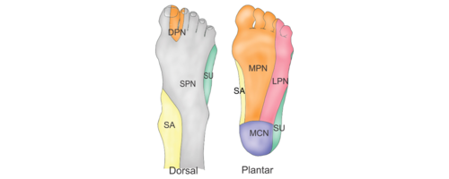
- The first branch of the lateral plantar nerve (Baxter’s nerve)
- Medial calcaneal branch of posterior tibial nerve or nerve to abductor digiti quinti muscle (see image).
- S1 radiculopathy
- Tarsal tunnel syndrome
- Peripheral neuropathy

Plantar heel pain is one of the most common lower limb musculoskeletal conditions that affects both sedentary and physically active people.[3][4] It can have a significant impact on health-related quality of life,[5] including work and activities.[6]
- It accounted for approximately one million physician consultations per year in the United States of America between 1995 and 2000.[7]
- 11-18 % of people continue to report symptoms beyond 1 year following conservative management.[6]
- Plantar heel pain was found to be most common in middle-aged women.[8]
- The reasons for the high incidence in women are not known, however, it has been linked to early menopause. The hormonal changes are believed to attribute to a weakening of the fascia and maybe increased stress.

There are a number of risk factors found to be associated with plantar heel pain, including:[9][2]
- Prolonged standing
- Recent changing of shoe wear
- Stress
- Excessive running or suddenly increasing running distance
- Pes planus
- Limited ankle dorsiflexion and foot strength deficits[10]
- Obesity
Patients often use the internet for education on plantar heel pain, advice and remedies for their symptoms.
- Some patients we see at the clinic may have tried strapping, rolling the foot on the ice bottle, rolling it on golf balls, and doing various stretches.
- Often they would say these treatments have been particularly painful, but they haven't made a difference to the pain.
- This drives patients to look for professional advice.
If we look at the literature, there are various studies showing that physiotherapy can have positive benefits on plantar heel pain.[11] However, when it comes to clinical practice, different measures should be considered to apply research findings properly and see positive results.
- Often, the treatment that works for a woman might differ from the treatment that works for a man.
- The level of activity is an important factor, as well as the patient's history.
- Considering individual factors and using clinical reasoning skills are mandatory when it comes to the treatment of plantar heel pain and other muscuoloskeletal issues.
Nerve Entrapment[edit | edit source]
- Entrapment of the lateral plantar nerve (see image), a branch of the Tibial nerve, represents 15–20% of the chronic plantar heel pain presentations.[12] This can result from compression between the abductor hallucis and quadratus plantae muscles.[13]
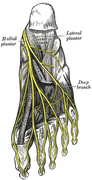
- The close proximity of this nerve to the calcaneal tuberosity suggests the possibility of entrapment, resulting in plantar heel pain.[2]

3. The medial calcaneal nerve (MCN) provides sensory innervation to most of the heel fat pad and to the superficial tissues overlying the inferior part of the calcaneus. MCN entrapment only occurred in 5 out of 200 surgical cases in one study.[14]
4. Tarsal tunnel syndrome (TTS) can also contribute to plantar heel pain (the tunnel lies posterior to the medial malleolus of the ankle, beneath the flexor retinaculum).[15]
Presentation[edit | edit source]
- Pain[16]: usually burning, sharp, shooting, shock-like, electric, and occasionally described as a dull aching. Pain can be local or radiating proximally or distally. Symptoms tend to be worse during or after activities involving weight-bearing, but this pain improves with rest. Progressively, the pain can occur with rest and in non weight-bearing positions. If the symptoms are caused by nerve entrapment, patients might experience night pain as a result of venostasis (slowing of venous outflow) and venous engorgement (local congestion an distension with blood).[2]
- Post-static dyskinesia: pain when a patient first stands after periods of rest. It is most commonly associated with nerve-related plantar heel pain. Patients report severe pain in the morning after rising from the bed. If the pain is due to neural compression it tends to ease with ambulation and movement. However, it tends to worsen if the pain is caused by plantar fasciitis.[2]
- Paraesthesiae and neurological changes such as numbness, sensory changes, pins and needles around the medial and plantar aspect of the heel.[2]
Assessment[edit | edit source]
Subjective Assessment[edit | edit source]
Pain Location. Physiotherapists are skilled at figuring out the source of the pain - i.e. the pain driver.
Pain Behaviour: It is important to understand the nature of the pain to get an idea of the main contributing factor.
- The pain can be neural or mechanical.
- Ask questions to investigate the pain pattern across the day and the aggravating and easing factors.
- Allowing patients to talk about their pain can be very insightful and can help us recognise catastrophising behavior, which is common in chronic pain patients.
- One useful motivational interviewing tip is to give your patients time to reflect on what they said. Try to summarise what they told you to understand the impact on the quality of their life.[17]
Objective Assessment[edit | edit source]
Observe and Assess:
- Foot, lower leg and the entire kinetic chain.
- Posture-related problems such as leaning backward and shifting the weight to the ankles during gait or if the weight is mostly on the toes. The shape of the foot e.g. collapsed arch on one side. You can link the history of trauma from the subjective assessment to the findings on observation. Also, compare the foot shape with the other foot; bilateral collapsed arches will be treated differently from somebody who comes in with steep thick high arched feet. The pes planus (see image) foot is not going to respond to the same treatment strategies as a very rigid pes cavus foot. A study by Im Yi et al.[9] found excessive pronation increased the stress on plantar fascia resulting in collapsed arch. The pronatory effect is believed to increase with age and is related to limited ankle dorsiflexion.
- Orthotics and footwear. The shape of footwear and the level of comfort they provide are an important assessment measure. One study reported that people with chronic plantar heel pain have greater difficulties with footwear comfort, fit and choice.[18] Another found a link between toe flexor weakness and foot-wear difficulties in people with plantar heel pain.[19]
- Neural examination is also recommended. Some patients might describe various "bizarre" pain sensations which might be in different locations and are often disregarded because the main concern is the foot pain. Therefore, neural dynamics should be included in your assessment.[8][2]
- Gait assessment
- See also Biomechanical Assessment of Foot and Ankle
.jpg)
Things to observe during gait:
- Overstriding
- Short strides
- Pounding or heavy throbbing on one side
- Slapping with the foot
- Walking with inverted foot/feet
Clinical Tests:[edit | edit source]

Palpation: over the abductor hallucis and/or on the medial calcaneal tuberosity for reproduced symptoms.
The presence of maximal tenderness over the lateral plantar nerve suggests that it is entrapped.
To diagnose medial calcaneal nerve entrapment, look for the following findings:
- Maximal tenderness over the medial anterior part of the heel fat pad and abductor hallucis
- Distal radiating pain with pressure on the nerve
- Minimal tenderness over the plantar fascia origin
In medial plantar nerve entrapment, the tenderness is typically located over the plantar aspect of the medial arch around the navicular tuberosity.
Dorsiflexion-eversion neurodynamic test: With this test, all metatarsophalangeal joints are passively extended while the ankle is held in dorsiflexion and eversion. This test is aimed mainly at reproducing symptoms in tarsal tunnel syndrome patients. By extending the metatarsophalangeal joints, we increase strain on the medial plantar and tibial nerves. Adding hip flexion with knee extension to ankle dorsiflexion can further increase the strain on the tibial and plantar nerves at the ankle and foot without increasing the tension in the plantar fascia.[20]
Tinel’s test: tapping along the course of a nerve. If tapping produces tingling along the nerve distribution, the test is considered positive.[2]
Differential Diagnosis[edit | edit source]

- Tibial nerve
- Fat pad: fat pad atrophy and limited shock absorption capacity was found in patients older than 40 years. Wearing hard-soled shoes and walking on hard surfaces usually aggravate pain in people with a soft and thin heel fat pad. On palpation, tenderness tends to be localised to the fat pad and does not radiate to the medial calceneal tuberosity and plantar fascia.[2]
- Kinetic chain problems
Case Study[edit | edit source]
In this section, we discuss a case presentation of a patient who had plantar heel pain and the successful management plan that she followed.
Our patient is a 50-year-old professional woman who travels for work from her city to another city on a weekly basis. She wears high heels and regularly runs in her high heels from one meeting to the next. She is a competitive cyclist and previously, she would train at the weekend when she was home.
However, she has recently decided to take up marathon running as well. In order to achieve her training goals, she has add treadmill running during the week to her exercise regime. She continues to cycle long distances at the weekend.

At one conference, she decided to wear flat shoes because she knew she would be walking a lot. Two days later, she started to get pain in her feet. She reported a burning pain in her heels that felt like electric shocks when she put her feet on the floor in the mornings.
She went to see a physiotherapist who did some shockwave therapy which she didn't find helpful. She was then referred to an orthopaedic specialist who took some x-rays and found a heel spur. The specialist felt that this spur explained the patient's pain. She was, therefore, put in a boot during waking hours for six to eight weeks and asked to stop exercising. At night, a splint was advised. In addition, it was recommended that she roll her foot on a PVC pipe to ease the pain.
Eight weeks later, she was still unable to walk. She was frustrated and felt unfit. So she presented for a second opinion to a physiotherapist.[8]
Remember: The spur is not the cause of the symptoms!
The spur is only an indication that there are some traction forces on the plantar fascia. Identifying these traction forces and the reason these forces generated a spur should guide our thinking to help address the symptoms.[8]
Also, remember the plantar fascia is integral to the Achilles tendon complex. By unloading and immobilising the fascia, the tendons are also unloaded and thus, they are no longer able to assist the plantar fascia. Thus, more stress is applied to the plantar fascia, which results in more pain. Typically, the Achilles and tibialis posterior tendons are responsible for unloading the foot when it is in contact with the ground. Unloading these tendons will make these tissues weaker and, consequently, further increase the stress on the plantar fascia.[8]
Addressing the pain[edit | edit source]
In this patient's case, it was essential to try and address her morning pain as this was her main complaint. Starting the day with pain affected the quality of her whole day.
Stretching the Plantar Fascia in a non-weight bearing position is recommended by Lim et al.[16] and is a great way to warm up and ease fascial pain at the start of the day.
Managing load:[16] The patient still has to be mobile and active at work and it was important to help her to return to running in the evenings on the treadmill. However, while treadmills are great in the short term, they can be problematic because they increase the tension and load on the Achilles tendon. By running thirty minutes to an hour on a treadmill, the load on the soft tissue structures is significantly increased. The patient was, therefore, advised to switch to stationary cycling while away for work during the week and to run during the weekends.
Instead of running on the treadmill, the patient was encouraged to do trail running, across undulating, different terrains, which provide more cushioning. It was also advised that she complete shorter runs twice a day. All these strategies were introduced to manage her load over a 24 hour period. In cases of tendon issues, a response to the training load can be seen over 24 hours. So in this case, the patient was asked to monitor her response to the load over 24 hours.[8]
This patient also had rigid feet from wearing high heels for years. Manual mobilisation for stiff feet can be beneficial.[24][25] Manual mobilisation and myofascial release were used to release the foot, and make it more pliable and shock absorbent. Other manual techniques include:
- Calf muscle release
- Mobilising the calcaneus so that it sits neatly under the talus
- Releasing the plantar fascia[26]
- Improving the mobility of the first metatarsal phalangeal joint
Improve the capacity of the plantar fascia: Stretching can be helpful, but it won't be enough to improve the tolerance of the tissue for running. The principals of tendinopathy treatment can be used to manage plantar fasciopathy pain. By combining the windlass mechanism with isometric exercises we can load the plantar fascia effectively and build up capacity gradually.
The patient was asked to do isometric sustained holds to reduce the pain for that first two weeks by extending her toes over a rolled-up towel to induce and load the foot in the windlass mechanism. Then, instead of doing more repetitions, she did 5 isometric holds holding each for 45 seconds with her heels just slightly off the ground then lowering down very slowly. This was followed by a one- to two-minute rest. This exercise was started bilaterally. As she improved, it was performed unilaterally.[30] A 2014 study by Rathleff et al.[31] compared a high load strength-training programme to a standard plantar specific stretching programme in the treatment of plantar fasciopathy. This study reported positive outcomes in terms of pain and function, i.e. quicker reduction in pain and improvements in function.[31] The study participants performed unilateral heel-raises with a towel inserted under the toes to activate the windlass-mechanism (similar to the isometric holds done by our patient in this case study). The exercise was performed every second day for three months. This exercise slowly progressed to include weights (backpack or books) and the number of repetitions was decreased. The results of this study showed improvement on the short term but on the long term, there was no difference between high load strengthening and plantar stretching programmes.
[32] The patient was also encouraged to do Pilates and/or yoga to release and mobilise her overall neural system. Tibial nerve mobilisation can also be prescribed, but working on overall flexibility and mobility is important to counteract stationary postures commonly adopted at work / throughout the day.

Foot-wear was also addressed: Clearly, high heels were contributing to this patient's pain as they put a lot of load and stress on the metatarsal heads. Thus, the patient was advised to wear a pair of wedges (to avoid hyper-extension of the toes and putting the plantar fascia in a stretched position). A rocker-bottom heel or sole can also be utilised. So this patient was able to off-load her without compromising her work dress code.[8] Load can also be managed by wearing appropriate foot orthotics.[16][33]
The patient agreed to keep a diary of her symptoms, modified the load as necessary, and increased the load gradually. Within two weeks, her pain was shifting and she was improving.
Plantar heel pain is not, however, a simple problem. This patient was also going through menopause, so she was advised to check with her doctor if these hormonal changes were contributing to her symptoms.
There are also other methods to desensitise the area and improve the motor control that can be used such as brushing the feet at night after a tough day and mirror therapy.[8]
References[edit | edit source]
- ↑ Riel H, Cotchett M, Delahunt E, Rathleff MS, Vicenzino B, Weir A, Landorf KB. Is 'plantar heel pain' a more appropriate term than 'plantar fasciitis'? Time to move on. Br J Sports Med. 2017;51(22):1576-7.
- ↑ 2.0 2.1 2.2 2.3 2.4 2.5 2.6 2.7 2.8 Alshami AM, Souvlis T, Coppieters MW. A review of plantar heel pain of neural origin: differential diagnosis and management. Manual therapy. 2008 Apr 1;13(2):103-11.
- ↑ Thomas MJ, Whittle R, Menz HB, Rathod-Mistry T, Marshall M, Roddy E. Plantar heel pain in middle-aged and older adults: population prevalence, associations with health status and lifestyle factors, and frequency of healthcare use. BMC Musculoskelet Disord. 2019;20(1):337.
- ↑ Harvey HD, Game C, Walsh TP, Wearing SC, Platt SR. Are models of plantar heel pain suitable for competitive runners? A narrative review. J Orthop. 2022;33:9-14.
- ↑ Cotchett M, Rathleff MS, Dilnot M, Landorf KB, Morrissey D, Barton C. Lived experience and attitudes of people with plantar heel pain: a qualitative exploration. J Foot Ankle Res. 2020;13(1):12.
- ↑ 6.0 6.1 Sullivan J, Pappas E, Adams R, Crosbie J, Burns J. Determinants of footwear difficulties in people with plantar heel pain. Journal of Foot and Ankle Research. 2015 Dec;8(1):1-7.
- ↑ Riddle DL, Schappert SM. Volume of ambulatory care visits and patterns of care for patients diagnosed with plantar fasciitis: a national study of medical doctors. Foot & ankle international. 2004 May;25(5):303-10.
- ↑ 8.0 8.1 8.2 8.3 8.4 8.5 8.6 8.7 Simpson H. Plantar Heel Pain Course. Plus2020
- ↑ 9.0 9.1 Im Yi T, Lee GE, Seo IS, Huh WS, Yoon TH, Kim BR. Clinical characteristics of the causes of plantar heel pain. Annals of rehabilitation medicine. 2011 Aug;35(4):507.
- ↑ Sullivan J, Pappas E, Burns J. Role of mechanical factors in the clinical presentation of plantar heel pain: Implications for management. Foot (Edinb). 2020;42:101636.
- ↑ Grim C, Kramer R, Engelhardt M, John SM, Hotfiel T, Hoppe MW. Effectiveness of Manual Therapy, Customised Foot Orthoses and Combined Therapy in the Management of Plantar Fasciitis—A RCT. Sports. 2019 Jun;7(6):128.
- ↑ Watson TS, Anderson RB, Davis WH, Kiebzak GM. Distal tarsal tunnel release with partial plantar fasciotomy for chronic heel pain: an outcome analysis. Foot & ankle international. 2002 Jun;23(6):530-7.
- ↑ May TJ, Judy TA, Conti M, Cowan JE. Current treatment of plantar fasciitis. Current sports medicine reports. 2002 Oct;1(5):278-84.
- ↑ Schon LC, Glennon TP, Baxter DE. Heel pain syndrome: electrodiagnostic support for nerve entrapment. Foot & ankle. 1993 Mar;14(3):129-35.
- ↑ Lau JT, Stavrou P. Posterior tibial nerve--primary. Foot and ankle clinics. 2004 Jun;9(2):271-85.
- ↑ 16.0 16.1 16.2 16.3 Lim AT, How CH, Tan B. Management of plantar fasciitis in the outpatient setting. Singapore medical journal. 2016 Apr;57(4):168.
- ↑ Roscher M. Motivational Interviewing course. Plus2019
- ↑ Irving DB, Cook JL, Young MA, Menz HB. Impact of chronic plantar heel pain on health-related quality of life. Journal of the American Podiatric Medical Association. 2008 Jul 1;98(4):283-9.
- ↑ Sullivan J, Pappas E, Adams R, Crosbie J, Burns J. Determinants of footwear difficulties in people with plantar heel pain. Journal of Foot and Ankle Research. 2015 Dec;8(1):1-7.
- ↑ Coppieters MW, Alshami AM, Babri AS, Souvlis T, Kippers V, Hodges PW. Strain and excursion of the sciatic, tibial, and plantar nerves during a modified straight leg raising test. Journal of Orthopaedic Research. 2006 Sep;24(9):1883-9.
- ↑ Dorsiflexion Eversion Test . Available from:https://www.youtube.com/watch?v=Z2LsxI8JWBE[last accessed 30/06/2020]
- ↑ Tinel's test (sign) for Tarsal Tunnel Syndrome . Available from:https://www.youtube.com/watch?v=xWi1tX7yrOA[last accessed 30/06/2020]
- ↑ Bay Podiatry. Plantar Fascia Stretch for Plantar Fasciitis (aka the DiGiovanni stretch). Available from: https://www.youtube.com/watch?v=pKx7swh47Uc [last accessed 6/9/2022]
- ↑ Mischke JJ, Jayaseelan DJ, Sault JD, Emerson Kavchak AJ. The symptomatic and functional effects of manual physical therapy on plantar heel pain: a systematic review. Journal of Manual & Manipulative Therapy. 2017 Jan 1;25(1):3-10.
- ↑ Pollack Y, Shashua A, Kalichman L. Manual therapy for plantar heel pain. The Foot. 2018 Mar 1;34:11-6.
- ↑ Ajimsha MS, Binsu D, Chithra S. Effectiveness of myofascial release in the management of plantar heel pain: a randomized controlled trial. The Foot. 2014 Jun 1;24(2):66-71.
- ↑ Sports Massage - Plantar Fasciitis. Available from:https://www.youtube.com/watch?v=9SIGxc4IFGc[last accessed 30/06/2020]
- ↑ Subtalar Joint Mobilisation Techniques. Available from:https://www.youtube.com/watch?v=JzOZl2YbzeA[last accessed 30/06/2020]
- ↑ Big Toe 1st MTP joint Mobilisation. Available from:https://www.youtube.com/watch?v=YeS9NmhWfvw[last accessed 30/06/2020]
- ↑ Rathleff MS, Thorborg K. ‘Load me up, Scotty’: mechanotherapy for plantar fasciopathy (formerly known as plantar fasciitis).
- ↑ 31.0 31.1 Rathleff MS, Mølgaard CM, Fredberg U, et al. High-load strength training improves outcome in patients with plantar fasciitis: A randomized controlled trial with 12-month follow-up. Scand J Med Sci Spor 2014:n/a-n/a doi: 10.1111/sms.12313[published Online First: Epub Date]|.
- ↑ High Load Strength Training Plantar Fasciitis | Chris Johnson PT. Available from:https://www.youtube.com/watch?v=wUua8tI3m5s[last accessed 30/06/2020]
- ↑ Whittaker GA, Munteanu SE, Menz HB, Tan JM, Rabusin CL, Landorf KB. Foot orthoses for plantar heel pain: a systematic review and meta-analysis. British journal of sports medicine. 2018 Mar 1;52(5):322-8.

