Pelvis: Difference between revisions
Kim Jackson (talk | contribs) (Updated page links) |
Leana Louw (talk | contribs) No edit summary |
||
| Line 1: | Line 1: | ||
<div class="editorbox"> '''Original Editor '''- [[User:User Name|User Name]] '''Top Contributors''' - {{Special:Contributors/{{FULLPAGENAME}}}}</div> | <div class="editorbox"> '''Original Editor '''- [[User:User Name|User Name]] '''Top Contributors''' - {{Special:Contributors/{{FULLPAGENAME}}}}</div> | ||
== Description == | |||
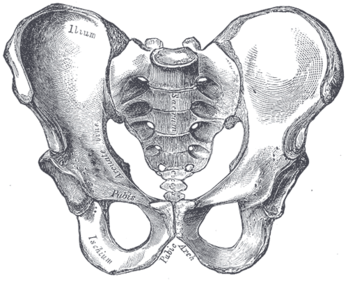
[[File:Gray241.png|alt=Bones of the pelvis|thumb|348x348px]]The pelvis consists of the sacrum, the coccyx,the ischium, the ilium, and the pubis. <ref name=":1">White | [[File:Gray241.png|alt=Bones of the pelvis|thumb|348x348px]]The pelvis consists of the sacrum, the coccyx, the ischium, the ilium, and the pubis.<ref name=":1">White TD, Black MT, Folkens PA. [https://books.google.co.za/books?hl=en&lr=&id=oCSG2mYlD90C&oi=fnd&pg=PP1&dq=+.+Human+osteology.+&ots=VnpEcJPCWD&sig=Ful9Jtenlzzw4M4nniLS00LstnM#v=onepage&q=.%20Human%20osteology.&f=false Human osteology.] Academic press; 2011.</ref><ref name=":0">Lewis CL, Laudicina NM, Khuu A, Loverro KL. [https://anatomypubs.onlinelibrary.wiley.com/doi/pdf/10.1002/ar.23552 The human pelvis: Variation in structure and function during gait.] The Anatomical Record 2017;300(4):633-42.</ref> The structure of the pelvis supports the contents of the abdomen while also helping to transfer the weight from the spine to the lower limbs.<ref name=":2">Magee DJ. [https://books.google.co.za/books?hl=en&lr=&id=cxu0BQAAQBAJ&oi=fnd&pg=PP1&dq=Orthopedic+physical+assessment.&ots=mqGLvPFASn&sig=Jf3KLqp1lfiYQYoeVU2fMhEhhvs#v=onepage&q=Orthopedic%20physical%20assessment.&f=false Orthopedic physical assessment.] Elsevier Health Sciences; 2013.</ref> During [[gait]], the joints within the pelvis work together to decrease the amount of force transferred from the ground and lower extremities to the spine and upper extremities.<ref name=":2" /> | ||
{{#ev:youtube|3v5AsAESg1Q|300}}<ref>Anatomy Zone. Bones of the Pelvis. Available from: https://youtu.be/3v5AsAESg1Q [last accessed 27/01/2020]</ref> | {{#ev:youtube|3v5AsAESg1Q|300}}<ref>Anatomy Zone. Bones of the Pelvis. Available from: https://youtu.be/3v5AsAESg1Q [last accessed 27/01/2020]</ref> | ||
== Osteology | == Osteology == | ||
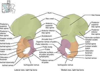
[[File:Innominate bone.jpg|thumb|349x349px]] | [[File:Innominate bone.jpg|thumb|349x349px]] | ||
* | * Sacrum | ||
* | * Coccyx | ||
* | * Two innominate bones, which consist of the: | ||
** | ** Ischium | ||
** | ** Ilium | ||
** | ** Pubis<ref name=":1" /> | ||
=== Joint Articulations | === Joint Articulations === | ||
There are three articulations within the pelvis: | There are three articulations within the pelvis: | ||
* | * Inferiorly between the sacrum and the coccyx | ||
* | * Posteriorly between the sacrum and each ilium ([[Sacroiliac joint|sacroiliac (SI) joint]]) | ||
* | * Anteriorly between the pubic bodies (pubic symphysis).<ref name=":0" /> | ||
'''Other articulations:''' | '''Other articulations:''' | ||
The pelvis and femur articulate via the acetabulum<ref name=":1" /> | The pelvis and [[femur]] articulate via the acetabulum<ref name=":1" /> | ||
=== Ligaments | === Ligaments === | ||
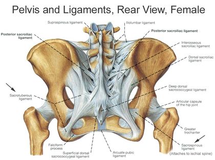
[[File:Bony-pelvis-2.jpg|thumb|420x420px]] | [[File:Bony-pelvis-2.jpg|thumb|420x420px]] | ||
| Line 46: | Line 46: | ||
* Posterior pubic ligament | * Posterior pubic ligament | ||
=== Muscles | === Muscles === | ||
There are 36 muscles that attach to the sacrum or innominates. The purpose of these muscles is primarily to provide stability to the joint not to produce movement.<ref>Calvillo O | There are 36 muscles that attach to the sacrum or innominates. The purpose of these muscles is primarily to provide stability to the joint not to produce movement.<ref>Calvillo O, Skaribas I, Turnipseed J. [https://link.springer.com/article/10.1007/s11916-000-0019-1 Anatomy and pathophysiology of the sacroiliac joint.] Current review of pain 2000;4(5):356-61.</ref> | ||
Muscles that attach to the sacrum or innominates are: | [[Muscle|Muscles]] that attach to the sacrum or innominates are: | ||
{| class="wikitable" style="text-align: center;" | {| class="wikitable" style="text-align: center;" | ||
|Adductor brevis | |[[Adductor Brevis|Adductor brevis]] | ||
|Adductor longus | |Adductor longus | ||
|[[Adductor Magnus|Adductor magnus]] | |[[Adductor Magnus|Adductor magnus]] | ||
| Line 63: | Line 63: | ||
|[[Gluteus Minimus|Gluteus minimus]] | |[[Gluteus Minimus|Gluteus minimus]] | ||
|- | |- | ||
|Gracilis | |[[Gracilis]] | ||
|[[Iliacus]] | |[[Iliacus]] | ||
|Inferior gemellus | |[[Gemellus Inferior|Inferior gemellus]] | ||
|[[Internal Abdominal Oblique|Internal oblique]] | |[[Internal Abdominal Oblique|Internal oblique]] | ||
|[[Latissimus Dorsi Muscle|Latissimus dorsi]] | |[[Latissimus Dorsi Muscle|Latissimus dorsi]] | ||
| Line 71: | Line 71: | ||
|[[Pelvic Floor Anatomy|Levator ani]] | |[[Pelvic Floor Anatomy|Levator ani]] | ||
|Multifidus | |Multifidus | ||
|Obturator internus | |[[Obturator Internus|Obturator internus]] | ||
|Obturator externus | |[[Obturator Externus|Obturator externus]] | ||
|Pectineus | |[[Pectinus Muscle|Pectineus]] | ||
|- | |- | ||
|[[Pelvic Floor Anatomy|Levator ani]] | |[[Pelvic Floor Anatomy|Levator ani]] | ||
|[[Piriformis]] | |[[Piriformis]] | ||
|[[Psoas Minor|Psoas minor]] | |[[Psoas Minor|Psoas minor]] | ||
|Pyramidalis | |[[Pyramidalis muscle|Pyramidalis]] | ||
|Quadratus femoris | |[[Quadratus Femoris|Quadratus femoris]] | ||
|- | |- | ||
|[[Quadratus Lumborum|Quadratus lumborum]] | |[[Quadratus Lumborum|Quadratus lumborum]] | ||
|[[Rectus Abdominis|Rectus abdominis]] | |[[Rectus Abdominis|Rectus abdominis]] | ||
|Rectus femoris | |[[Rectus Femoris|Rectus femoris]] | ||
|Sartorius | |[[Sartorius]] | ||
|Semimembranosus | |[[Semimembranosus]] | ||
|- | |- | ||
|Semitendonosus | |[[Semitendinosus|Semitendonosus]] | ||
|[[Pelvic Floor Anatomy|Sphincter urethrae]] | |[[Pelvic Floor Anatomy|Sphincter urethrae]] | ||
|[[Pelvic Floor Anatomy|Superficial transverse perineal ischiocavernous]] | |[[Pelvic Floor Anatomy|Superficial transverse perineal ischiocavernous]] | ||
| | |[[Gemellus Superior|Superior gemellus]] | ||
|[[Tensor Fascia Lata|Tensor fascia lata]] | |[[Tensor Fascia Lata|Tensor fascia lata]] | ||
|- | |- | ||
| Line 100: | Line 100: | ||
|} | |} | ||
== Sex-specific differences | == Sex-specific differences == | ||
The female pelvis consists of a wider sacrum and a wider subpubic angle when compared to males. The female pelvis’ ischial spines are also less prominent than the male’s ischial spines.<ref name=":3">Kurki HK. Pelvic dimorphism in relation to body size and body size dimorphism in humans. Journal of Human Evolution | The female pelvis consists of a wider sacrum and a wider subpubic angle when compared to males. The female pelvis’ ischial spines are also less prominent than the male’s ischial spines.<ref name=":3">Kurki HK. [https://www.sciencedirect.com/science/article/abs/pii/S0047248411001679 Pelvic dimorphism in relation to body size and body size dimorphism in humans.] Journal of Human Evolution 2011;61(6):631-43.</ref><ref name=":4">Meindl RS, Lovejoy CO, Mensforth RP, Carlos LD. [https://s3.amazonaws.com/academia.edu.documents/48786250/Accuracy_and_Direction_of_Error_in_the_S20160912-8250-ox8gbo.pdf?response-content-disposition=inline%3B%20filename%3DAccuracy_and_direction_of_error_in_the_s.pdf&X-Amz-Algorithm=AWS4-HMAC-SHA256&X-A Accuracy and direction of error in the sexing of the skeleton: implications for paleodemography.] American Journal of Physical Anthropology 1985;68(1):79-85.</ref><ref name=":5">Tague RG. [https://onlinelibrary.wiley.com/doi/abs/10.1002/ajpa.1330880102 Sexual dimorphism in the human bony pelvis, with a consideration of the Neandertal pelvis from Kebara Cave, Israel.] American Journal of Physical Anthropology 1992;88(1):1-21.</ref> The male pelvis’ sacrum is generally longer and more curved with a narrower sub-pubic arch.<ref name=":5" /> In females a wider pelvic aperture is needed as it functions as the birth canal during labour.<ref name=":3" /><ref name=":4" /><ref>Rosenberg KR. [https://onlinelibrary.wiley.com/doi/pdf/10.1002/ajpa.1330350605 The evolution of modern human childbirth.] American Journal of Physical Anthropology.1992;35(S15):89-124.</ref><ref>Lovejoy CO. [https://www.researchgate.net/profile/Owen_Lovejoy/publication/51367382_The_natural_history_of_human_gait_and_posture_Part_2_Hip_and_thigh/links/5cd6ee9592851c4eab969302/The-natural-history-of-human-gait-and-posture-Part-2-Hip-and-thigh.pdf The natural history of human gait and posture: part 2. Hip and thigh.] Gait & posture 2005;21(1):113-24.</ref><ref>Abitbol MM. [https://europepmc.org/article/med/8728076 The shapes of the female pelvis. Contributing factors.] The Journal of reproductive medicine 1996;41(4):242-50.</ref> | ||
== Clinical Examination | == Clinical Examination == | ||
=== Assessment | === Assessment === | ||
Prior to the assessment of the sacroiliac joint both the [[Lumbar Examination|lumbar spine]] and [[Hip Anatomy|hip]] should be assessed and any underlying pathologist should be ruled out. | |||
=== Special Tests | === Special Tests === | ||
==== SI Joint stress tests ==== | ==== SI Joint stress tests ==== | ||
* Anterior | * Anterior gapping test | ||
* [[Sacroiliac Distraction Test|Sacroiliac | * [[Sacroiliac Distraction Test|Sacroiliac distraction test]] | ||
* Sacrotuberous | * Sacrotuberous ligament stress test | ||
* Sacral | * Sacral compression test | ||
* Rotational | * Rotational stress test | ||
==== [[Leg Length Discrepancy|Leg Length tests]] ==== | ==== [[Leg Length Discrepancy|Leg Length tests]] ==== | ||
| Line 125: | Line 125: | ||
* Seated Flexion test (Piedallu's Sign) | * Seated Flexion test (Piedallu's Sign) | ||
* [[Supine Long Sitting Test|Supine long sitting test]] | * [[Supine Long Sitting Test|Supine long sitting test]] | ||
* [[Sign of the Buttock]] | * [[Sign of the Buttock|Sign of the buttock]] | ||
* [[Posterior pelvic pain provocation test (aka Thigh Thrust aka Posterior Shear)|Posterior | * [[Posterior pelvic pain provocation test (aka Thigh Thrust aka Posterior Shear)|Posterior pelvic pain provocation test]] | ||
* [[Gaenslen Test|Gaenslen test]] | * [[Gaenslen Test|Gaenslen test]] | ||
* Yeoman's test | * Yeoman's test | ||
* [[FABER Test|FABER (Figure-Four) test]] | * [[FABER Test|FABER (Figure-Four) test]] | ||
* Fortin Finger Test | * Fortin Finger Test | ||
* [[Straight Leg Raise Test|Straight | * [[Straight Leg Raise Test|Straight leg raise]] | ||
* [[Stork Test|Gillet's test (Stork test)]] | * [[Stork Test|Gillet's test (Stork test)]] | ||
| Line 139: | Line 139: | ||
== Pathology/Injury == | == Pathology/Injury == | ||
*[[Pubic Symphysis Dysfunction|Pubic | *[[Pubic Symphysis Dysfunction|Pubic symphysis dysfunction]] | ||
*[[Spondyloarthritis]] | *[[Spondyloarthritis]] | ||
*[[Pregnancy Related Pelvic Pain|Pregnancy | *[[Pregnancy Related Pelvic Pain|Pregnancy related pelvic pain]] | ||
*[[Pelvic Fractures|Pelvic | *[[Pelvic Fractures|Pelvic fractures]] | ||
*[[Sacroiliitis]] | *[[Sacroiliitis]] | ||
Revision as of 15:57, 31 May 2020
Description[edit | edit source]
The pelvis consists of the sacrum, the coccyx, the ischium, the ilium, and the pubis.[1][2] The structure of the pelvis supports the contents of the abdomen while also helping to transfer the weight from the spine to the lower limbs.[3] During gait, the joints within the pelvis work together to decrease the amount of force transferred from the ground and lower extremities to the spine and upper extremities.[3]
Osteology[edit | edit source]
- Sacrum
- Coccyx
- Two innominate bones, which consist of the:
- Ischium
- Ilium
- Pubis[1]
Joint Articulations[edit | edit source]
There are three articulations within the pelvis:
- Inferiorly between the sacrum and the coccyx
- Posteriorly between the sacrum and each ilium (sacroiliac (SI) joint)
- Anteriorly between the pubic bodies (pubic symphysis).[2]
Other articulations:
The pelvis and femur articulate via the acetabulum[1]
Ligaments[edit | edit source]
Ligaments of the Pelvis[edit | edit source]
- Iliolumbar ligament
- Lateral lumbosacral ligament
- Sacrotuberous ligament
- Sacrospinous ligament
Sacroiliac Ligaments[edit | edit source]
- Ventral/Anterior sacroiliac ligament
- Dorsal/Posterior sacroiliac ligament
- Interosseous sacroiliac ligament
Sacrococcygeal Ligaments[edit | edit source]
- Ventral/Anterio sacrococcygeal ligament
- Dorsal sacrococcygeal ligament
- Lateral sacrococcygeal ligament
Pubic Symphysis Ligaments[edit | edit source]
- Superior pubic ligament
- Inferior pubic ligament
- Anterior pubic ligament
- Posterior pubic ligament
Muscles[edit | edit source]
There are 36 muscles that attach to the sacrum or innominates. The purpose of these muscles is primarily to provide stability to the joint not to produce movement.[5]
Muscles that attach to the sacrum or innominates are:
Sex-specific differences[edit | edit source]
The female pelvis consists of a wider sacrum and a wider subpubic angle when compared to males. The female pelvis’ ischial spines are also less prominent than the male’s ischial spines.[6][7][8] The male pelvis’ sacrum is generally longer and more curved with a narrower sub-pubic arch.[8] In females a wider pelvic aperture is needed as it functions as the birth canal during labour.[6][7][9][10][11]
Clinical Examination[edit | edit source]
Assessment[edit | edit source]
Prior to the assessment of the sacroiliac joint both the lumbar spine and hip should be assessed and any underlying pathologist should be ruled out.
Special Tests[edit | edit source]
SI Joint stress tests[edit | edit source]
- Anterior gapping test
- Sacroiliac distraction test
- Sacrotuberous ligament stress test
- Sacral compression test
- Rotational stress test
Leg Length tests[edit | edit source]
- Prone test
- Standing leg length test
- Functional leg length test
Other Special Tests[edit | edit source]
- Seated Flexion test (Piedallu's Sign)
- Supine long sitting test
- Sign of the buttock
- Posterior pelvic pain provocation test
- Gaenslen test
- Yeoman's test
- FABER (Figure-Four) test
- Fortin Finger Test
- Straight leg raise
- Gillet's test (Stork test)
Outcome Measures[edit | edit source]
Pathology/Injury[edit | edit source]
- Pubic symphysis dysfunction
- Spondyloarthritis
- Pregnancy related pelvic pain
- Pelvic fractures
- Sacroiliitis
Resources:[edit | edit source]
- ↑ 1.0 1.1 1.2 White TD, Black MT, Folkens PA. Human osteology. Academic press; 2011.
- ↑ 2.0 2.1 Lewis CL, Laudicina NM, Khuu A, Loverro KL. The human pelvis: Variation in structure and function during gait. The Anatomical Record 2017;300(4):633-42.
- ↑ 3.0 3.1 Magee DJ. Orthopedic physical assessment. Elsevier Health Sciences; 2013.
- ↑ Anatomy Zone. Bones of the Pelvis. Available from: https://youtu.be/3v5AsAESg1Q [last accessed 27/01/2020]
- ↑ Calvillo O, Skaribas I, Turnipseed J. Anatomy and pathophysiology of the sacroiliac joint. Current review of pain 2000;4(5):356-61.
- ↑ 6.0 6.1 Kurki HK. Pelvic dimorphism in relation to body size and body size dimorphism in humans. Journal of Human Evolution 2011;61(6):631-43.
- ↑ 7.0 7.1 Meindl RS, Lovejoy CO, Mensforth RP, Carlos LD. Accuracy and direction of error in the sexing of the skeleton: implications for paleodemography. American Journal of Physical Anthropology 1985;68(1):79-85.
- ↑ 8.0 8.1 Tague RG. Sexual dimorphism in the human bony pelvis, with a consideration of the Neandertal pelvis from Kebara Cave, Israel. American Journal of Physical Anthropology 1992;88(1):1-21.
- ↑ Rosenberg KR. The evolution of modern human childbirth. American Journal of Physical Anthropology.1992;35(S15):89-124.
- ↑ Lovejoy CO. The natural history of human gait and posture: part 2. Hip and thigh. Gait & posture 2005;21(1):113-24.
- ↑ Abitbol MM. The shapes of the female pelvis. Contributing factors. The Journal of reproductive medicine 1996;41(4):242-50.


